Podcast
Questions and Answers
What is the correct sequence of zones in intracartilagenous ossification?
What is the correct sequence of zones in intracartilagenous ossification?
- Proliferation, resting cartilage, hypertrophy, and ossification
- Hypertrophy, proliferation, resting cartilage, and ossification
- Resting cartilage, proliferation, hypertrophy, calcification, and ossification (correct)
- Hypertrophy, proliferation, calcification, resting cartilage, and ossification
What is the composition of a triad in muscle cells?
What is the composition of a triad in muscle cells?
- One T-tubule and two terminal cisternae (correct)
- Composed of actin, myosin, and tropomyosin
- Two T-tubules and one terminal cisternum
- Repeated at every Z line
Which type of cell is known as a 'big eater' due to its phagocytic activity?
Which type of cell is known as a 'big eater' due to its phagocytic activity?
- Fibroblasts
- Plasma cells
- Lymphocytes
- Fixed macrophages (correct)
During allergic reactions, which cells are responsible for secreting histamine?
During allergic reactions, which cells are responsible for secreting histamine?
Which connective tissue cells are primarily found along the long axis of blood capillaries?
Which connective tissue cells are primarily found along the long axis of blood capillaries?
Which type of connective tissue is predominantly present during embryonic life?
Which type of connective tissue is predominantly present during embryonic life?
Which of the following is the primary cell type responsible for synthesizing the extracellular matrix in connective tissue?
Which of the following is the primary cell type responsible for synthesizing the extracellular matrix in connective tissue?
Which cell type is characterized by a large, eccentric nucleus with a 'clock-face' appearance of its chromatin?
Which cell type is characterized by a large, eccentric nucleus with a 'clock-face' appearance of its chromatin?
What is the function of the sarcoplasmic reticulum in skeletal muscle fibers?
What is the function of the sarcoplasmic reticulum in skeletal muscle fibers?
All of the following statements are false regarding hyaline cartilage, EXCEPT:
All of the following statements are false regarding hyaline cartilage, EXCEPT:
What histological feature primarily characterizes skeletal muscle fibers?
What histological feature primarily characterizes skeletal muscle fibers?
Which of the following is NOT a function of the perichondrium?
Which of the following is NOT a function of the perichondrium?
Which connective tissue layer directly surrounds individual muscle fibers?
Which connective tissue layer directly surrounds individual muscle fibers?
Which of the following characteristics is FALSE regarding fibrocartilage?
Which of the following characteristics is FALSE regarding fibrocartilage?
Tendons, which connect muscles to bones, are primarily composed of which type of connective tissue?
Tendons, which connect muscles to bones, are primarily composed of which type of connective tissue?
Which of the following statements is TRUE regarding the periosteum?
Which of the following statements is TRUE regarding the periosteum?
Appositional growth in cartilage is accomplished by which of the following cells?
Appositional growth in cartilage is accomplished by which of the following cells?
What does the formation of cell nests characterize in cartilage?
What does the formation of cell nests characterize in cartilage?
All of the following statements are true regarding osteoblasts, EXCEPT:
All of the following statements are true regarding osteoblasts, EXCEPT:
Which of the following statements is TRUE regarding osteocytes?
Which of the following statements is TRUE regarding osteocytes?
Which of the following statements is TRUE regarding the haversian system?
Which of the following statements is TRUE regarding the haversian system?
Flashcards
Haversian system
Haversian system
The structural organization of bone featuring concentric layers.
Intracartilaginous ossification order
Intracartilaginous ossification order
The sequence: resting cartilage, proliferation, hypertrophy, calcification, ossification.
Sarcomere
Sarcomere
The segment of myofibril between two Z lines; basic unit of muscle contraction.
Triad in muscle fibers
Triad in muscle fibers
Signup and view all the flashcards
Big eaters (cells)
Big eaters (cells)
Signup and view all the flashcards
Cells secreting histamine
Cells secreting histamine
Signup and view all the flashcards
Connective tissue in embryonic life
Connective tissue in embryonic life
Signup and view all the flashcards
Collagen fibers properties
Collagen fibers properties
Signup and view all the flashcards
Hyaline cartilage
Hyaline cartilage
Signup and view all the flashcards
Perichondrium
Perichondrium
Signup and view all the flashcards
Fibrocartilage
Fibrocartilage
Signup and view all the flashcards
Osteoblasts
Osteoblasts
Signup and view all the flashcards
Osteocytes
Osteocytes
Signup and view all the flashcards
Osteoclasts
Osteoclasts
Signup and view all the flashcards
Cancellous bone
Cancellous bone
Signup and view all the flashcards
Principal connective tissue cells
Principal connective tissue cells
Signup and view all the flashcards
Signet-ring appearance
Signet-ring appearance
Signup and view all the flashcards
Nucleus of a plasma cell
Nucleus of a plasma cell
Signup and view all the flashcards
Skeletal muscle fibers
Skeletal muscle fibers
Signup and view all the flashcards
Dark band in skeletal muscle
Dark band in skeletal muscle
Signup and view all the flashcards
Connective tissue covering muscle bundle
Connective tissue covering muscle bundle
Signup and view all the flashcards
Composition of tendon
Composition of tendon
Signup and view all the flashcards
Formation of cell nests
Formation of cell nests
Signup and view all the flashcards
Study Notes
Histology Midterm Study Notes
-
Principal Connective Tissue Cells: The principal connective tissue cells are fibroblasts, reticular cells, and macrophages.
-
Signet-Ring Appearance: Plasma cells have a signet-ring appearance.
-
Plasma Cell Nucleus: Plasma cell nuclei are segmented into many lobes, not kidney-shaped or "horse shoe-shaped".
-
Skeletal Muscle Fiber Characteristics: Skeletal muscle fibers are cylindrical, have acidophilic cytoplasm, are striated, and have a single nucleus. Skeletal muscle fibers are surrounded by basal lamina. Sarcoplasmic reticulum is rough endoplasmic reticulum.
-
Dark Band in Skeletal Muscle: The dark band in skeletal muscle is the A band.
-
Muscle Bundle Covering: The connective tissue covering a muscle bundle is the perimysium.
-
Tendon Composition: Tendons are composed mainly of dense, regularly arranged collagen fibers.
-
Chondroblasts: Chondroblasts are mesenchymal cells responsible for appositional growth of cartilage.
-
Cell Nest Formation: The formation of cell nests in chondrocytes.
-
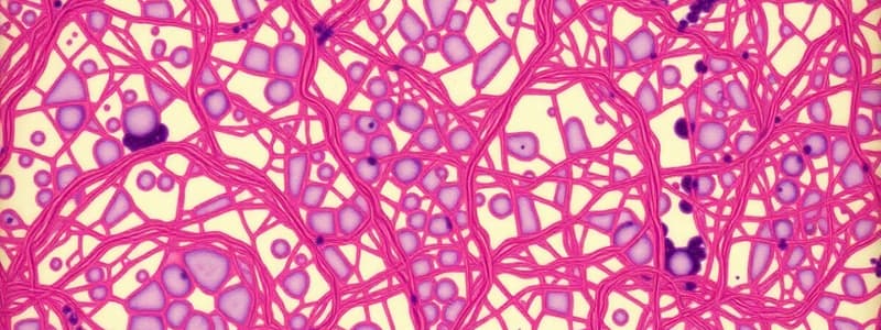
Hyaline Cartilage Histological Structure: Hyaline cartilage consists of irregularly arranged collagen fibers.
-
Hyaline Cartilage Characteristics: Hyaline cartilage is surrounded by perichondrium (except at articular cartilage), it is opaque and appears yellowish-white in color, does not contain collagen type I in its matrix and it is not calcified.
-
Perichondrium Characteristics: The perichondrium is composed of an outer fibrous layer and inner chondriogenic layer. The perichondrium helps with nutrient delivery and attachment to muscles and ligaments. It does not help with interstitial growth.
-
Fibrocartilage Characteristics: Fibrocartilage lacks perichondrium, contains type I collagen, is white and opaque; and chondrocytes are arranged in rows.
-
Periosteum Characteristics: The periosteum contains osteogenic cells, and is vascular.
-
Osteoblast Characteristics: Osteoblasts are bone-building cells derived from osteogenic cells, and have a deep, basophilic cytoplasm.
-
Osteocytes/Osteoclasts: Osteocytes form cell nests inside lacunae (not osteoblasts), and the cell processes of adjacent cells are connected by gap junctions ; Osteocytes are not dividing cells.
-
Cancellous Bone: Cancellous bone has a spongy appearance, is made of irregularly arranged bone trabeculae, contains active red bone marrow, and has bone marrow spaces. It does not have a Haversian system.
-
Sarcomere Definition: A sarcomere is the region of a myofibril lying between two successive Z lines.
-
Triad Structure: A triad is formed of one T-tubule and two terminal cisternae.
Studying That Suits You
Use AI to generate personalized quizzes and flashcards to suit your learning preferences.


