Podcast
Questions and Answers
Why is the preparation process often not feasible?
Why is the preparation process often not feasible?
- Because it can cause slight structural distortions and the removal of cellular lipid. (correct)
- Because it always results in the complete loss of cellular structure.
- Because the process only works on certain cell types.
- Because it requires extensive training to perform.
What is the relationship between tissues and organs?
What is the relationship between tissues and organs?
- Tissues and organs are interchangeable in the body.
- Tissues and organs are developed independently of each other.
- Tissues are formed by a combination of different types of organs.
- Organs are formed by an orderly combination of different types of tissues. (correct)
What role do advances in other scientific fields play in the study of histology?
What role do advances in other scientific fields play in the study of histology?
- They are essential for a deeper understanding of histology. (correct)
- They are primarily used to make microscopic tools.
- They are only useful for identifying new types of cells.
- They are unnecessary for studying histology.
What is the main reason for using fixation in tissue preparation?
What is the main reason for using fixation in tissue preparation?
Which of the followings best describes why histology depends on microscopes?
Which of the followings best describes why histology depends on microscopes?
What is the primary purpose of fixation in tissue preparation?
What is the primary purpose of fixation in tissue preparation?
In the process of tissue dehydration, what is the final solution used?
In the process of tissue dehydration, what is the final solution used?
What is the purpose of 'clearing' in histological tissue processing?
What is the purpose of 'clearing' in histological tissue processing?
What is the temperature range typically used in the process of tissue infiltration with paraffin?
What is the temperature range typically used in the process of tissue infiltration with paraffin?
What instrument is used to cut paraffin-embedded tissue into thin sections for light microscopy?
What instrument is used to cut paraffin-embedded tissue into thin sections for light microscopy?
What material is used to embed tissue for Transmission Electron Microscopy (TEM)?
What material is used to embed tissue for Transmission Electron Microscopy (TEM)?
In tissue sectioning using a microtome, what is the typical range of thickness advanced by each turn of the drive wheel?
In tissue sectioning using a microtome, what is the typical range of thickness advanced by each turn of the drive wheel?
After dehydration, what type of solvents are used?
After dehydration, what type of solvents are used?
What is the purpose of the 'clearing' step in tissue processing?
What is the purpose of the 'clearing' step in tissue processing?
Which embedding medium requires lower temperatures, thus avoiding tissue distortion?
Which embedding medium requires lower temperatures, thus avoiding tissue distortion?
What is the primary function of a microtome in tissue preparation?
What is the primary function of a microtome in tissue preparation?
What type of tissue components are primarily stained by basic dyes?
What type of tissue components are primarily stained by basic dyes?
Which of the following best describes the staining characteristics of hematoxylin?
Which of the following best describes the staining characteristics of hematoxylin?
Which cellular structures or tissues are stained pink by eosin in H&E staining?
Which cellular structures or tissues are stained pink by eosin in H&E staining?
What is the primary purpose of embedding tissues before sectioning?
What is the primary purpose of embedding tissues before sectioning?
Which of the following describes the process of dehydrating a fixed tissue?
Which of the following describes the process of dehydrating a fixed tissue?
What is stained purple or dark blue by hematoxylin in H&E staining?
What is stained purple or dark blue by hematoxylin in H&E staining?
What is the typical thickness of paraffin sections for light microscopy?
What is the typical thickness of paraffin sections for light microscopy?
Why are tissue sections typically stained after being prepared?
Why are tissue sections typically stained after being prepared?
What type of chemical interaction is primarily responsible for staining tissues?
What type of chemical interaction is primarily responsible for staining tissues?
Which type of microscopy primarily uses paraffin as an embedding material?
Which type of microscopy primarily uses paraffin as an embedding material?
Which embedding material is suitable for use in both light and electron microscopy?
Which embedding material is suitable for use in both light and electron microscopy?
What role does ethanol concentration play in tissue dehydration?
What role does ethanol concentration play in tissue dehydration?
What is the main purpose of fixing tissue before processing?
What is the main purpose of fixing tissue before processing?
Why is it necessary to cut tissues into small fragments before fixation?
Why is it necessary to cut tissues into small fragments before fixation?
Which of the following is a fixative commonly used for light microscopy?
Which of the following is a fixative commonly used for light microscopy?
What is the function of osmium tetroxide in electron microscopy?
What is the function of osmium tetroxide in electron microscopy?
What is the purpose of embedding tissue in paraffin?
What is the purpose of embedding tissue in paraffin?
What is the key difference between sectioning for light microscopy and transmission electron microscopy (TEM), described in the text?
What is the key difference between sectioning for light microscopy and transmission electron microscopy (TEM), described in the text?
What is the purpose of trimming a paraffin block before sectioning on a microtome?
What is the purpose of trimming a paraffin block before sectioning on a microtome?
In vascular perfusion, how are fixatives introduced into the body?
In vascular perfusion, how are fixatives introduced into the body?
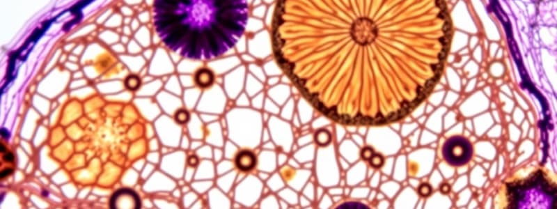
In the context of the provided micrographs, what is the primary purpose of using the PAS reaction?
In the context of the provided micrographs, what is the primary purpose of using the PAS reaction?
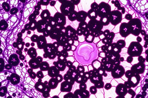
What does the purple staining in the H&E stained micrograph (a) primarily indicate?
What does the purple staining in the H&E stained micrograph (a) primarily indicate?
What is the role of hematoxylin in the PAS-stained tissue?
What is the role of hematoxylin in the PAS-stained tissue?
According to the passage, what is the final step before observation of a slide using a microscope?
According to the passage, what is the final step before observation of a slide using a microscope?
What is the approximate total magnification of tissue in image b given an objective lens of 10x and an ocular lens of 30x?
What is the approximate total magnification of tissue in image b given an objective lens of 10x and an ocular lens of 30x?
What cellular feature is primarily highlighted by staining with the PAS reaction?
What cellular feature is primarily highlighted by staining with the PAS reaction?
Based on the context, which cell type is most prominently associated with the staining pattern observed in the PAS-stained micrograph?
Based on the context, which cell type is most prominently associated with the staining pattern observed in the PAS-stained micrograph?
Why are tissue samples mounted with a glass coverslip before microscopic observation?
Why are tissue samples mounted with a glass coverslip before microscopic observation?
Flashcards
Tissue Preparation
Tissue Preparation
The process of preparing biological tissue for examination under a microscope.
Fixation
Fixation
A step in tissue preparation that preserves structures and prevents degradation by enzymes.
Tissues
Tissues
Groups of cells that work together to perform a specific function in an organism.
Microscopy
Microscopy
Signup and view all the flashcards
Histology
Histology
Signup and view all the flashcards
Dehydration
Dehydration
Signup and view all the flashcards
Clearing
Clearing
Signup and view all the flashcards
Infiltration
Infiltration
Signup and view all the flashcards
Embedding
Embedding
Signup and view all the flashcards
Microtome
Microtome
Signup and view all the flashcards
Transmission Electron Microscopy (TEM)
Transmission Electron Microscopy (TEM)
Signup and view all the flashcards
Paraffin block
Paraffin block
Signup and view all the flashcards
Sectioning
Sectioning
Signup and view all the flashcards
Staining
Staining
Signup and view all the flashcards
Ethyl Alcohol Series
Ethyl Alcohol Series
Signup and view all the flashcards
Dyes
Dyes
Signup and view all the flashcards
Acidic and Basic Dyes
Acidic and Basic Dyes
Signup and view all the flashcards
Electrostatic Linkages
Electrostatic Linkages
Signup and view all the flashcards
Trimming
Trimming
Signup and view all the flashcards
Fixatives
Fixatives
Signup and view all the flashcards
Vascular Perfusion
Vascular Perfusion
Signup and view all the flashcards
Glutaraldehyde
Glutaraldehyde
Signup and view all the flashcards
Osmium Tetroxide
Osmium Tetroxide
Signup and view all the flashcards
Ultra-microtome
Ultra-microtome
Signup and view all the flashcards
Basophilic
Basophilic
Signup and view all the flashcards
Acidophilic
Acidophilic
Signup and view all the flashcards
Hematoxylin
Hematoxylin
Signup and view all the flashcards
Eosin
Eosin
Signup and view all the flashcards
Embedding Medium
Embedding Medium
Signup and view all the flashcards
Section Thickness
Section Thickness
Signup and view all the flashcards
H&E Staining
H&E Staining
Signup and view all the flashcards
PAS Reaction
PAS Reaction
Signup and view all the flashcards
Basophilic Cells
Basophilic Cells
Signup and view all the flashcards
Oligosaccharides
Oligosaccharides
Signup and view all the flashcards
Goblet Cells
Goblet Cells
Signup and view all the flashcards
Microvilli
Microvilli
Signup and view all the flashcards
Mounting
Mounting
Signup and view all the flashcards
Light Microscope
Light Microscope
Signup and view all the flashcards
Study Notes
Histology & Its Methods of Study
- Histology studies tissues and how they form organs.
- Tissues have cells and extracellular matrix (ECM). ECM supports cells and transports nutrients/waste.
- Cells and ECM work together to form functional tissues then organs.
- Microscopes and molecular methods essential for studying histology.
- Biochemistry, molecular biology, physiology, immunology, and pathology vital for understanding tissue biology.
Preparation of Tissues for Study
- Tissue preparation involves creating thin sections for light microscopy.
- Ideal preparation preserves original tissue structure.
- Basic steps: fixation, dehydration, clearing, infiltration, embedding, and trimming.
- Fixation: Preserves tissue structure with chemicals that cross-link proteins, inactivate enzymes.
- Dehydration: Uses increasing alcohol concentrations to remove water.
- Clearing: Removes alcohol with organic solvents, making tissue translucent.
- Infiltration: Tissue is placed in melted paraffin until it is fully infiltrated.
- Embedding: Placing paraffin-infiltrated tissue in a mold for hardening.
- Trimming: Removing excess paraffin to expose tissue.
Embedding and Sectioning
- Embedding materials like paraffin (light microscopy) and plastic resins (both light and electron microscopy).
- Dehydration removes water via increasing ethanol solutions.
- Clearing replaces ethanol with an organic solvent.
- Infiltration involves placing the tissue in melted paraffin, replacing the clearing solvent and embedding in paraffin.
- Trimming the embedded block in a microtome (instrument) to prepare for slicing.
Staining
- Staining dyes tissues to make cellular components visible.
- Dyes act like acidic or basic compounds forming salt linkages.
- Basophilic components (negatively charged) bound to basic dyes.
- Acidophilic components (positively charged) bind to acidic dyes.
- Hematoxylin (basic dye) stains nuclei, RNA. Eosin stains other parts of the cell.
- H&E (hematoxylin & eosin) stains are the most commonly used combination.
- PAS (periodic acid-Schiff) stains carbohydrate-rich areas distinct colors.
- Techniques like enzyme digestion, pretreatment with enzymes (ex: RNase for cytoplasm) to identify the role of substances like RNA.
- Lipid-rich structures visible with lipid-soluble dyes (ex: Sudan black).
Light Microscopy
- Bright-field microscopy uses ordinary light to view stained tissue.
- Resolving power (minimum distance between structures seen as separate) is approximately 0.2 µm.
- Magnification of 1000-1500x possible.
- Higher magnification lenses have higher resolving power.
- Virtual microscopy digitizes bright-field slides.
Studying That Suits You
Use AI to generate personalized quizzes and flashcards to suit your learning preferences.