Podcast
Questions and Answers
What is the primary origin of connective tissue?
What is the primary origin of connective tissue?
- Neuroectoderm
- Endoderm
- Mesoderm (correct)
- Ectoderm
Which characteristic is NOT associated with connective tissue?
Which characteristic is NOT associated with connective tissue?
- Penetrated by blood vessels
- Contains a large amount of extracellular matrix
- Connects and supports other tissues
- Composed of closely packed cells (correct)
Which type of connective tissue is considered hard?
Which type of connective tissue is considered hard?
- Bone (correct)
- Adipose
- Blood
- Cartilage
Which type of cell is classified as a fixed (resident) cell in connective tissue?
Which type of cell is classified as a fixed (resident) cell in connective tissue?
What distinguishes wandering (transient) cells from fixed (resident) cells in connective tissue?
What distinguishes wandering (transient) cells from fixed (resident) cells in connective tissue?
Which of the following is NOT a function of undifferentiated mesenchymal cells (UMCs)?
Which of the following is NOT a function of undifferentiated mesenchymal cells (UMCs)?
What is the primary function of pericytes in connective tissue?
What is the primary function of pericytes in connective tissue?
What is a key histological feature of undifferentiated mesenchymal cells (UMCs)?
What is a key histological feature of undifferentiated mesenchymal cells (UMCs)?
What characteristic is specific to multilocular fat cells?
What characteristic is specific to multilocular fat cells?
Which type of cell is primarily responsible for antibody synthesis and secretion?
Which type of cell is primarily responsible for antibody synthesis and secretion?
What feature is associated with macrophages?
What feature is associated with macrophages?
What type of granules are found in the cytoplasm of mast cells?
What type of granules are found in the cytoplasm of mast cells?
What visual characteristic distinguishes the nucleus of plasma cells?
What visual characteristic distinguishes the nucleus of plasma cells?
What is the primary function of unilocular fat cells?
What is the primary function of unilocular fat cells?
Which type of fat staining method is used to identify specific fat cells?
Which type of fat staining method is used to identify specific fat cells?
What is a significant role of macrophages besides phagocytosis?
What is a significant role of macrophages besides phagocytosis?
What color do granules of mast cells appear when stained with toluidine blue due to their content of heparin?
What color do granules of mast cells appear when stained with toluidine blue due to their content of heparin?
Which chemical mediator released by mast cells is primarily responsible for smooth muscle contraction?
Which chemical mediator released by mast cells is primarily responsible for smooth muscle contraction?
What is the origin of pigment cells that store melanin?
What is the origin of pigment cells that store melanin?
What condition is potentially life-threatening and involves an exaggerated allergic reaction?
What condition is potentially life-threatening and involves an exaggerated allergic reaction?
Which component of the ground substance in connective tissue consists of large protein molecules with branched oligosaccharide chains?
Which component of the ground substance in connective tissue consists of large protein molecules with branched oligosaccharide chains?
What is the predominant effect of leukotrienes released by mast cells?
What is the predominant effect of leukotrienes released by mast cells?
What term is used for the excessive accumulation of interstitial fluid in connective tissue?
What term is used for the excessive accumulation of interstitial fluid in connective tissue?
Which of the following is a characteristic feature of protein-secreting cells like mast cells?
Which of the following is a characteristic feature of protein-secreting cells like mast cells?
What is the primary function of pericytes in tissues?
What is the primary function of pericytes in tissues?
Which structures are characteristic features of fibroblasts?
Which structures are characteristic features of fibroblasts?
How do active fibroblasts differ from inactive fibrocytes?
How do active fibroblasts differ from inactive fibrocytes?
What is a distinguishing feature of unilocular fat cells?
What is a distinguishing feature of unilocular fat cells?
What is the cytoplasmic characteristic of inactive fibrocytes?
What is the cytoplasmic characteristic of inactive fibrocytes?
Pericytes primarily regulate blood flow through which structure?
Pericytes primarily regulate blood flow through which structure?
Which of the following statements regarding fibroblasts is false?
Which of the following statements regarding fibroblasts is false?
Which feature is common to both unilocular and multilocular fat cells?
Which feature is common to both unilocular and multilocular fat cells?
Flashcards
Connective Tissue Origin
Connective Tissue Origin
Connective tissue originates from the mesoderm.
Connective Tissue Cells
Connective Tissue Cells
Connective tissues contain widely separated cells embedded within a large amount of extracellular matrix.
Connective Tissue Structure Components
Connective Tissue Structure Components
Connective tissue has three main components; cells, ground substance, and fibers.
Undifferentiated Mesenchymal Cells (UMCs)
Undifferentiated Mesenchymal Cells (UMCs)
Signup and view all the flashcards
UMCs Locations
UMCs Locations
Signup and view all the flashcards
Fixed/Resident Connective Tissue Cells
Fixed/Resident Connective Tissue Cells
Signup and view all the flashcards
Wandering/Transient Connective Tissue Cells
Wandering/Transient Connective Tissue Cells
Signup and view all the flashcards
Connective Tissue Classification by Matrix
Connective Tissue Classification by Matrix
Signup and view all the flashcards
Pericyte Origin
Pericyte Origin
Signup and view all the flashcards
Pericyte Function in Injury
Pericyte Function in Injury
Signup and view all the flashcards
Fibroblast Origin
Fibroblast Origin
Signup and view all the flashcards
Fibroblast Function
Fibroblast Function
Signup and view all the flashcards
Fibrocyte vs. Fibroblast
Fibrocyte vs. Fibroblast
Signup and view all the flashcards
Adipocyte Origin
Adipocyte Origin
Signup and view all the flashcards
Unilocular vs. Multilocular Adipocytes
Unilocular vs. Multilocular Adipocytes
Signup and view all the flashcards
White Adipose Tissue
White Adipose Tissue
Signup and view all the flashcards
Mast cell granules
Mast cell granules
Signup and view all the flashcards
Mast cell function
Mast cell function
Signup and view all the flashcards
What happens when mast cells degranulate?
What happens when mast cells degranulate?
Signup and view all the flashcards
Heparin function
Heparin function
Signup and view all the flashcards
Ground substance
Ground substance
Signup and view all the flashcards
What are GAGs?
What are GAGs?
Signup and view all the flashcards
Interstitial fluid
Interstitial fluid
Signup and view all the flashcards
Edema
Edema
Signup and view all the flashcards
Unilocular Fat Cells
Unilocular Fat Cells
Signup and view all the flashcards
Multilocular Fat Cells
Multilocular Fat Cells
Signup and view all the flashcards
Macrophage Function
Macrophage Function
Signup and view all the flashcards
Plasma Cell Structure
Plasma Cell Structure
Signup and view all the flashcards
Plasma Cell Function
Plasma Cell Function
Signup and view all the flashcards
Mast Cell Location
Mast Cell Location
Signup and view all the flashcards
Mast Cell Structure
Mast Cell Structure
Signup and view all the flashcards
What is the function of brown adipose tissue?
What is the function of brown adipose tissue?
Signup and view all the flashcards
Study Notes
Histology - Connective Tissue (Part 1)
- Connective tissue (CT) originates from the mesoderm.
- CT is composed of widely separated cells with a significant amount of extracellular matrix.
- CT is penetrated by blood vessels, nerves, and lymphatic vessels.
- CT connects, supports, and protects other tissues and organs.
Objectives
- List the characteristics of connective tissue (CT).
- Recognize the histological structure of CT.
- Classify the types of CT cells.
- Describe the histological structure and functions of CT cells.
- List the components of the ground substance of CT
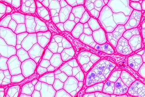
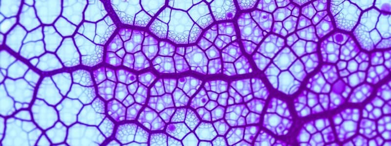
Connective Tissue Structure
- Cells: Blood vessels, ground substance, protein fibers (elastic, collagen, reticular).
- Extracellular Matrix: Fibers, ground substance.
- Fibers: Protein fibers (elastic, collagen, reticular).
- Ground Substance: Glycoproteins, glycosaminoglycans, proteoglycans.
Types of Connective Tissue (CT) According to Matrix Consistency
- Soft: CT proper
- Rubbery: Cartilage
- Hard: Bone
- Fluid: Blood
Types of CT Cells
- Fixed (Resident or Permanent Cells):
- Originate locally within the CT.
- Stable and long-lived cells.
- Undifferentiated mesenchymal cells
- Pericytes
- Fibroblasts & fibrocytes
- Adipocytes
- Wandering (Transient or Temporary Cells):
- Originate from stem cells in bone marrow.
- Circulate in blood then move into connective tissue to perform their functions.
- Motile and short-lived cells.
- Macrophages
- Mast cells
- Plasma cells
- Leucocytes
Undifferentiated Mesenchymal Cells (UMCs)
- Origin: The mother cells of all types of CT.
- Sites:
- Present mainly in embryos as stem cells.
- In adults, some cells remain after birth around blood capillaries in the form of pericytes.
- Structure:
- Shape: Small stellate-shaped cells with cytoplasmic processes (branched).
- Nucleus: Large, pale, with prominent nucleolus.
- Cytoplasm: Basophilic.
- (EM) Nucleus: Euchromatic
- (EM) Cytoplasm: Contains many free ribosomes.
- Function:
- They can differentiate into different cells of all types of connective tissue (CT proper, cartilage, and bone).
- Give rise to blood elements.
- Some cells remain after birth around blood capillaries (pericytes).
Pericytes
- Origin: UMCs.
- Sites: Present around blood capillaries.
- Structure:
- LM Shape: Branched cells with long cytoplasmic processes.
- Cytoplasm: Basophilic.
- Nucleus: Central and oval.
- EM Cytoplasm: contains network of actin and myosin
- Function:
- In case of tissue injury, pericytes can differentiate into fibroblasts, endothelial cells, and smooth muscle cells.
- They have contractile ability to regulate blood flow through capillaries.
Fibroblasts
- Origin: UMCs and pericytes.
- Sites: Most common type of CT cells.
- Structure:
- LM Shape: Flat branched cells with cytoplasmic processes.
- Nucleus: Large, pale, with prominent nucleolus.
- Cytoplasm: Deep basophilic.
- EM
- Well-developed rough endoplasmic reticulum (rER).
- Well-developed Golgi apparatus.
- Mitochondria.
- Euchromatic nucleus.
- No secretory granules in the cytoplasm.
- Function:
- Synthesis of CT matrix (fibers and ground substance).
- Synthesis of growth factors that enhance cell growth.
- Healing of wounds.
Fibrocytes
- Smaller and spindle-shaped with fewer processes.
- Smaller and darker nucleus.
- Pale basophilic cytoplasm (less rER).
- Maintenance of CT matrix.
Adipocytes (Fat Cells)
-
Origin: UMCs.
-
Types: Unilocular and multilocular fat cells.
-
Unilocular fat cells
- Large oval cells with peripheral nucleus.
- Cytoplasm with large fat droplet (by H&E, the fat droplet dissolves).
- Thin rim of cytoplasm.
- Signet ring appearance.
-
Multilocular fat cells
- Small rounded cells with central nucleus.
- Cytoplasm contains many fat droplets of various sizes.
- Numerous mitochondria.
-
Special Stain:
- Unilocular fat cells: Fat stains orange with Sudan III or black with Sudan black B and osmic acid.
- Multilocular fat cells: Function: Heat generation (Brown fat). Energy storage (White fat)
Macrophages
- Origin: Blood monocytes.
- Structure:
- LM Shape: Large irregular cells.
- Nucleus: Eccentric and kidney-shaped.
- EM Contains many lysosomes
- Has many cell processes called pseudopodia.
- Function: Phagocytosis of foreign bodies, microorganisms, and dead cells, act as antigen-presenting cells(to T-lymphocytes), secrete growth factors important for tissue repair.
Plasma Cells
- Origin: B-lymphocytes.
- Site: Abundant in lymphoid tissue.
- Structure: Oval shape, eccentric nucleus (cart-wheel), heterochromatin alternating with euchromatin, deep basophilic cytoplasm, well-developed Golgi apparatus.
- EM: Well-developed rER, well-developed Golgi apparatus, mitochondria, no secretory granules in cytoplasm.
- Function: Synthesis and secretion of antibodies (humoral immunity).
Mast Cells
- Origin: Hematopoietic stem cells.
- Sites: Along blood vessels and mucosa of the GIT and respiratory tract.
- Structure: Oval or rounded shape, central nucleus, cytoplasm containing basophilic granules.
- Special stain: Granules are metachromatically stained by toluidine blue (appear purple or red).
- EM: RER, Golgi apparatus, mitochondria, electron-dense secretory granules.
- Function: Release chemical mediators (e.g. heparin, histamine, leukotrienes) resulting in an allergic reaction, carry IgE surface receptors. Binding of antigens to IgE leads to degranulation.
Ground Substance of CT Matrix
- Highly hydrated material.
- Composed of 3 macromolecules:
- Glycosaminoglycans (GAGs): Polysaccharides (e.g., hyaluronic acid, chondroitin sulfate)
- Proteoglycans: GAGs attached to a core protein
- Multiadhesive glycoproteins: Large protein molecules (e.g., fibronectin) with branched oligosaccharide chains.
- Water in the ground substance is called interstitial fluid.
- Edema is excessive interstitial fluid accumulation in CT, often due to inflammation.
Pigment Cells
- Origin: UMCs
- Long branching processes
- Melanin storing cells
- Site: Iris of the eye, dermis of skin
Studying That Suits You
Use AI to generate personalized quizzes and flashcards to suit your learning preferences.