Podcast
Questions and Answers
What is the primary purpose of histology?
What is the primary purpose of histology?
- To study the behaviors of living organisms
- To analyze the chemical composition of cells
- To determine the genetic makeup of tissues
- To examine the microscopic structure of normal tissues (correct)
Which component is essential for light-transmission microscopy?
Which component is essential for light-transmission microscopy?
- Visible light (correct)
- Electron beams
- Fluorescent dyes
- Ultraviolet radiation
What magnification is achieved using objective lenses with X40 and eyepiece lenses X10?
What magnification is achieved using objective lenses with X40 and eyepiece lenses X10?
- X400 (correct)
- X40
- X100
- X60
What is the first step in preparing tissue for microscopic examination?
What is the first step in preparing tissue for microscopic examination?
What is the purpose of the dehydration step in tissue preparation?
What is the purpose of the dehydration step in tissue preparation?
Why is clearing important in the tissue preparation process?
Why is clearing important in the tissue preparation process?
During which step is the tissue placed in a small mold with melted paraffin?
During which step is the tissue placed in a small mold with melted paraffin?
What is the typical size range of a cell examined in histology?
What is the typical size range of a cell examined in histology?
What is the main function of epithelial tissue?
What is the main function of epithelial tissue?
Which domain of epithelial cells is exposed to the lumen or external environment?
Which domain of epithelial cells is exposed to the lumen or external environment?
What type of connective tissue features fixed and wandering cells within an abundant matrix?
What type of connective tissue features fixed and wandering cells within an abundant matrix?
Which type of epithelial tissue forms glands that secrete substances?
Which type of epithelial tissue forms glands that secrete substances?
What is the main function of the basement membrane?
What is the main function of the basement membrane?
Which of the following components is NOT part of the basement membrane?
Which of the following components is NOT part of the basement membrane?
What characteristic differentiates muscle tissue from other types of tissue?
What characteristic differentiates muscle tissue from other types of tissue?
Which statement correctly describes the differences between epithelial and connective tissues?
Which statement correctly describes the differences between epithelial and connective tissues?
What is the primary purpose of staining sections in microscopy?
What is the primary purpose of staining sections in microscopy?
Which stain is most commonly used for examining biological material?
Which stain is most commonly used for examining biological material?
Why does water freeze quickly when using liquid nitrogen for tissue preparation?
Why does water freeze quickly when using liquid nitrogen for tissue preparation?
What characteristic makes cytoplasm eosinophilic?
What characteristic makes cytoplasm eosinophilic?
What type of microscopy is used when observing untainted samples?
What type of microscopy is used when observing untainted samples?
Which method is confined to the examination of blood and bone marrow cells?
Which method is confined to the examination of blood and bone marrow cells?
In the context of immunochemistry, what does an epitope refer to?
In the context of immunochemistry, what does an epitope refer to?
What phenomenon occurs in phase-contrast microscopy when light waves are out of phase?
What phenomenon occurs in phase-contrast microscopy when light waves are out of phase?
What type of proteins transport vesicles along the cytoskeleton?
What type of proteins transport vesicles along the cytoskeleton?
Which shape of cells is more likely to differentiate into osteoblasts?
Which shape of cells is more likely to differentiate into osteoblasts?
What is the main difference between prokaryotic and eukaryotic ribosomes?
What is the main difference between prokaryotic and eukaryotic ribosomes?
Which component is crucial for integrins to function effectively?
Which component is crucial for integrins to function effectively?
What effect does blebbistatin have on cells?
What effect does blebbistatin have on cells?
How does the stiffness of the matrix influence cell differentiation?
How does the stiffness of the matrix influence cell differentiation?
What is the primary function of intermediate filaments in a cell?
What is the primary function of intermediate filaments in a cell?
Which approach is NOT considered a method of tissue engineering?
Which approach is NOT considered a method of tissue engineering?
What is the main function of importins in the cell?
What is the main function of importins in the cell?
Which of the following statements is true about euchromatin?
Which of the following statements is true about euchromatin?
What role does the H1 histone play in relation to nucleosomes?
What role does the H1 histone play in relation to nucleosomes?
How do histone acetylation and methylation differ in their effects on gene expression?
How do histone acetylation and methylation differ in their effects on gene expression?
What is true about chromosomes during cellular division?
What is true about chromosomes during cellular division?
Which component of the nucleosome serves as the core around which DNA is wrapped?
Which component of the nucleosome serves as the core around which DNA is wrapped?
What structure is responsible for the initial organization of DNA into chromatin?
What structure is responsible for the initial organization of DNA into chromatin?
What are the two arms of a chromosome called?
What are the two arms of a chromosome called?
What primarily increases the surface area of the inner mitochondrial membrane?
What primarily increases the surface area of the inner mitochondrial membrane?
What does the endosymbiotic theory suggest about the origin of mitochondria?
What does the endosymbiotic theory suggest about the origin of mitochondria?
How do metabolites like pyruvate and fatty acids enter the mitochondria?
How do metabolites like pyruvate and fatty acids enter the mitochondria?
What is a result of the movement of electrons through the inner membrane's electron-transport system?
What is a result of the movement of electrons through the inner membrane's electron-transport system?
What role does ATP synthase play in ATP production?
What role does ATP synthase play in ATP production?
What do peroxisomes primarily function in?
What do peroxisomes primarily function in?
What byproduct is formed in peroxisomes during the oxidation of fatty acids?
What byproduct is formed in peroxisomes during the oxidation of fatty acids?
What type of DNA do mitochondria possess that supports the endosymbiotic theory?
What type of DNA do mitochondria possess that supports the endosymbiotic theory?
Flashcards
What is histology?
What is histology?
The study of the microscopic structure of normal tissues. It reveals how tissues are organized to perform their functions.
What are tissues?
What are tissues?
Discrete collections of cells with similar morphology (shape) and function. Example: epithelial, muscular, nervous, connective.
What is a microscope?
What is a microscope?
The instrument used to magnify small objects, like cells, beyond what the human eye can see.
How does light-transmission microscopy work?
How does light-transmission microscopy work?
Signup and view all the flashcards
What is tissue preparation?
What is tissue preparation?
Signup and view all the flashcards
What is fixation in tissue preparation?
What is fixation in tissue preparation?
Signup and view all the flashcards
What is dehydration in tissue preparation?
What is dehydration in tissue preparation?
Signup and view all the flashcards
What is embedding in tissue preparation?
What is embedding in tissue preparation?
Signup and view all the flashcards
H&E Staining
H&E Staining
Signup and view all the flashcards
Antibody-Antigen Binding
Antibody-Antigen Binding
Signup and view all the flashcards
Antibody Variable Domain
Antibody Variable Domain
Signup and view all the flashcards
Phase-Contrast Microscopy
Phase-Contrast Microscopy
Signup and view all the flashcards
Tissue Embedding
Tissue Embedding
Signup and view all the flashcards
Staining (Histology)
Staining (Histology)
Signup and view all the flashcards
PAS Staining
PAS Staining
Signup and view all the flashcards
May-Grünwald-Giemsa Staining
May-Grünwald-Giemsa Staining
Signup and view all the flashcards
Integrins
Integrins
Signup and view all the flashcards
Intermediate filaments
Intermediate filaments
Signup and view all the flashcards
Motor Proteins
Motor Proteins
Signup and view all the flashcards
Kinesin
Kinesin
Signup and view all the flashcards
Dynein
Dynein
Signup and view all the flashcards
Cell Shape and Differentiation
Cell Shape and Differentiation
Signup and view all the flashcards
Matrix Stiffness and Differentiation
Matrix Stiffness and Differentiation
Signup and view all the flashcards
Tissue Engineering
Tissue Engineering
Signup and view all the flashcards
Cristae
Cristae
Signup and view all the flashcards
Mitochondrial Matrix
Mitochondrial Matrix
Signup and view all the flashcards
Endosymbiotic Theory
Endosymbiotic Theory
Signup and view all the flashcards
Mitochondrial DNA
Mitochondrial DNA
Signup and view all the flashcards
Acetyl CoA Production
Acetyl CoA Production
Signup and view all the flashcards
Electron Transport Chain
Electron Transport Chain
Signup and view all the flashcards
ATP Synthase
ATP Synthase
Signup and view all the flashcards
Peroxisomes
Peroxisomes
Signup and view all the flashcards
What is the function of covering/lining epithelium?
What is the function of covering/lining epithelium?
Signup and view all the flashcards
What is the function of secretory/glandular epithelium?
What is the function of secretory/glandular epithelium?
Signup and view all the flashcards
What is the apical domain of an epithelial cell?
What is the apical domain of an epithelial cell?
Signup and view all the flashcards
What is the lateral domain of an epithelial cell?
What is the lateral domain of an epithelial cell?
Signup and view all the flashcards
What is the basal domain of an epithelial cell?
What is the basal domain of an epithelial cell?
Signup and view all the flashcards
What is the basement membrane?
What is the basement membrane?
Signup and view all the flashcards
What is the basal lamina?
What is the basal lamina?
Signup and view all the flashcards
Nucleus
Nucleus
Signup and view all the flashcards
Nucleolus
Nucleolus
Signup and view all the flashcards
Chromatin
Chromatin
Signup and view all the flashcards
Euchromatin
Euchromatin
Signup and view all the flashcards
Heterochromatin
Heterochromatin
Signup and view all the flashcards
Nucleosome
Nucleosome
Signup and view all the flashcards
Histone acetylation
Histone acetylation
Signup and view all the flashcards
Histone methylation
Histone methylation
Signup and view all the flashcards
Study Notes
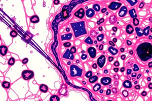
Histology
- Histology is the study of the microscopic structure of normal tissues. It helps understand how tissues are built to perform their functions.
- Tissues are collections of cells with similar characteristics.
- Cell size ranges from 10-30 micrometers.
- Microscopes are used to study cells.
Light Transmission Microscopy
- Uses visible light to examine a sample.
- Objective lenses enlarge and project the image to the eyepiece.
- Common magnifications are X4, X10, and X40.
- The sample needs to be transparent.
Tissue Preparation
- Fixation: Preserves tissue structure using chemicals that cross-link proteins and inactivate enzymes.
- Dehydration: Removes water from the tissue using alcohol solutions (e.g., moving from 65% to 100% alcohol).
- Clearing: Tissue is transferred to organic solvents that are miscible with both alcohol and paraffin to make it transparent.
- Infiltration: Tissue is immersed in melted paraffin until fully infiltrated (absorbed by paraffin).
- Embedding: The tissue is placed in molds filled with melted paraffin. Once solidified the specimen forms paraffin block.
- Trimming: The block is trimmed to expose the tissue for sectioning. Freezing with liquid nitrogen is an alternative to paraffin embedding for tissue sectioning.
- Staining: Highlights specific tissue structures and components. H&E staining is common (nuclei stain blue, cytoplasm stains pink/red). PAS stain is used for carbohydrates.
Microscopy Techniques
- Phase-contrast microscopy: Used to observe unstained samples, useful for live cells that would be killed under staining.
- Confocal microscopy: Allows observation of a single plane of a sample, useful for 3D-imaging.
- Stereomicroscopy: A microscope that magnifies an object without thin-sectioning the specimen.
- Electron microscopy: Uses electrons, in two kinds of configurations, Transmission electron microscope (TEM) or Scanning electron microscope (SEM). TEM can give a 3D picture (in a 2D arrangement) while SEM gives a 3D surface rendering.
Immunochemistry
- Uses antibodies (produced by B-lymphocytes) to identify specific proteins/molecules.
- Antibodies have a variable domain (paratope) that attaches to the specific protein/molecule (antigen).
- The epitope is the part of the antigen that the antibody recognizes.
The Cell
- The basic unit of life, and composed of plasma membrane, the encircling membrane surrounding the cell.
- Phospholipids create a bi-layer with hydrophobic tails facing inward and hydrophilic heads facing outward.
- Cholesterol is found between the phospholipids affecting membrane fluidity and flexibility.
- Proteins perform various functions, some face outward, some inward, and others span the membrane.
- Glycocalyx: a sugar layer surrounding the membrane, bonded with proteins and phospholipids. It plays a role in recognition and cell-cell interactions.
- Hyaluronic acid: creates a jelly-like layer around cells, which retains water.
Membrane Transport
- Simple diffusion: Lipophilic molecules and small, uncharged molecules pass through easily.
- Channel/facilitated diffusion: Membrane proteins act as channels to allow passage of specific molecules(usually charged).
- Carrier/pump: Larger molecules/charged molecules transported against their concentration gradient, requiring energy., often using ATP as energy source.
Cytoskeleton
- Microfilaments: Composed of actin.
- Microtubules: Composed of tubulin required for mitosis and other cellular needs.
- Intermediate filaments: Provide structural support.
Organelles
- Ribosome: Synthesize proteins based on instructions from mRNA. They have three sites (A, P, and E) to perform protein synthesis.
- Endoplasmic Reticulum (ER): Rough ER (RER) has ribosomes and synthesizes proteins for transport/secretion. Smooth ER (SER) synthesizes lipids and other molecules.
- Golgi Apparatus: Modifies and packages proteins and lipids for secretion or use within the cell.
- Lysosomes: Contain digestive enzymes to break down waste and molecules.
- Mitochondria: Produce ATP through cellular respiration.
- Peroxisomes: Contain enzymes to break down fatty acids into smaller molecules.
- Cellular inclusion: Accumulation of substances like lipids or glycogen, storing energy in some cells.
- Nucleus: Contains DNA, controls cellular activities, including protein production, and contains the nucleolus where ribosomes assemble.
Cell Cycle
- Interphase: period of cell growth and DNA replication. Divided in G1, S, and G2 phases.
Mitochondria
- Produce ATP
- Metabolism of amino acids and lipids
- Act as a sensor for the health of the cell
- Cells can undergo apoptosis (programmed cell death)
Cell Division
- Mitosis: Process of cell division, resulting in two identical daughter cells.
- Cytokinesis: Division of the cytoplasm.
- Stem cells: Undifferentiated cells that can divide indefinitely and differentiate into specialized cell types.
- Types of stem cells include embryonic and adult stem cells.
Types of tissues
- Epithelial: Covers body surfaces, lines cavities, forms glands responsible for secretions.
- Connective: Supports, connects, and separates different tissues and cells. Forms extracellular matrix.
- Muscular: Responsible for movement, contracts.
- Nervous: Transmits information via nerve impulses.
Bone
- Compact bone: Dense bone tissue organized into osteons. Each osteon is around a central canal.
- Spongy bone: A framework of trabeculae.
- Epiphysis: Ends of the long bone.
- Diaphysis: Shaft of the bone.
- Osteoblasts: Bone-forming cells.
- Osteocytes: Mature bone cells.
- Osteoclasts: Bone-resorbing cells. -Periosteum: Membrane covering bone. Contains cells that produce new bone tissue, which has a key role in bone growth and fracture healing. -Endosteum: Membrane lining the internal surfaces of the bone. Contains cells that produce new bone tissue, which has a key role in bone growth and bone remodeling.
Blood
- Consists of a suspension of cells (red blood cells, white blood cells, platelets) within a fluid matrix called plasma.
- Function is to transport substances throughout the body.
Connective Tissue
- Connective tissue is characterized by an abundance of extracellular matrix that is largely composed of protein fibers (collagen, elastic) embedded in a ground substance.
- Types include loose (areolar) connective tissues, dense regular connective tissue, dense irregular connective tissue, reticular connective tissue.
- Specialized connective tissues include cartilage, bone, and blood.
Glands
- Glands are epithelial tissue; they produce and secrete substances. Glands can be exocrine (secrete through ducts) or endocrine (secrete into the bloodstream).
Special epithelia structures
- Cilia: hair-like extensions of the cell membrane that can beat, move substances.
- Stereocilia: (also called static cilia), extensions of the cell membrane that do not beat or move.
- Microvilli: finger-like projections that increase surface area for absorption.
Studying That Suits You
Use AI to generate personalized quizzes and flashcards to suit your learning preferences.