Podcast
Questions and Answers
Which of the following techniques is specifically used to highlight the presence of glycoproteins and polysaccharides in the small intestinal epithelium?
Which of the following techniques is specifically used to highlight the presence of glycoproteins and polysaccharides in the small intestinal epithelium?
- PAS Reaction (correct)
- Transmission Electron Microscopy
- Immunofluorescence Staining
- H&E Staining
What is the main purpose of counterstaining with hematoxylin in the PAS reaction?
What is the main purpose of counterstaining with hematoxylin in the PAS reaction?
- To enhance the visibility of cell nuclei (correct)
- To increase the contrast between the cytoplasm and the cell membrane
- To visualize the microvilli on the cell surface
- To differentiate between different types of carbohydrates
What is the likely function of the goblet cells in the small intestine?
What is the likely function of the goblet cells in the small intestine?
- Digestion of carbohydrates
- Production of digestive enzymes
- Secretion of mucus (correct)
- Absorption of nutrients
In the micrograph stained with H&E, what color are the cell nuclei?
In the micrograph stained with H&E, what color are the cell nuclei?
What component of the small intestine is most prominently stained with the PAS reaction?
What component of the small intestine is most prominently stained with the PAS reaction?
Which of the following is NOT a factor influencing the duration of slide preparation?
Which of the following is NOT a factor influencing the duration of slide preparation?
What is the purpose of the coverslip mounted on the slide after preparation?
What is the purpose of the coverslip mounted on the slide after preparation?
How is the total magnification of a light microscope achieved?
How is the total magnification of a light microscope achieved?
What is the primary reason the preparatory process can remove cellular lipid?
What is the primary reason the preparatory process can remove cellular lipid?
Which type of methods are essential for advancing histology study?
Which type of methods are essential for advancing histology study?
What is the function of fixation in tissue preparation?
What is the function of fixation in tissue preparation?
Why is histology dependent on the use of microscopes?
Why is histology dependent on the use of microscopes?
What results from an orderly combination of tissues in an organ?
What results from an orderly combination of tissues in an organ?
What is the primary purpose of fixation in tissue preparation?
What is the primary purpose of fixation in tissue preparation?
During which step is alcohol removed from the tissue?
During which step is alcohol removed from the tissue?
What is one of the key differences in tissue preparation for transmission electron microscopy compared to light microscopy?
What is one of the key differences in tissue preparation for transmission electron microscopy compared to light microscopy?
What is the purpose of the drive wheel in the sectioning process?
What is the purpose of the drive wheel in the sectioning process?
What concentration of alcohol is used to completely remove water during the dehydration step?
What concentration of alcohol is used to completely remove water during the dehydration step?
Which step directly follows dehydration in the tissue preparation process?
Which step directly follows dehydration in the tissue preparation process?
What type of knife is typically used for sectioning paraffin-embedded tissues?
What type of knife is typically used for sectioning paraffin-embedded tissues?
What is the primary function of embedding in the tissue processing sequence?
What is the primary function of embedding in the tissue processing sequence?
What are the two primary interacting components of tissues?
What are the two primary interacting components of tissues?
What is the primary role of the extracellular matrix (ECM) in relation to cells?
What is the primary role of the extracellular matrix (ECM) in relation to cells?
Which method is most commonly used in histologic research for examining tissues?
Which method is most commonly used in histologic research for examining tissues?
What is the primary focus of histology?
What is the primary focus of histology?
Why must tissue sections be thin when preparing for microscopic examination?
Why must tissue sections be thin when preparing for microscopic examination?
Which microscopy technique is best for visualizing cellular structures in detail?
Which microscopy technique is best for visualizing cellular structures in detail?
How do cells and the extracellular matrix (ECM) interact within tissues?
How do cells and the extracellular matrix (ECM) interact within tissues?
What technique allows for the visualization of specific molecules within tissues?
What technique allows for the visualization of specific molecules within tissues?
What is the significance of preserving tissue during preparation for examination?
What is the significance of preserving tissue during preparation for examination?
Which method is primarily utilized for sectioning tissues before microscopic examination?
Which method is primarily utilized for sectioning tissues before microscopic examination?
What role do specific cell surface receptors play in the interaction with the ECM?
What role do specific cell surface receptors play in the interaction with the ECM?
How does the specialization of cells occur during development?
How does the specialization of cells occur during development?
Which microscopy technique uses fluorescent dyes to visualize specimens?
Which microscopy technique uses fluorescent dyes to visualize specimens?
What is the role of staining in histology?
What is the role of staining in histology?
Which of the following techniques helps in visualizing the surface structure of specimens?
Which of the following techniques helps in visualizing the surface structure of specimens?
Which technique involves culturing cells in a controlled environment?
Which technique involves culturing cells in a controlled environment?
What process involves placing tissue in melted paraffin until infiltration is complete?
What process involves placing tissue in melted paraffin until infiltration is complete?
What is the purpose of using fixatives in tissue processing?
What is the purpose of using fixatives in tissue processing?
Which fixative is commonly used for light microscopy?
Which fixative is commonly used for light microscopy?
During the paraffin embedding process, what happens to the tissue block after melting?
During the paraffin embedding process, what happens to the tissue block after melting?
What technique is described for preparing thinner sections for transmission electron microscopy (TEM)?
What technique is described for preparing thinner sections for transmission electron microscopy (TEM)?
What is a common practice to improve fixative penetration in large organs?
What is a common practice to improve fixative penetration in large organs?
What is the role of glutaraldehyde in electron microscopy preparation?
What is the role of glutaraldehyde in electron microscopy preparation?
What is the purpose of vascular perfusion during fixation?
What is the purpose of vascular perfusion during fixation?
Flashcards
Histology
Histology
The study of body tissues and their organization in organs.
Fixation
Fixation
A process to preserve tissue samples for microscopic study.
Staining
Staining
A technique used to enhance contrast in tissue samples for better visualization under a microscope.
Embedding
Embedding
Signup and view all the flashcards
Sectioning
Sectioning
Signup and view all the flashcards
Light Microscopy
Light Microscopy
Signup and view all the flashcards
Electron Microscopy
Electron Microscopy
Signup and view all the flashcards
Immunohistochemistry
Immunohistochemistry
Signup and view all the flashcards
Tissue Biology
Tissue Biology
Signup and view all the flashcards
Extracellular Matrix (ECM)
Extracellular Matrix (ECM)
Signup and view all the flashcards
Collagen Fibrils
Collagen Fibrils
Signup and view all the flashcards
Microscopic Examination
Microscopic Examination
Signup and view all the flashcards
Histologic Research
Histologic Research
Signup and view all the flashcards
Tissue Sections
Tissue Sections
Signup and view all the flashcards
Cell Surface Receptors
Cell Surface Receptors
Signup and view all the flashcards
Functional Specialization
Functional Specialization
Signup and view all the flashcards
Tissue Preparation
Tissue Preparation
Signup and view all the flashcards
Microscope Dependence
Microscope Dependence
Signup and view all the flashcards
Tissue Structure
Tissue Structure
Signup and view all the flashcards
Molecular Methods
Molecular Methods
Signup and view all the flashcards
Dehydration
Dehydration
Signup and view all the flashcards
Clearing
Clearing
Signup and view all the flashcards
Infiltration
Infiltration
Signup and view all the flashcards
Paraffin block
Paraffin block
Signup and view all the flashcards
Microtome
Microtome
Signup and view all the flashcards
Epoxy resins
Epoxy resins
Signup and view all the flashcards
Fixation for TEM
Fixation for TEM
Signup and view all the flashcards
Histological preparation
Histological preparation
Signup and view all the flashcards
Microvilli
Microvilli
Signup and view all the flashcards
Trimming
Trimming
Signup and view all the flashcards
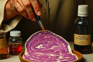
Goblet Cells
Goblet Cells
Signup and view all the flashcards
Fixatives
Fixatives
Signup and view all the flashcards
H&E Staining
H&E Staining
Signup and view all the flashcards
Vascular perfusion
Vascular perfusion
Signup and view all the flashcards
PAS Reaction
PAS Reaction
Signup and view all the flashcards
Oligosaccharides
Oligosaccharides
Signup and view all the flashcards
Glutaraldehyde
Glutaraldehyde
Signup and view all the flashcards
Mucin
Mucin
Signup and view all the flashcards
Osmium tetroxide
Osmium tetroxide
Signup and view all the flashcards
Counterstaining
Counterstaining
Signup and view all the flashcards
TEM sections
TEM sections
Signup and view all the flashcards
Total Magnification
Total Magnification
Signup and view all the flashcards
Study Notes
Histology and Its Methods of Study
- Histology is the study of bodily tissues and how they form organs. It examines how cell structure optimizes organ function.
- Tissues consist of cells and extracellular matrix (ECM). ECM contains complex molecules (e.g., collagen fibrils), supporting cells and transporting nutrients/wastes.
- Cells and ECM interact in a coordinated manner. Cells produce ECM and are influenced by it.
- Tissues specialize during development, forming basic tissue types with specific structures.
- Organs are formed by a specific combination of tissues, enabling organ and organismal function.
- Histology relies on microscopes and molecular methods. Understanding advancements in biochemistry, molecular biology, physiology, immunology, and pathology is crucial.
Preparation of Tissues for Study
- Histological research commonly involves preparing thin tissue sections for light microscopy.
- Preserved tissue sections on slides should have the same structure as in the body.
- Steps in tissue preparation for light microscopy:
- Fixation: Preserves tissue structure using chemical solutions that crosslink proteins and inactivate enzymes. Small tissue pieces facilitate penetration. Formalin is a common fixative.
- Dehydration: Removal of water using progressively concentrated alcohol solutions.
- Clearing: Removal of alcohol with organic solvents.
- Infiltration: Tissue is placed in melted paraffin until completely infiltrated.
- Embedding: Tissue is placed in a mold with paraffin to harden.
- Trimming: Excess paraffin is removed to expose the tissue for sectioning.
- Electron microscopy requires special fixatives, dehydration solutions, and embedding in epoxy resins, resulting in very thin sections.
Embedding and Sectioning
- Embedding materials (e.g., paraffin, plastic resins) ensure firm tissue consistency for sectioning.
- Before embedding, tissue undergoes dehydration, clearing, and infiltration with the embedding medium.
- Paraffin sections are typically 3-10 µm thick for light microscopy. Electron microscopy needs sections less than 1 µm thick. (1 µm = 1/1000 mm)
Staining
- Most cells and ECM are colorless and require staining for microscopic study.
- Dyes stain tissue components selectively. Dyes behave as acids or bases, forming bonds with ionized macromolecules.
- Basophilic components (e.g., nucleic acids) are attracted to basic dyes (e.g., hematoxylin).
- Acidophilic components (e.g., proteins) are attracted to acidic dyes (e.g., eosin).
- H&E staining (hematoxylin and eosin) is a common method. Hematoxylin stains DNA/RNA-rich parts, while eosin stains other cytoplasmic components.
- The PAS (periodic acid-Schiff) reaction stains carbohydrate-rich tissues (purple/magenta).
- Other special staining techniques visualize specific tissues or molecules (e.g., enzymes).
Light Microscopy
- Bright-field microscopy uses ordinary light to study stained tissue sections.
- Microscopy involves using an optical system to view and focus on the specimen. Components include:
- Condenser
- Objective lens
- Eyepiece/ocular lens
- Resolving power is the smallest distance to distinguish two separate objects. (0.2 µm or less). Higher magnification is useful only if resolution is also high.
- Virtual microscopy digitizes microscopic images for study/storage and digital transmission.
Studying That Suits You
Use AI to generate personalized quizzes and flashcards to suit your learning preferences.