Podcast
Questions and Answers
Which technique is commonly used to study the distribution of specific molecules within cells and tissues, utilizing antibodies to bind to target antigens?
Which technique is commonly used to study the distribution of specific molecules within cells and tissues, utilizing antibodies to bind to target antigens?
- Immunohistochemistry (correct)
- Autoradiography
- Enzyme Histochemistry
- Hybridization Techniques
Which type of microscopy uses a beam of electrons to create images of the internal structure of cells and tissues?
Which type of microscopy uses a beam of electrons to create images of the internal structure of cells and tissues?
- Phase-Contrast Microscopy
- Fluorescence Microscopy
- Bright-Field Microscopy
- Transmission Electron Microscopy (correct)
What type of microscopy employs a special condenser to enhance the contrast between different structures with varying refractive indices, enabling visualization of unstained specimens?
What type of microscopy employs a special condenser to enhance the contrast between different structures with varying refractive indices, enabling visualization of unstained specimens?
- Phase-Contrast Microscopy (correct)
- Bright-Field Microscopy
- Confocal Microscopy
- Polarizing Microscopy
Which technique involves the use of radioactive isotopes to label specific molecules, enabling their detection and localization within cells and tissues?
Which technique involves the use of radioactive isotopes to label specific molecules, enabling their detection and localization within cells and tissues?
Which type of microscopy is used to study the distribution of materials that emit light when excited by a specific wavelength of light?
Which type of microscopy is used to study the distribution of materials that emit light when excited by a specific wavelength of light?
What type of microscopy utilizes a laser beam to scan a specimen, creating a three-dimensional image by collecting light emitted from different focal planes?
What type of microscopy utilizes a laser beam to scan a specimen, creating a three-dimensional image by collecting light emitted from different focal planes?
Which microscopy technique is particularly useful for studying birefringent materials, such as muscle fibers and bone, that exhibit different refractive indices in different directions?
Which microscopy technique is particularly useful for studying birefringent materials, such as muscle fibers and bone, that exhibit different refractive indices in different directions?
Why can it often be difficult to accurately visualize the structure of cells in tissues using light microscopy?
Why can it often be difficult to accurately visualize the structure of cells in tissues using light microscopy?
What is the primary purpose of fixation in tissue preparation for light microscopy?
What is the primary purpose of fixation in tissue preparation for light microscopy?
Which of the following is NOT a factor that contributes to the complexity of studying tissues and their components using light microscopy?
Which of the following is NOT a factor that contributes to the complexity of studying tissues and their components using light microscopy?
Which of the following statements accurately reflects the concept of tissue organization in the human body?
Which of the following statements accurately reflects the concept of tissue organization in the human body?
In the context of tissue preparation for light microscopy, why is it important to consider the potential for lipid removal during processing?
In the context of tissue preparation for light microscopy, why is it important to consider the potential for lipid removal during processing?
Which of the following statements is NOT true about the use of microscopes in histology?
Which of the following statements is NOT true about the use of microscopes in histology?
Based on the provided content, which of the following statements best describes the relationship between cell structure and tissue function?
Based on the provided content, which of the following statements best describes the relationship between cell structure and tissue function?
What is the significance of studying the arrangement of tissues within organs?
What is the significance of studying the arrangement of tissues within organs?
Which of the following fields of study is NOT considered essential for a comprehensive understanding of histology?
Which of the following fields of study is NOT considered essential for a comprehensive understanding of histology?
What is the primary purpose of dehydration in tissue preparation for light microscopy?
What is the primary purpose of dehydration in tissue preparation for light microscopy?
What is the purpose of using a microtome during the tissue preparation process?
What is the purpose of using a microtome during the tissue preparation process?
Which of the following steps in tissue preparation is essential for producing thin, uniform slices of tissue for microscopic observation?
Which of the following steps in tissue preparation is essential for producing thin, uniform slices of tissue for microscopic observation?
What is the main function of the fixative used in tissue preparation?
What is the main function of the fixative used in tissue preparation?
What is the most likely reason why epoxy resins are used for embedding in transmission electron microscopy (TEM) instead of paraffin?
What is the most likely reason why epoxy resins are used for embedding in transmission electron microscopy (TEM) instead of paraffin?
What is the main reason for using alcohol during the dehydration process in tissue preparation?
What is the main reason for using alcohol during the dehydration process in tissue preparation?
Which of the following statements is TRUE about the relationship between the tissue preparation methods for light microscopy and transmission electron microscopy (TEM)?
Which of the following statements is TRUE about the relationship between the tissue preparation methods for light microscopy and transmission electron microscopy (TEM)?
What is the primary purpose of clearing in tissue preparation?
What is the primary purpose of clearing in tissue preparation?
Why is it essential to have a controlled advancement of the tissue block in a microtome?
Why is it essential to have a controlled advancement of the tissue block in a microtome?
How does the process of fixation help prepare tissue for microscopic analysis?
How does the process of fixation help prepare tissue for microscopic analysis?
What is the primary purpose of embedding tissues in materials like paraffin or plastic resins?
What is the primary purpose of embedding tissues in materials like paraffin or plastic resins?
Why is dehydration a crucial step before embedding tissues?
Why is dehydration a crucial step before embedding tissues?
What is the main reason for staining tissue sections?
What is the main reason for staining tissue sections?
What is the primary mechanism by which most dyes stain tissue components?
What is the primary mechanism by which most dyes stain tissue components?
Which of the following is NOT a typical embedding material used for tissue preparation?
Which of the following is NOT a typical embedding material used for tissue preparation?
What is the primary purpose of using a series of increasing ethanol solutions during dehydration?
What is the primary purpose of using a series of increasing ethanol solutions during dehydration?
Why are plastic resins suitable for both light and electron microscopy?
Why are plastic resins suitable for both light and electron microscopy?
What is the main advantage of using paraffin as an embedding medium?
What is the main advantage of using paraffin as an embedding medium?
Why is it important to have a thorough understanding of the staining process in histology?
Why is it important to have a thorough understanding of the staining process in histology?
How do different dyes selectively stain specific tissue components?
How do different dyes selectively stain specific tissue components?
Based on the provided information, which of the following statements accurately reflects the role of clearing reagents in tissue preparation?
Based on the provided information, which of the following statements accurately reflects the role of clearing reagents in tissue preparation?
Why is tissue embedding in plastic resin advantageous over embedding in paraffin for certain applications?
Why is tissue embedding in plastic resin advantageous over embedding in paraffin for certain applications?
Which of the following tissue components are specifically targeted by hematoxylin during the H&E staining process?
Which of the following tissue components are specifically targeted by hematoxylin during the H&E staining process?
Based on the provided information, what is the primary function of eosin in the H&E staining process, considering it is a type of acidic dye?
Based on the provided information, what is the primary function of eosin in the H&E staining process, considering it is a type of acidic dye?
Considering the mentioned characteristics of basic and acidic dyes, which of the following tissue components would MOST LIKELY exhibit a strong affinity for eosin staining?
Considering the mentioned characteristics of basic and acidic dyes, which of the following tissue components would MOST LIKELY exhibit a strong affinity for eosin staining?
Flashcards
Histology
Histology
The study of the tissues of the body and their arrangement.
Fixation
Fixation
The process of preserving tissue structure for microscopic examination.
Embedding
Embedding
Inserting tissue samples in a medium for easier sectioning.
Staining
Staining
Signup and view all the flashcards
Light Microscopy
Light Microscopy
Signup and view all the flashcards
Electron Microscopy
Electron Microscopy
Signup and view all the flashcards
Immunohistochemistry
Immunohistochemistry
Signup and view all the flashcards
Dehydration
Dehydration
Signup and view all the flashcards
Clearing
Clearing
Signup and view all the flashcards
Infiltration
Infiltration
Signup and view all the flashcards
Paraffin block
Paraffin block
Signup and view all the flashcards
Microtome
Microtome
Signup and view all the flashcards
Sectioning
Sectioning
Signup and view all the flashcards
Transmission Electron Microscopy (TEM)
Transmission Electron Microscopy (TEM)
Signup and view all the flashcards
Chemical cross-linking
Chemical cross-linking
Signup and view all the flashcards
Tissues
Tissues
Signup and view all the flashcards
Organ formation
Organ formation
Signup and view all the flashcards
Microscopy
Microscopy
Signup and view all the flashcards
Cellular lipid
Cellular lipid
Signup and view all the flashcards
Molecular methods
Molecular methods
Signup and view all the flashcards
Enzyme degradation
Enzyme degradation
Signup and view all the flashcards
Structural features
Structural features
Signup and view all the flashcards
Basophilic
Basophilic
Signup and view all the flashcards
Acidophilic
Acidophilic
Signup and view all the flashcards
Hematoxylin and Eosin (H&E)
Hematoxylin and Eosin (H&E)
Signup and view all the flashcards
Paraffin
Paraffin
Signup and view all the flashcards
Plastic Resins
Plastic Resins
Signup and view all the flashcards
Ionizable Radicals
Ionizable Radicals
Signup and view all the flashcards
Dyes
Dyes
Signup and view all the flashcards
Microscopic Study
Microscopic Study
Signup and view all the flashcards
Study Notes
Histology & Its Methods of Study
- Histology: Study of body tissues and their arrangement in organs. Focuses on cell structure, arrangement, and organ function
- Tissue Components: Cells and extracellular matrix (ECM)
- ECM: Consists of macromolecules (e.g., collagen fibrils) supporting cells, transporting nutrients/wastes, and coordinating with cells. Cells produce and are influenced by ECM components.
- Tissue Development: Specialized cells and ECM form tissues. Tissues combine to form organs, enabling organism function.
- Histology Methods: Microscopy and molecular approaches are crucial for small cell/matrix study. Requires knowledge of biochemistry, molecular biology, immunology, and pathology.
- Tissue Preparation: Common procedure: thin tissue sections for light microscopy examination. Preserves tissue structure as similar to in vivo state
Preparation of Tissues for Study
-
Fixation: Preserves tissue structure from degradation by enzymes/microorganisms. Uses cross-linking chemical solutions; important for light & electron microscopy. Typically achieved within hours of tissue removal. Small tissue fragments placed in solutions to allow for better penetration and preservation. Can be induced via blood vessels. Commonly uses formalin (buffered isotonic solution of 37% formaldehyde) for light microscopy and glutaraldehyde for electron microscopy
-
Dehydration: Removes water using increasing ethanol concentrations (e.g., to 100%).
-
Clearing: Removes alcohol with organic solvents compatible with embedding media. Tissues become translucent.
-
Infiltration: Tissue is placed in melted paraffin, allowing for replacement of the clearing agent with paraffin.
-
Embedding: Paraffin-infiltrated tissue is set in a mold with melted paraffin and allowed to harden.
-
Trimming: Paraffin block trimmed to expose tissue for sectioning
-
Special preparation for EM: Includes specific fixatives and dehydrating solutions for smaller samples, using epoxy resins
-
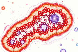
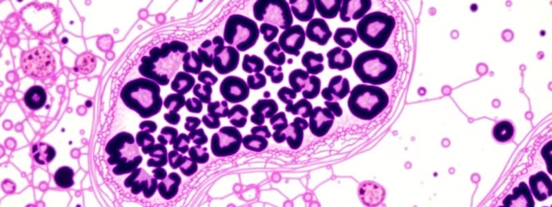
Microtome sectioning: Sectioning process using a microtome. Paraffin sections (~3-10 μm) for Light Microscopy. Thin sections (<1μm) are used for Electron Microscopy (Ultra-microtome-glass or diamond knife)
Embedding & Sectioning
- Embedding Materials: Paraffin (light microscopy), plastic resins (both light and electron microscopy)
- Dehydration: Gradual ethanol transfer to remove water.
- Clearing: Replaces alcohol with organic solvent.
- Infiltration: Tissue is fully immersed in melted paraffin.
- Embedding: Tissue in mold with melted paraffin, allowed to harden.
- Trimming: Hardened block trimmed for sectioning.
- Section Thickness: Typically ~3-10 µm for light microscopy; <1 μm for electron microscopy.
Staining
-
General Approach: Staining procedures make cell/extracellular components visible and distinguishable.
-
Basis of Staining: Dyes react with ionizable components (macromolecules with ionizable groups).
-
Basophilic: Affinity for basic dyes; common in DNA/RNA-rich areas.
-
Acidophilic: Affinity for acidic dyes; targets proteins with many ionized amino groups
-
Example Dyes: Hematoxylin (basic), eosin (acidic), toluidine blue, alcian blue, methylene blue
-
Common stain: H&E (hematoxylin and eosin) stain - hematoxylin stains basophilic structures (e.g., DNA, RNA) in dark blue/purple, and eosin stains acidophilic structures, often in pink.
-
Other stains: PAS (periodic acid-Schiff), Feulgen reactions, Sudan black
Light Microscopy
- Bright-field Microscopy: Uses ordinary light to examine stained tissue sections. Components include condenser, objective lens, and eyepiece.
- Resolution: Determined by objective lens; best attainable with light microscopy is roughly 0.2 µm. This limits magnification for detailed cellular structures
- Magnification: Calculated from the power of the objective and eyepiece lenses
- Virtual Microscopy: Conversion of bright-field images for easy storage and access
Studying That Suits You
Use AI to generate personalized quizzes and flashcards to suit your learning preferences.