Podcast
Questions and Answers
Which of the following describes the resting position of the hip joint?
Which of the following describes the resting position of the hip joint?
- Hip flexion of 30 degrees, abduction of 30 degrees, and slight external rotation. (correct)
- Hip extension of 15 degrees, adduction of 15 degrees, and slight internal rotation.
- Hip flexion of 15 degrees, abduction of 15 degrees, and slight external rotation.
- Hip extension of 30 degrees, adduction of 30 degrees, and slight internal rotation.
During hip distraction with caudal glide, where should the therapist place their hands on the patient?
During hip distraction with caudal glide, where should the therapist place their hands on the patient?
- Distal to the iliac crest, over the belt.
- Proximal to the iliac crest, under a belt.
- Distal to the malleoli, over the belt.
- Proximal to the malleoli, under the belt. (correct)
What is the primary indication for performing a hip posterior glide mobilization?
What is the primary indication for performing a hip posterior glide mobilization?
- To decrease hip abduction and external rotation.
- To increase hip abduction and external rotation.
- To increase hip flexion and internal rotation. (correct)
- To decrease hip flexion and internal rotation.
In which patient position is the hip anterior glide mobilization performed?
In which patient position is the hip anterior glide mobilization performed?
What is the resting position of the tibiofemoral joint?
What is the resting position of the tibiofemoral joint?
The treatment plane for tibiofemoral joint mobilization is best described as which of the following?
The treatment plane for tibiofemoral joint mobilization is best described as which of the following?
What is the recommended hand placement for performing a tibiofemoral distraction?
What is the recommended hand placement for performing a tibiofemoral distraction?
A therapist is about to perform a tibiofemoral posterior glide. Which patient position is MOST appropriate for this technique?
A therapist is about to perform a tibiofemoral posterior glide. Which patient position is MOST appropriate for this technique?
What is the direction of force application for a tibiofemoral anterior glide mobilization technique?
What is the direction of force application for a tibiofemoral anterior glide mobilization technique?
What is the action at the patellofemoral joint during a distal glide?
What is the action at the patellofemoral joint during a distal glide?
When performing a patellofemoral medial glide on a supine patient with the knee extended, where should you stand?
When performing a patellofemoral medial glide on a supine patient with the knee extended, where should you stand?
A patient's ankle has limited dorsiflexion. Which of the following mobilizations would be MOST appropriate to address this restriction?
A patient's ankle has limited dorsiflexion. Which of the following mobilizations would be MOST appropriate to address this restriction?
What is the resting position of the talocrural joint?
What is the resting position of the talocrural joint?
During a talocrural distraction, where are the therapist's thumbs placed in relation to the foot?
During a talocrural distraction, where are the therapist's thumbs placed in relation to the foot?
Which spinal structures make up the anterior pillar of the spine?
Which spinal structures make up the anterior pillar of the spine?
Which statement best describes the location for palpation during a unilateral palpatation physical examination?
Which statement best describes the location for palpation during a unilateral palpatation physical examination?
Which of the following features is assessed using active cervical spine movement assessment?
Which of the following features is assessed using active cervical spine movement assessment?
A therapist is considering using a central postero-anterior (PA) mobilization technique on a patient's cervical spine. What patient presentation would MOST appropriately indicate the use of THIS technique?
A therapist is considering using a central postero-anterior (PA) mobilization technique on a patient's cervical spine. What patient presentation would MOST appropriately indicate the use of THIS technique?
A therapist performs cervical traction. Which grip type and patient position is MOST appropriate?
A therapist performs cervical traction. Which grip type and patient position is MOST appropriate?
For which patient would a central posterior anterior cervical mobilization be MOST beneficial?
For which patient would a central posterior anterior cervical mobilization be MOST beneficial?
When performing a unilateral postero-anterior glide to the cervical spine, the clinician uses the paraspinals to expose what?
When performing a unilateral postero-anterior glide to the cervical spine, the clinician uses the paraspinals to expose what?
A physical therapist is using cervical lateral glides. What is the patient position for that?
A physical therapist is using cervical lateral glides. What is the patient position for that?
How does the orientation of the thoracic spinous processes influence the approach for thoracic spine mobilization?
How does the orientation of the thoracic spinous processes influence the approach for thoracic spine mobilization?
What is MOST important when palpating the thoracic spine for a central posteroanterior assessment?
What is MOST important when palpating the thoracic spine for a central posteroanterior assessment?
What are the signs/symptoms of Thoracic outlet syndrome?
What are the signs/symptoms of Thoracic outlet syndrome?
A therapist is performing a mobilization technique on the first rib. What is the MOST likely rationale for this treatment?
A therapist is performing a mobilization technique on the first rib. What is the MOST likely rationale for this treatment?
The mobilization force of the first rib should be applied in which direction?
The mobilization force of the first rib should be applied in which direction?
When assessing active movement of the lumbar spine, which movement is tested.
When assessing active movement of the lumbar spine, which movement is tested.
When assessing central posterior-anterior intervertebral movement, a therapist would most likely apply what?
When assessing central posterior-anterior intervertebral movement, a therapist would most likely apply what?
For patients that have unilateral pain during posterior-anterior intervertebral assessment of the lumbar spine, a therapist should use what technique?
For patients that have unilateral pain during posterior-anterior intervertebral assessment of the lumbar spine, a therapist should use what technique?
Why must the assessment method of transverse glide be used with caution?
Why must the assessment method of transverse glide be used with caution?
What should the clinician do after reporting pain to the left when using the transverse glide method of the lumbar?
What should the clinician do after reporting pain to the left when using the transverse glide method of the lumbar?
Which of the following mobilizations would target the lower limb and spine?
Which of the following mobilizations would target the lower limb and spine?
Which joint mobilization would focus in increasing lumbar flexion?
Which joint mobilization would focus in increasing lumbar flexion?
A patient has hip pain due to limitation in the acetabulum and femur condyles. Which treatment can be used to stabilize the patient?
A patient has hip pain due to limitation in the acetabulum and femur condyles. Which treatment can be used to stabilize the patient?
A patient is recommended Tibiofemoral Distraction, Long-Axis Traction. Which choice is a contradiction to this recommendation?
A patient is recommended Tibiofemoral Distraction, Long-Axis Traction. Which choice is a contradiction to this recommendation?
Which joint has 10 plantar flexion?
Which joint has 10 plantar flexion?
Flashcards
Hip joint: Resting position
Hip joint: Resting position
Describes the position where hip flexion 30, abduction 30 and slight external rotation.
Hip Distraction Indications
Hip Distraction Indications
Testing; initial treatment; pain control; general mobility.
Hip Distraction patient position
Hip Distraction patient position
Supine, to allow for hip relaxation and access for mobilizing.
Tibiofemoral Articulation
Tibiofemoral Articulation
Signup and view all the flashcards
Knee Joint Resting Position
Knee Joint Resting Position
Signup and view all the flashcards
Treatment plane knee joint.
Treatment plane knee joint.
Signup and view all the flashcards
Tibiofemoral Distraction Indications
Tibiofemoral Distraction Indications
Signup and view all the flashcards
Tibiofemoral Distraction patient position
Tibiofemoral Distraction patient position
Signup and view all the flashcards
Hand placement for knee distraction
Hand placement for knee distraction
Signup and view all the flashcards
Mobilizing force
Mobilizing force
Signup and view all the flashcards
Patient position Tibiofemoral Posterior Glide
Patient position Tibiofemoral Posterior Glide
Signup and view all the flashcards
Therapist position for Tibiofemoral Posterior Glide
Therapist position for Tibiofemoral Posterior Glide
Signup and view all the flashcards
Mobilizing force for tibia posterior glide
Mobilizing force for tibia posterior glide
Signup and view all the flashcards
Tibiofemoral Anterior Glide: Indication
Tibiofemoral Anterior Glide: Indication
Signup and view all the flashcards
Tibiofemoral Anterior Glide pt position
Tibiofemoral Anterior Glide pt position
Signup and view all the flashcards
Talocrural articulation
Talocrural articulation
Signup and view all the flashcards
Talocrural Resting Position
Talocrural Resting Position
Signup and view all the flashcards
Talocrural Distraction Indications
Talocrural Distraction Indications
Signup and view all the flashcards
Talocrural Distraction patient position
Talocrural Distraction patient position
Signup and view all the flashcards
Talocrural Posterior Glide mobilizing force
Talocrural Posterior Glide mobilizing force
Signup and view all the flashcards
Ventral (anterior) Glide: Indication
Ventral (anterior) Glide: Indication
Signup and view all the flashcards
Components of the Spine
Components of the Spine
Signup and view all the flashcards
What is the anterior pillar
What is the anterior pillar
Signup and view all the flashcards
The posterior pillar contains:
The posterior pillar contains:
Signup and view all the flashcards
Cervical Spine Inspection
Cervical Spine Inspection
Signup and view all the flashcards
Examination B- Palpation
Examination B- Palpation
Signup and view all the flashcards
What movements should be assessed during mobilization
What movements should be assessed during mobilization
Signup and view all the flashcards
Postero-anterior Central V Pressure
Postero-anterior Central V Pressure
Signup and view all the flashcards
Body Position During Cervical Traction
Body Position During Cervical Traction
Signup and view all the flashcards
Force used during Cervical Traction
Force used during Cervical Traction
Signup and view all the flashcards
Central Postero-Anterior glide of neck indications
Central Postero-Anterior glide of neck indications
Signup and view all the flashcards
Using a unilateral glide
Using a unilateral glide
Signup and view all the flashcards
When to perform a transverse Glide
When to perform a transverse Glide
Signup and view all the flashcards
Thoracic outlet syndrome
Thoracic outlet syndrome
Signup and view all the flashcards
How to mobilise the thoracic spine.
How to mobilise the thoracic spine.
Signup and view all the flashcards
Force while Intervertebral Assessment
Force while Intervertebral Assessment
Signup and view all the flashcards
Transverse Glides of Thoracic spinous process
Transverse Glides of Thoracic spinous process
Signup and view all the flashcards
Study Notes
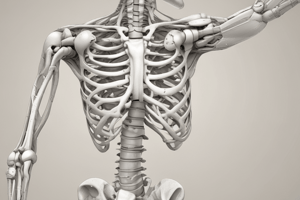
Hip Joint
- The concave acetabulum receives the convex femoral.
- Resting Position: hip flexion 30, abduction 30, and slight external rotation.
- Stabilization: fixate the pelvis to the treatment table with belts.
Hip Distraction of the Weight-Bearing Surface, Caudal Glide
- Indications: testing, initial treatment, pain control and general mobility.
- Patient Position: supine, with the hip in resting position and the knee extended.
- Therapist Position and Hand Placement: stand at the end of the treatment table, place a belt around the trunk, then cross the belt over the patient's foot and ankle, hands proximal to the malleoli, under the belt.
- The belt allows use of body weight to apply mobilizing force.
- Mobilizing Force: apply long-axis traction by pulling on the leg while leaning backward.
- Because of the deep configuration, traction applied perpendicular to the treatment plane causes lateral glide of the superior, weight-bearing surface.
- Caudal glide is used to obtain separation of the weight-bearing surface.
Hip Posterior Glide
- Indications: to increase flexion and internal rotation.
- Patient Position: supine, with hips at the end of the table.
- The patient stabilizes the pelvis/lumbar spine by flexing the opposite hip and holding the thigh against the chest with hands.
- The hip is positioned in resting position initially, then progress to the end of the range.
- Therapist Position and Hand Placement: stand on the medial side of the patient's thigh, place a belt around your shoulder and under the patient's thigh to hold the weight of the lower extremity.
- Place distal hand under the belt and distal thigh, and proximal hand on the anterior surface of the proximal thigh.
- Mobilizing Force: keep elbows extended and flex knees; apply the force through proximal hand in a posterior direction.
Hip Anterior Glide
- Indications: to increase extension and external rotation.
- Patient Position: prone, with the trunk resting on the table and hips over the edge, with the opposite foot on the floor.
- Therapist Position and Hand Placement: stand on the medial side of the patient's thigh, place a belt around shoulder and the patient's thigh to help support the weight of the leg.
- Hold the patient's leg with distributor hand, and place the proximal hand posteriorly on the proximal thigh just below the buttock.
- Alternate Position: patient side-lying with the thigh comfortably flexed and supported by pillows.
- Stand posterior to the patient and stabilize the pelvis across the anterior superior iliac spine with your superior hand.
- Mobilizing Force: keep your elbow extended and flex your knees, apply the force through your proximal hand in an anterior direction.
Knee and Leg
- The concave tibial plateaus articulate on the convex femoral condyles in the tibiofemoral articulation.
- Resting Position: the resting position of the knee is 25 degrees flexion.
- Treatment Plane: the treatment plane is along the surface of the tibial plateaus, move with the tibia as the knee angle changes.
- Stabilization: the femur is stabilized with a belt or by the table in most cases.
Tibiofemoral Distraction, Long-Axis Traction
- Indications: testing, initial treatment, pain control and general mobility.
- Patient Position: sitting, supine, or prone beginning with the knee in the resting position, can progress to the limit of flexion or extension.
- Hand Placement: grasp the distal leg, proximal to the malleoli with both hands.
- Mobilizing Force: pull on the long axis of the tibia to separate the joint surfaces.
Tibiofemoral Posterior Glide
- Indications: testing and to increase flexion.
- Patient Position: supine, with the foot resting on the table, position for the drawer test to mobilize the tibia either anteriorly or posteriorly.
- No grade I distraction can be applied with the glides.
- Therapist Position and Hand Placement: sit on the table with thigh fixating the patient's foot, and grasp around the tibia with both hands and fingers pointing posteriorly, thumbs anteriorly.
- Mobilizing Force: extend elbows and lean body weight forward; push the tibia posteriorly with thumbs.
Tibiofemoral Anterior Glide
- Indication: to increase extension.
- Patient Position: prone, beginning with the knee in resting position; progress to the end of the available range.
- The tibia can also be positioned in lateral rotation, a small pad can be placed under the distal femur to prevent patellar compression.
- Hand Placement: grasp the distal tibia with the hand that is closer to it, and place the palm of the proximal hand on the posterior aspect of the proximal tibia.
- Mobilizing Force: force with the hand on the proximal tibia in an anterior direction.
- Direct the force to the lateral or medial tibial plateau to isolate one side of the joint.
- Alternate Position: if the patient cannot be positioned prone, position them supine with a pad under the tibia.
- Mobilizing force is placed against the femur in a posterior direction.
- Drawer test position can also be used, the mobilizing force comes from the fingers on the posterior tibia as you lean backward.
Patellofemoral Joint, Distal Glide
- Indication: to increase patellar mobility for knee flexion.
- Patient Position: supine, with knee extended; progress to positioning the knee at the end of the available range in flexion.
- Hand Placement: stand next to the patient's thigh, facing the patient's feet.
- Place the web space of the hand that is closer to the thigh around the superior border of the patella, use the other hand for reinforcement.
- Mobilizing Force: glide the patella in a caudal direction, parallel to the femur.
- Precaution: do not compress the patella into the femoral condyles while performing this technique.
Patellofemoral Medial-Lateral Glide
- Indication: to increase patellar mobility.
- Patient Position: supine with the knee extended, side-lying may be used to apply a medial glide.
- Hand Placement: place the heel of your hand along either the medial or lateral aspect of the patella.
- Stand on the opposite side of the table to position your hand along the medial border and on the same side of the table to position your hand along the lateral border; place the other hand under the femur to stabilize it.
- Mobilizing Force: glide the patella in a medial or lateral direction, against the restriction.
Ankle and Foot Joints Talocrural Joint (Upper Ankle Joint)
- The convex talus articulates with the concave mortise made up of the tibia and fibula.
- Resting Position: 10 plantar flexion.
- Treatment Plane: in the mortise, in an anterior posterior direction with respect to the leg.
- Stabilization: the tibia is strapped or held against the table.
Talocrural Distraction
- Indications: testing, initial treatment, pain control and general mobility.
- Patient Position: supine, with the lower extremity extended, begin with the ankle in resting position, progress to the end of the available range of dorsiflexion or plantar flexion.
- Therapist Position and Hand Placement: stand at the end of the table and wrap the fingers of both hands over the dorsum of the patient’s foot, just distal to the mortise.
- Place thumbs on the plantar surface of the foot to hold it in resting position.
- Mobilization Force: pull the foot away from the long axis of the leg in a distal direction by leaning backward.
Talocrural Dorsal (Posterior) Glide
- Indication: to increase dorsiflexion.
- Patient Position: supine, with the leg supported on the table and the heel over the edge.
- Therapist Position and Hand Placement: stand to the side of the patient and stabilize the leg with your superior hand, or use a belt to secure the leg to the table.
- Place the palmar aspect of the web space of your other hand over the talus just distal to the mortise.
- Wrap fingers and thumb around the foot to maintain the ankle in resting position.
- Grade I distraction force is applied in a caudal direction.
- Mobilizing Force: glide the talus posteriorly with respect to the tibia by pushing against the talus.
Mobilization of Spine Functional Components
- Functionally, the spine is divided into anterior and posterior pillars.
- The anterior pillar is made up of the vertebral bodies and intervertebral disks.
- The posterior pillar, or vertebral arch, is made up of the articular processes and facet joints.
- The two transverse processes and spinous process, to which the muscles attach, are also part of the posterior pillar.
- Orientation of the facets in the sagittal plane:
- C for cervical vertebrae at 45 degrees.
- T for thoracic vertebrae at 60 degrees.
- L for lumbar vertebrae at 90 degrees.
Physical Examination
- Inspection: observe head position, any lateral flexion, FHP, neck contour, shoulder level, and arm movement.
- Palpation:
- Central: spinous process in midline.
- Unilateral: about 2.3 cm from midline will be apophyseal joints.
- Special Tests:
- Segmental traction and compression tests are technically very difficult to perform and are used only by the most skilled manual therapists.
Assessment of Active Movements of Cervical Spine
- Cervical flexion, extension, lateral flexion, and rotation
- Upper cervical extension and lower cervical flexion overpressure
- Upper cervical flexion and lower cervical extension overpressure
- Combined movement Flexion, side bending and rotation
Mobilization for Cervical Spine
- Postero-anterior Central V Pressure: used in case of Bilateral pain.
- Postero-anterior Unilateral V.Pressure: used in case of Unilateral pain.
Cervical Traction
- Usually provided in a supine position.
- Use of a chin-cradle grip with equal force anterior and posterior.
- If the patient has posterior TMJ disorder, apply force using a towel.
- The clinician supplies a light traction force, and the patient's symptoms are assessed for change.
- The force is held for approximately one minute, and the patient is reevaluated.
Central Postero-Anterior Glide
- This glide is beneficial for patients who demonstrate bilateral or midline pain.
- The patient lies in prone with the neck positioned in neutral and resting.
- The clinician palpates the C2 spinous process using the tips of the thumb.
- A gentle downward force is applied up to the first point of the patient's complaint of pain, and the pain response is assessed via thumb-to-thumb application.
Unilateral Postero-Anterior Glide
- A combined extension-based movement applied with rotation to the same side the examination is applied.
- Useful in unilateral pain.
- Patient is in prone, with neck placed in neutral.
- The clinician pulls the paraspinals lie to the side of the spinous process medially to expose the articular pillars (facets).
- To isolate the articular pillar, the clinician finds the spinous process of C2, slides approximately a thumb-width lateral, and moves his or her thumb in a caudal and cephalic direction.
- The raised area felt under the thumb is the articular pillar, the targeted region for mobilization.
- Clinician applies gentle downward force up to the first point of the patient's complaint of pain with a thumb-to-thumb application.
Cervical Lateral or Transverse Glides
- Applies a lateral glide to the lateral aspects of the spinous processes of the spine.
- The patient is positioned in prone or side-lying, with the neck is placed in neutral.
- The clinician palpates the C2 spinous process using the tips of the thumb.
- A very broad and deep contact is applied to the side of the spinous process using one thumb pad.
Assessment of Active Movements Of Thoracic Spine
- Thoracic flexion overpressure overpressure
- Thoracic extension
- Thoracic rotation overpressure
- Thoracic lateral flexion overpressure
- Thoracic flexion and rotation overpressure
- Thoracic lateral flexion and rotation to same side overpressure
- Thoracic lateral flexion and rotation to opposite sides overpressure
- Thoracic extension and rotation to overpressure
Mobilization for Thoracic Spine Central Posterior Anterior (CPA)
- The spinous processes of the thoracic spine are angled inferiorly, a posterior to anterior motion to the thoracic spine may promote extension of the specific segments.
- Patient assumes a prone lying position and resting symptoms are assessed.
- The clinician palpates the targeted level feeling for the first thoracic spinous processes.
- A PA force using a pisiform contact to the first point of reported pain is applied, and pain is assessed to determine if concordant.
Unilateral Posterior Anterior to the Facet
- Patient assumes a prone lying position.
- Clinician palpates the targeted level feeling for the deficit just lateral to the spinous processes.
- Clinician uses a thumb to thumb contact point, since the facets are not as pronounced as the spinous processes.
- Applies a PA force to the first point of reported pain and assesses pain to determine if concordant.
Transverse Glides
- A form of assessment technique that applies a lateral glide to the spinous processes of the spine.
- Patient is positioned in prone or sidelying, and the neck is placed in neutral.
- The clinician palpates the targeted spinous process using the tips of th thumb.
- Applies a very broad and deep contact to the side of the spinous process and more force to the base of the spinous process than the posterior tip using one thumb pad.
Mobilization of the First Rib
- Can be a cause in thoracic outlet syndrome, which is a combination of symptoms in which cervico-brachial nerve-related pain occurs because of compression or traction.
- Elevation of the first rib may lead to entrapment or injury of the nerve.
- Patient is placed in a supine position and resting symptoms are assessed.
- The clinician places the patient's head in side flexion to the same side and rotation away, reduces the stress of the scalene muscles on the first rib and allows the rib to "drop" during mobilization.
- Applies force toward the anterior, contralateral hip anterior superior iliac spine (ASIS).
- The technique can be performed in a sitting position.
Assessment of Active Movements Of Lumbar Spine
- Lumbar flexion overpressure
- Lumbar extension overpressure
- Lumbar lateral flexion overpressure
- Lumbar rotation overpressure
- Lumbar rotation with flexion and extension overpressure
- Lumbar rotation with Lateral flexion overpressure
- Lumbar rotation with Lateral flexion overpressure
Mobilization of Lumbar Spine Central Posterior-Anterior
- Assessment using a central posterior-anterior (CPA) is beneficial when patients report midline or bilateral symptoms.
- The technique is applied using a pisiform contact to the spinous process of the lumbar spine.
- A Thumb to thumb technique can be used
- Light force should be applied first and then followed by progressing intensities.
Unilateral Posterior - Anterior
- Involves a three-point movement targeted to the facet, lamina, or transverse process of the targeted segment.
- A gentle force perpendicular to the targeted transverse process of the lumbar spine (right or left) using thumb pad to thumb nail contact is applied..
- Asking for the reproduction of the concordant sign is recommended.
- Repeated movements or sustained holds help determine appropriateness of the technique.
- A segment can only be cleared if a significant amount of PA force is applied.
Transverse Glide
- A combined translation and rotation assessment method is achieved.
- Identified by the patient as a greater stimulator than a UPA or CPA.
- Should be used with caution and might lead to soreness and easier reproduction of symptoms.
- Using a thumb pad, the clinician hooks the targeted spinous process, removing the slack associated with soft tissue.
- If the pain of reported on the left, the a move to the right and should push the spinous process patient to the left.
- Placing the lateral force on the spinous process elicits rotation.
Studying That Suits You
Use AI to generate personalized quizzes and flashcards to suit your learning preferences.