Podcast
Questions and Answers
Parasympathetic stimulation increases heart rate and contractility.
Parasympathetic stimulation increases heart rate and contractility.
False (B)
The 'lub-dub' sound of the heart is produced by the opening of the heart valves.
The 'lub-dub' sound of the heart is produced by the opening of the heart valves.
False (B)
Adrenaline and noradrenaline can increase heart rate and blood pressure during stress.
Adrenaline and noradrenaline can increase heart rate and blood pressure during stress.
True (A)
Baroreceptors primarily detect changes in oxygen levels in the blood.
Baroreceptors primarily detect changes in oxygen levels in the blood.
Passive immunity provides immediate, specific protection against pathogens.
Passive immunity provides immediate, specific protection against pathogens.
The renal cortex is the innermost layer of the kidney.
The renal cortex is the innermost layer of the kidney.
Renal pyramids are situated in the renal medulla of the kidney.
Renal pyramids are situated in the renal medulla of the kidney.
Filtration in the kidneys is a passive process driven solely by gravity.
Filtration in the kidneys is a passive process driven solely by gravity.
The urinary tract includes only the urethra and the urinary bladder.
The urinary tract includes only the urethra and the urinary bladder.
Nephrons are responsible for both filtration and reabsorption in the kidneys.
Nephrons are responsible for both filtration and reabsorption in the kidneys.
The resting membrane potential of neurons is maintained by an equal distribution of ions inside and outside the neuron.
The resting membrane potential of neurons is maintained by an equal distribution of ions inside and outside the neuron.
Potassium ions (K+) are more permeable than sodium ions (Na+) at the resting membrane potential.
Potassium ions (K+) are more permeable than sodium ions (Na+) at the resting membrane potential.
Depolarization in a neuron results in an increase in the negative charge inside the neuron.
Depolarization in a neuron results in an increase in the negative charge inside the neuron.
The threshold level for triggering an action potential in neurons is typically around -55 millivolts.
The threshold level for triggering an action potential in neurons is typically around -55 millivolts.
Action potentials are propagated along the axon due to the opening of voltage-gated potassium channels.
Action potentials are propagated along the axon due to the opening of voltage-gated potassium channels.
The influx of sodium ions during depolarization can trigger further depolarization in adjacent regions of the neuron's membrane.
The influx of sodium ions during depolarization can trigger further depolarization in adjacent regions of the neuron's membrane.
Resting membrane potential refers to the neuron's activity while it is actively sending signals.
Resting membrane potential refers to the neuron's activity while it is actively sending signals.
When a neuron is at rest, it has a greater concentration of sodium ions (Na+) inside compared to outside.
When a neuron is at rest, it has a greater concentration of sodium ions (Na+) inside compared to outside.
The action potential can travel in a backward direction along the axon once it is initiated.
The action potential can travel in a backward direction along the axon once it is initiated.
The rapid change in membrane potential during action potential generation is primarily due to the movement of potassium ions.
The rapid change in membrane potential during action potential generation is primarily due to the movement of potassium ions.
Flashcards are hidden until you start studying
Study Notes
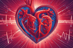
Heart Rate Regulation
- Sympathetic nervous system stimulation increases heart rate and contractility.
- Parasympathetic nervous system stimulation decreases heart rate.
- Adrenaline and noradrenaline released by adrenal glands during stress or exercise increase heart rate and blood pressure.
- Baroreceptors in blood vessel walls detect blood pressure changes and send signals to the brain to regulate heart rate and blood vessel diameter for blood pressure homeostasis.
- Local factors like tissue metabolism, oxygen levels, and carbon dioxide levels influence blood flow to specific tissues by causing vasodilation or vasoconstriction.
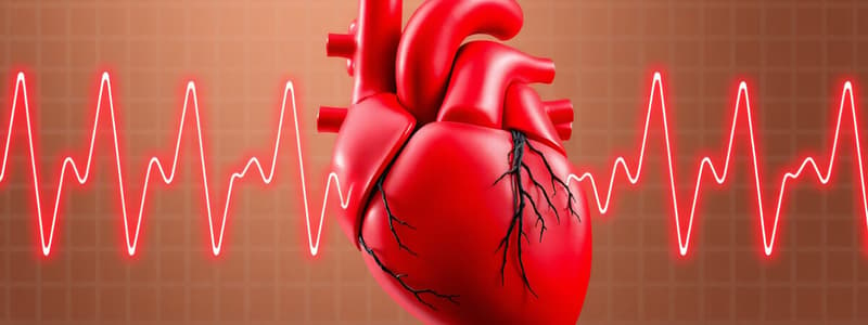
"Lub-Dub" Heart Sounds
- Heart sounds are caused by the closing of heart valves during the cardiac cycle.
- The "lub" sound is caused by the closure of the atrioventricular (AV) valves (mitral and tricuspid) at the start of systole (ventricular contraction).
- The "lub" sound marks the beginning of ventricular contraction and blood ejection into the pulmonary artery and aorta.
- The "dub" sound is caused by the closure of the semilunar valves (pulmonary and aortic valves) at the end of systole, as ventricles relax (diastole).
- The "dub" sound indicates the end of ventricular contraction and prevents blood backflow into the ventricles.
Passive (Innate) Immunity
- Passive immunity is the body's first line of defense against pathogens.
- It provides immediate, non-specific protection and is present at birth.
- Key components include:
- Physical barriers: Skin and mucous membranes prevent pathogen entry.
- Skin acts as a barrier against bacteria, viruses, and substances.
- Mucous membranes line respiratory, digestive, and reproductive tracts, producing mucus to trap pathogens.
- Chemical barriers: Body chemicals act as defenses.
- Physical barriers: Skin and mucous membranes prevent pathogen entry.
Excretory System
- The excretory system removes metabolic waste and maintains homeostasis by regulating water and electrolyte balance.
- Key organs include:
- Kidneys: Primary organ of the excretory system.
- Filter waste products from the blood to form urine.
- Have three regions:
- Renal cortex: Outer granulated layer dipping into the renal medulla.
- Renal medulla: Consists of cone-shaped renal pyramids.
- Renal pelvis: Central space, continuous with the ureter.
- Contain nephrons, the functional units for urine formation. -Nephrons filter blood to remove waste products, excess ions, and water.
- Urinary tract: Consists of ureters, bladder, and urethra.
- Transports urine from the kidneys through the ureters to the bladder for storage and elimination through the urethra.
- Kidneys: Primary organ of the excretory system.
Kidney Structure and Function
- Kidneys perform multiple functions beyond waste excretion:
- Filtration: Blood is filtered in the renal corpuscle to form filtrate.
- Occurs across glomerular capillaries, where blood pressure forces water, ions, and small molecules out of the blood into the renal tubule.
- Reabsorption: Useful substances (glucose, ions, water) are reabsorbed back into the bloodstream.
- Occurs selectively in different segments of the renal tubule.
- Secretion: Substances are transferred from the blood into the renal tubule for excretion in urine.
- Helps eliminate additional waste products and maintain electrolyte balance.
- Filtration: Blood is filtered in the renal corpuscle to form filtrate.
Neuron Membrane Potential
- Neurons maintain a resting membrane potential, an electrical charge difference across their membranes.
- The resting membrane potential is maintained by the unequal distribution of ions inside and outside the neuron.
- At rest, the neuron's membrane is more permeable to potassium ions (K+) than sodium ions (Na+).
- Potassium ions diffuse out of the neuron, leaving the inside negatively charged relative to the outside.
Neuron Depolarization
- Stimulation triggers depolarization, making the membrane less negative (or positive).
- Depolarization occurs when gated ion channels open, allowing sodium ions (Na+) to enter the neuron.
- This influx of positive sodium ions neutralizes the negative charge inside, causing a change in membrane potential.
Action Potential Generation
- If depolarization reaches the threshold level (-55 mV), an action potential is triggered.
- Voltage-gated sodium channels open rapidly, allowing massive sodium influx.
- This rapid depolarization creates a positive feedback loop, further depolarizing neighboring regions.
- The action potential propagates along the axon.
Action Potential Propagation
- The action potential travels down the axon as a self-regenerating wave of depolarization.
- This occurs because the action potential triggers the opening of voltage-gated sodium channels in the adjacent region of the axon membrane.
Cutaneous Mechanoreceptors
- Cutaneous (skin) mechanoreceptors are classified based on structure, location, and function:
- Merkel cells (Tactile Discs): Found in the epidermis, associated with slowly adapting (SA) mechanoreceptors.
- Respond to sustained pressure and fine touch, detecting tactile features like edges and shapes.
- Meissner's corpuscles: In the dermal papillae of hairless skin (fingertips, palms, lips, soles).
- Rapidly adapting (RA) mechanoreceptors for changes in skin indentation and dynamic touch.
- Involved in detecting light touch and low-frequency vibrations.
- Pacinian corpuscles: Located in the deeper layers of the dermis and hypodermis.
- RA mechanoreceptors sensitive to high-frequency vibrations and deep pressure.
- Respond to rapid skin indentation changes, detecting strong pressure and vibration stimuli.
- Ruffini endings (corpuscles): Located in the dermis and subcutaneous tissue.
- SA mechanoreceptors responding to skin stretch and sustained pressure.
- Involved in detecting skin distortion and contributing to proprioception and tactile perception.
- Merkel cells (Tactile Discs): Found in the epidermis, associated with slowly adapting (SA) mechanoreceptors.
Proprioceptors
- Proprioceptors are specialized mechanoreceptors in muscles, tendons, ligaments, and joints.
- They provide information about body position, movement, and limb orientation (proprioception).
- They play a vital role in coordinating voluntary movements, maintaining balance and posture, and preventing injury.
- Include:
- Muscle spindles: Within skeletal muscles, stretch receptors detecting changes in muscle length and contraction velocity.
- Provide feedback about muscle length and contribute to proprioceptive control of muscle tone and coordination.
- Muscle spindles: Within skeletal muscles, stretch receptors detecting changes in muscle length and contraction velocity.
Chemoreceptors
- Chemoreceptors detect changes in chemical composition of body fluids and transmit signals to the central nervous system for physiological regulation.
- Arterial chemoreceptors: Located in carotid bodies and aortic bodies, detect blood oxygen and carbon dioxide levels.
- Central chemoreceptors: In medulla oblongata, detect changes in cerebrospinal fluid pH and carbon dioxide levels.
- Together, they regulate respiratory drive and ensure adequate oxygenation and ventilation.
- Chemoreceptors in internal organs: Found in the heart, lungs, gastrointestinal tract, and kidneys.
- Detect changes in body fluid composition and regulate cardiovascular function, respiratory rate, gastrointestinal motility, and fluid balance.
- Chemoreceptors in the carotid bodies and aortic bodies: Detect changes in blood chemistry and influence heart rate, blood pressure, and blood flow distribution to vital organs.
- Functions of chemoreceptors:
- Detection of environmental cues: Chemoreceptors in the olfactory epithelium and taste buds detect chemical signals from the environment, allowing organisms to perceive odors and tastes.
- Regulation of respiratory function: Arterial and central chemoreceptors monitor blood and cerebrospinal fluid chemistry and regulate breathing to maintain blood gas homeostasis.
- Regulation of cardiovascular function: Chemoreceptors in the carotid bodies and aortic bodies detect changes in blood chemistry and influence heart rate, blood pressure, and blood flow distribution to vital organs.
- Control of homeostasis: Chemoreceptors in internal organs monitor changes in body fluid composition and help maintain internal homeostasis and organ system function.
Photoreceptors:
- Photoreceptors are specialized sensory cells in the retina of the eye that respond to light stimuli.
Studying That Suits You
Use AI to generate personalized quizzes and flashcards to suit your learning preferences.