Podcast
Questions and Answers
Which anatomical structure anchors and protects the heart?
Which anatomical structure anchors and protects the heart?
- Pericardium (correct)
- Thoracic cavity
- Pleural cavities
- Mediastinum
What is the function of the atrial syncytium?
What is the function of the atrial syncytium?
- To rapidly spread electrical signals over the atria, causing contraction (correct)
- To delay the electrical signal for ventricular filling
- To insulate the atria from ventricular electrical activity
- To filter the blood passing through the atria
What is the primary function of the cardiac valves?
What is the primary function of the cardiac valves?
- To regulate the speed of blood flow through the heart
- To ensure one-way blood flow through the heart (correct)
- To generate the heart's electrical impulses
- To provide structural support to the heart chambers
What event on an ECG is directly associated with ventricular systole?
What event on an ECG is directly associated with ventricular systole?
During ventricular diastole, what happens to the atrioventricular (AV) valves?
During ventricular diastole, what happens to the atrioventricular (AV) valves?
In a healthy individual, where would you typically palpate to most accurately assess heart rate?
In a healthy individual, where would you typically palpate to most accurately assess heart rate?
What best describes the role of the sympathetic nervous system on heart rate?
What best describes the role of the sympathetic nervous system on heart rate?
Which of the following best illustrates the relationship between end-diastolic volume (EDV), end-systolic volume (ESV), and stroke volume (SV)?
Which of the following best illustrates the relationship between end-diastolic volume (EDV), end-systolic volume (ESV), and stroke volume (SV)?
What is the origin and significance of the 'S1' heart sound?
What is the origin and significance of the 'S1' heart sound?
Where does the left atrium receive blood from?
Where does the left atrium receive blood from?
What is the role of the Purkinje fibers in the cardiac conduction system?
What is the role of the Purkinje fibers in the cardiac conduction system?
If a physician hears a 'swishing' sound during auscultation of the heart, what is the most likely cause?
If a physician hears a 'swishing' sound during auscultation of the heart, what is the most likely cause?
Where is the location of the heart inside the thoracic cavity?
Where is the location of the heart inside the thoracic cavity?
What is the sequence of blood flow after it leaves the right atrium?
What is the sequence of blood flow after it leaves the right atrium?
What causes the excitation of the ventricles in the heart, as seen on an ECG?
What causes the excitation of the ventricles in the heart, as seen on an ECG?
What determines the opening and closing of the heart valves?
What determines the opening and closing of the heart valves?
How does the parasympathetic nervous system affect the heart?
How does the parasympathetic nervous system affect the heart?
How many chambers does the heart have?
How many chambers does the heart have?
What is the definition of cardiac output?
What is the definition of cardiac output?
On an electrocardiogram (ECG), what does the T wave represent?
On an electrocardiogram (ECG), what does the T wave represent?
Flashcards
Heart Location
Heart Location
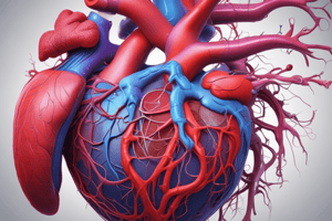
The heart is a hollow, muscular, cone-shaped organ positioned in the mediastinum and within the pericardial cavity.
Heart Chambers
Heart Chambers
The heart has four chambers: two superior atria and two inferior ventricles.
Right Atrium Input
Right Atrium Input
The right atrium receives deoxygenated blood from the superior vena cava, inferior vena cava, and coronary sinus.
Right Ventricle Output
Right Ventricle Output
Signup and view all the flashcards
Left Atrium Input
Left Atrium Input
Signup and view all the flashcards
Left Ventricle Output
Left Ventricle Output
Signup and view all the flashcards
Systemic Circulation
Systemic Circulation
Signup and view all the flashcards
Heart Syncytia
Heart Syncytia
Signup and view all the flashcards
SA Node Function
SA Node Function
Signup and view all the flashcards
ECG Function
ECG Function
Signup and view all the flashcards
Isoelectric Line
Isoelectric Line
Signup and view all the flashcards
Heart Valve Function
Heart Valve Function
Signup and view all the flashcards
AV Valves
AV Valves
Signup and view all the flashcards
Semilunar Valves
Semilunar Valves
Signup and view all the flashcards
Cardiac Output (CO)
Cardiac Output (CO)
Signup and view all the flashcards
Heart Rate Control
Heart Rate Control
Signup and view all the flashcards
Tachycardia
Tachycardia
Signup and view all the flashcards
Bradycardia
Bradycardia
Signup and view all the flashcards
Assessing Pulse
Assessing Pulse
Signup and view all the flashcards
Ventricular Diastole
Ventricular Diastole
Signup and view all the flashcards
Study Notes
Heart Location
- Located in the thoracic cavity as a hollow, muscular, cone-shaped organ
- The general location is in the mediastinum
- Found within the pericardial cavity and between the pleural cavities
- Enclosed by the pericardium, which anchors and protects it
- The esophagus and trachea are posterior to it
Heart's Internal Structure
- The heart has four chambers or compartments
- Two superior chambers, termed atria
- Two inferior chambers, termed ventricles
- The interatrial and interventricular septum prevent blood mixing between chambers
- Ventricular walls are thicker than atrial walls because they pump blood into the systemic and pulmonary circulations
Right Atrium
- Receives deoxygenated blood from the body through three vessels
- The three vessels are the superior vena cava, inferior vena cava, and coronary sinus
- From this chamber, blood flows to the right ventricle
Right Ventricle
- The right ventricle pumps deoxygenated blood into the pulmonary trunk
Left Atrium
- The left atrium receives oxygenated blood via the pulmonary veins
Left Ventricle
- The left ventricle pumps oxygenated blood into the aorta
- The left ventricle walls are thicker than the right requiring more force to send blood throughout the systemic circulation
Systemic Circulation
- Supplies the body's tissues and organs with oxygenated blood
- Newly deoxygenated blood returns to the right atrium via the superior vena cava, inferior vena cava, and coronary sinus
Heart at Microscopic Level
- There are two functional units (syncytia)
- These are the atrial syncytium and ventricular syncytium
- The atria then ventricles contract, and then the heart relaxes
Heart Cells
- Two different types of cells exist in the heart wall
- These are contracting cells and cells that generate an electrical signal
- Cells generating electrical signals form the cardiac conduction system in each heartbeat
Cardiac Conduction System
- Starts at the sinoatrial node (SA node)
- The SA node is part of the cardiac conduction system that generates an electrical signal most rapidly
- The SA node spreads signals over the entire atrial syncytium, causing atrial contraction
- Signals then spread to the atrioventricular node (AV node)
- From the AV node signals pass through the atrioventricular bundle (AV bundle)
- Signals arrive in the interventricular septum then pass through two bundle branches (right and left)
- At the apex, fibers branch extensively, forming Purkinje fibers
Electrocardiograms (ECGs)
- ECGs assess the cardiac conduction system
- Determines whether the electrical activity of the heart is working properly
- Electrodes are placed on the body in 2 upper limb leads, 2 lower limb leads, and 6 precordial leads
- Enables assessment of the heart from 12 different angles to pinpoint abnormality locations
Electrical Current & Waves
- The current arising from the SA node, called depolarization, is a positive current
- Positive current travels through the atrial walls
- A negative current then restores the electrical potential of the atrium to normal after the positive current passes, called repolarization
- The first wave is the P wave
- The second group of waves is the QRS complex consisting of the Q, R, and S waves
- The third wave is the T wave
- Isoelectric lines occur when there is no change occurring in the electrical state of the heart
- This happens between the P wave and QRS complex and between the QRS complex and T wave
Heart Valves
- Heart valves ensure one-way blood flow
- They are composed of dense, fibrous connective tissue which is covered in endocardium
- There are 4 valves organized as two pairs: atrioventricular (AV) and semilunar (SL)
Atrioventricular Valves (AV)
- The tricuspid and mitral valve are AV valves
Valve Function
- AV valves close when ventricles contract and the pressure in the ventricles exceeds pressure in the atria
- Chordae tendinae and papillary muscles contract along with ventricles, creating tension that prevents the free edges of the valves from swinging upward into the atria
- AV valves open after ventricular relaxation, when atrial pressure exceeds ventricular pressure
Semilunar Valves (SL)
- Blood passes from the right ventricle to the pulmonary trunk and from the left ventricle into the aorta through these valves
- The pulmonic and aortic valves are semilunar valves valves
- When closed, the cusps fall into the center of the pulmonary trunk and aorta to prevent backflow of blood from the vessel into the ventricle
- During ventricular contraction pressure increases, and when ventricular pressure exceeds pressure in the aorta and pulmonary trunk, the semilunar valves open
- During ventricular relaxation, pressure drops, and when ventricular pressure falls below the pressure in the aorta and pulmonary trunk, the semilunar valves close
Cardiac Cycle
- When the heart is relaxed, the semilunar valves are closed and the AV valves are open
- Blood returns to the right atrium through the superior and inferior vena cava and coronary sinus
- On the left side, blood returns to the heart from the pulmonary veins from the lungs
- During atrial contraction, the pressure in atria increases
- Atria relax during ventricular contraction when The pressure exceeds the pressure in the atria
- As ventricles contract ventricular pressure climbs, exceeding pressure in the aorta and pulmonary trunk
- Finally ventricles stop contracting and begin to relax
- When valves close, vibrations occur in the blood passing through the heart, producing heart sounds (sound one) that are carried to the body’s surface and can be heard with a stethoscope
Heart Sounds
- S1 (sound one) and S2 (sound two) are the heart sounds
- S2 (sound two) indicates start of ventricular diastole
Heart Position
- The heart is positioned deep to the sternum, slightly to the left of the midline in the chest cavity
- The point of maximal impulse is the most accurate location to check heart rate
- Palpating assesses the number of beats per minute
- Auscultation allows assessment of heart rate as well as heart sounds
Auscultation & Heart Rate
- Physicians often auscultate in multiple locations to assess heart sounds related to the aortic, pulmonic, tricuspid, and mitral valves
- A valve that does not close all the way makes a swishing sound
- A valve that does not open all the way makes a clicking sound
Electrical Changes during the Cardiac Cycle
- The electrical changes start at the SA node
- Excitation of the atria creates the P-wave on an ECG
- A signal is sent to the AV node, delayed for 1/10th of a second
- This then passes to the AV bundle which is the only electrical connection between the atrial and ventricular syncytium
- Excitation of the ventricles creates the QRS complex on an ECG
- The heart relaxes and ventricles repolarize, creating the T wave on the ECG
Pressure Changes during the Cardiac Cycle
- Pressure changes in the heart prevent backflow of blood, causing the valves to open and close during the cardiac cycle
- The P wave on an ECG is followed closely by increased atrial pressure during atrial contraction (atrial systole)
- After atrial systole, the atria contract, and pressure remains low
- The QRS complex on an ECG is followed almost immediately by increased ventricular pressure
- The period of ventricular contraction is called ventricular systole
- When ventricular pressure exceeds aortic pressure, the semilunar valves open
- Once this pressure peaks, the ventricles stop contracting, and pressure in ventricles falls below pressure in the aorta and pulmonary trunk until ventricular pressure continues to fall
Ventricular Volume
- Ventricular volume is fairly high during relaxation, or diastole
- During atrial systole a little more blood is pushed into the ventricles, slightly increasing volume
- Ventricles contract, pressure increases and volume decreases
- Blood is ejected to the aorta and pulmonary trunk
- AV valves open and ventricular volume begins to increase again
Assessing Pulse Demo & Heart Rate
- Pulse and heart rate are typically the same in a person with healthy cardiovascular function
- Pulse and heart rate may differ if someone has poor peripheral circulation or arterial disease
- Pulse is assessed in elastic arteries, which can distend and retract
Locations to Assess Pulse
- Carotid artery
- Brachial artery (at the antecubital fossa)
- Radial artery
- Femoral artery
- Dorsalis pedis
- Posterior tibial
Cardiac Output
- Cardiac output is the volume of blood ejected per minute
- CO is vital to maintain blood flow circulating through the body to supply O2 and nutrients to cells and carry away metabolic waste
- Cardiac output is influenced by heart rate (HR) and stroke volume (SV)
- Cardiac output can be calculated as HR x SV
- Stroke volume is the volume of blood ejected during a single heartbeat
- Stroke volume is calculated as End diastolic volume (EDV) - End systolic volume (ESV)
- Cardiac output can be increased or decreased to meet the needs of the body
Autonomic Nervous System
- Plays a role in regulating cardiac output through two branches: parasympathetic and sympathetic
Sympathetic Division
- In the heart, the sympathetic nervous system innervates the SA node, AV node, and contractile cells of the myocardium
- When activated, causes the SA node to depolarize more quickly and can shorten the delay at the AV node
- Tachycardia is defined as HR > 100 bpm
- Sympathetic activation also causes contractile cells of myocardium to contract more forcefully, leading to increased stroke volume
Parasympathetic Division
- In the heart, the parasympathetic nervous system innervates the SA node and the AV node
- Parasympathetic nervous system slows the SA node’s rate of self-excitation
- Parasympathetic signals are carried to the SA node by the vagus nerve
- Bradycardia is defined as HR < 60 bpm
Studying That Suits You
Use AI to generate personalized quizzes and flashcards to suit your learning preferences.


