Podcast
Questions and Answers
Which of the following structures is responsible for preventing blood from flowing back into the atria during ventricular contraction?
Which of the following structures is responsible for preventing blood from flowing back into the atria during ventricular contraction?
- Pulmonary veins
- Aorta
- Atrioventricular valves (correct)
- Semilunar valves
What is the primary function of the right ventricle in a heart box diagram?
What is the primary function of the right ventricle in a heart box diagram?
- Receive deoxygenated blood from the body
- Pump deoxygenated blood to the lungs (correct)
- Receive oxygenated blood from the lungs
- Pump oxygenated blood to the body
Which of the following statements correctly describes the relationship between the pulmonary artery and the pulmonary veins?
Which of the following statements correctly describes the relationship between the pulmonary artery and the pulmonary veins?
- The pulmonary artery carries oxygenated blood, and the pulmonary vein carries deoxygenated blood.
- The pulmonary artery carries deoxygenated blood, and the pulmonary vein carries oxygenated blood. (correct)
- Both carry oxygenated blood.
- Both carry deoxygenated blood.
What happens during the diastole phase of the cardiac cycle?
What happens during the diastole phase of the cardiac cycle?
Which of the following is NOT a limitation of the heart box diagram?
Which of the following is NOT a limitation of the heart box diagram?
What is the correct sequence of blood flow through the heart, as depicted in a heart box diagram?
What is the correct sequence of blood flow through the heart, as depicted in a heart box diagram?
The heart box diagram primarily focuses on which of the following aspects?
The heart box diagram primarily focuses on which of the following aspects?
Which of the following is a key concept highlighted by the heart box diagram?
Which of the following is a key concept highlighted by the heart box diagram?
Flashcards
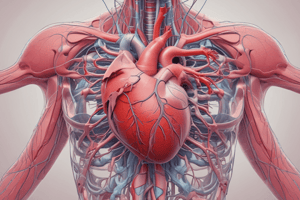
Heart Box Diagram
Heart Box Diagram
A simplified representation of the heart to visualize its structure and function.
Atria
Atria
Upper chambers of the heart that receive blood from the body or lungs.
Ventricles
Ventricles
Lower chambers of the heart responsible for pumping blood out.
AV Valves
AV Valves
Signup and view all the flashcards
Semilunar Valves
Semilunar Valves
Signup and view all the flashcards
Blood Flow Order
Blood Flow Order
Signup and view all the flashcards
Systole
Systole
Signup and view all the flashcards
Diastole
Diastole
Signup and view all the flashcards
Study Notes
Introduction
- A heart box diagram is a simplified representation of the heart, useful for visualizing its structure and function.
- It emphasizes the chambers and valves, often omitting smaller details like cardiac muscle.
- This diagram helps in understanding the flow of blood through the heart, demonstrating how blood is pumped.
Components of a Heart Box Diagram
- Atria (left and right): Upper chambers of the heart.
- Receive blood from the body or lungs.
- Ventricles (left and right): Lower chambers of the heart.
- Pump blood out to the body or lungs.
- Valves: Structures that direct blood flow.
- Atrioventricular (AV) valves: Between atria and ventricles (tricuspid on right, mitral on left).
- Prevent backflow into the atria.
- Semilunar valves: At the exits of the ventricles, near the major blood vessels (pulmonary and aortic).
- Prevent backflow from the arteries into the ventricles (pulmonary and aortic).
Blood Flow Through the Heart (Simplified)
- Superior/Inferior Vena Cava: Returns deoxygenated blood to the right atrium.
- Right Atrium: Receives deoxygenated blood from the body.
- Tricuspid Valve: Opens to allow blood flow to the right ventricle.
- Right Ventricle: Pumps deoxygenated blood to the lungs (via pulmonary artery).
- Pulmonary Artery: Carries deoxygenated blood to the lungs.
- Pulmonary Veins: Returns oxygenated blood from the lungs, entering the left atrium.
- Left Atrium: Receives oxygenated blood from the lungs.
- Mitral Valve (Bicuspid): Opens to allow blood flow to the left ventricle.
- Left Ventricle: Pumps oxygenated blood to the body (via aorta).
- Aorta: Carries oxygenated blood to the body's tissues.
Key Concepts Highlighted by a Heart Box Diagram
- One-way valves: Ensures blood flows in a singular direction through the heart.
- Atrial contraction (systole): Blood is pushed into the ventricles.
- Ventricular contraction (systole): Blood is pumped out of the ventricles.
- Diastole: Relaxation phase of the heart, allowing the chambers to fill with blood.
Limitations of the Heart Box Diagram
- Oversimplification: Omits intricate details like cardiac muscle structure, conducting system, and various cellular components.
- Static Representation: Does not depict the dynamic processes like changes in pressure, volume, and electrical changes throughout the cardiac cycle.
- Lack of detail: Doesn't show the complex anatomy of the heart valves or blood vessel connections.
Uses of Heart Box Diagrams
- Basic introduction to cardiac function.
- Teaching tool for understanding blood flow.
- Visual aid in medical education for beginners and quick review.
- Conceptual framework for complex cardiac concepts.
- Helps students visualize relationships among parts of the heart.
Studying That Suits You
Use AI to generate personalized quizzes and flashcards to suit your learning preferences.