Podcast
Questions and Answers
During ventricular systole, which event directly leads to the ejection phase?
During ventricular systole, which event directly leads to the ejection phase?
- The opening of the atrioventricular valves.
- Repolarization of the ventricles.
- Closure of the semilunar valves.
- Ventricular pressure exceeding aortic pressure. (correct)
What is the primary function of the AV node delay in the cardiac cycle?
What is the primary function of the AV node delay in the cardiac cycle?
- To initiate ventricular depolarization.
- To ensure the atria complete their contraction before ventricular systole. (correct)
- To allow the ventricles to fill completely before atrial contraction.
- To regulate heart rate based on blood pressure.
If the sinoatrial (SA) node is damaged, but the atrioventricular (AV) node is still functional, what would be the expected heart rate?
If the sinoatrial (SA) node is damaged, but the atrioventricular (AV) node is still functional, what would be the expected heart rate?
- The heart rate would decrease to a range of 40-60 bpm. (correct)
- The heart rate would decrease to approximately 35 bpm.
- The heart rate would increase above 100 bpm.
- The heart rate would remain normal (60-100 bpm).
What is the effect of increased venous return on stroke volume, according to the Frank-Starling mechanism?
What is the effect of increased venous return on stroke volume, according to the Frank-Starling mechanism?
Which of the following best describes the role of the pulmonary artery?
Which of the following best describes the role of the pulmonary artery?
During which phase of the cardiac cycle is the left ventricular volume at its minimum?
During which phase of the cardiac cycle is the left ventricular volume at its minimum?
Which event is directly responsible for the 'lub' sound during a normal cardiac cycle?
Which event is directly responsible for the 'lub' sound during a normal cardiac cycle?
Which of the following occurs during the T-P segment of an electrocardiogram (ECG)?
Which of the following occurs during the T-P segment of an electrocardiogram (ECG)?
Why does ventricular fibrillation lead to a low BP and cardiac output?
Why does ventricular fibrillation lead to a low BP and cardiac output?
What would directly cause a decreased glomerular filtration rate (GFR)?
What would directly cause a decreased glomerular filtration rate (GFR)?
In a healthy kidney, where does most of the isosmotic reabsorption occur?
In a healthy kidney, where does most of the isosmotic reabsorption occur?
How does antidiuretic hormone (ADH, vasopressin) contribute to water reabsorption in the kidneys?
How does antidiuretic hormone (ADH, vasopressin) contribute to water reabsorption in the kidneys?
What is the mechanism of action of the sodium-glucose cotransporter (SGLT) in the proximal tubule?
What is the mechanism of action of the sodium-glucose cotransporter (SGLT) in the proximal tubule?
Which of the following conditions would stimulate the release of renin from granular cells?
Which of the following conditions would stimulate the release of renin from granular cells?
What is the initial effect of angiotensin II on the glomerular capillaries?
What is the initial effect of angiotensin II on the glomerular capillaries?
Which of the following statements best describes the function of the vasa recta in the kidney?
Which of the following statements best describes the function of the vasa recta in the kidney?
Substances such as penicillin are secreted into the nephron. What is the advantage of secretion in addition to filtration for the excretion of these substances?
Substances such as penicillin are secreted into the nephron. What is the advantage of secretion in addition to filtration for the excretion of these substances?
If the concentration of a substance in the plasma is higher than its transport maximum (Tm) in the kidney, how will this affect its reabsorption and excretion?
If the concentration of a substance in the plasma is higher than its transport maximum (Tm) in the kidney, how will this affect its reabsorption and excretion?
How does severe dehydration lead to conservation of blood volume via influence on GFR?
How does severe dehydration lead to conservation of blood volume via influence on GFR?
What is the primary function of organic anion transporters (OATs) in the kidneys?
What is the primary function of organic anion transporters (OATs) in the kidneys?
In the countercurrent exchange system of the vasa recta, how does blood flow contribute to maintaining the medullary osmotic gradient?
In the countercurrent exchange system of the vasa recta, how does blood flow contribute to maintaining the medullary osmotic gradient?
If the afferent arteriole of a glomerulus constricts, what immediate change would occur in the glomerular filtration rate (GFR) and why?
If the afferent arteriole of a glomerulus constricts, what immediate change would occur in the glomerular filtration rate (GFR) and why?
What would occur if the aquaporins of the collecting duct were non-functional?
What would occur if the aquaporins of the collecting duct were non-functional?
What effect do beta-blockers have on cardiac contractility and why?
What effect do beta-blockers have on cardiac contractility and why?
Why is it important that blood flows passively through the heart?
Why is it important that blood flows passively through the heart?
How does atrial fibrillation lead to less effective ventricular filling?
How does atrial fibrillation lead to less effective ventricular filling?
If someone has a blockage between their left ventricle and atrium where would blood build up?
If someone has a blockage between their left ventricle and atrium where would blood build up?
What is the effect of increasing cytosolic $Ca^{2+}$ in contractile fibers?
What is the effect of increasing cytosolic $Ca^{2+}$ in contractile fibers?
In what scenario would kidney release renin?
In what scenario would kidney release renin?
Flashcards
Atria
Atria
Upper heart chambers that receive blood.
Ventricles
Ventricles
Lower heart chambers that pump blood out.
Atrioventricular Valve (AV)
Atrioventricular Valve (AV)
Prevents backflow from ventricles into atria.
Semilunar Valves
Semilunar Valves
Signup and view all the flashcards
Veins
Veins
Signup and view all the flashcards
Arteries
Arteries
Signup and view all the flashcards
Right Atrium
Right Atrium
Signup and view all the flashcards
Right Ventricle
Right Ventricle
Signup and view all the flashcards
Pulmonary Valve
Pulmonary Valve
Signup and view all the flashcards
Pulmonary Arteries
Pulmonary Arteries
Signup and view all the flashcards
Pulmonary Veins
Pulmonary Veins
Signup and view all the flashcards
Left Atrium
Left Atrium
Signup and view all the flashcards
Bicuspid Valve
Bicuspid Valve
Signup and view all the flashcards
Intrinsic Conduction System
Intrinsic Conduction System
Signup and view all the flashcards
Internodal Pathway
Internodal Pathway
Signup and view all the flashcards
Cardiac Cycle
Cardiac Cycle
Signup and view all the flashcards
Diastole
Diastole
Signup and view all the flashcards
Systole
Systole
Signup and view all the flashcards
Complete Heart Block
Complete Heart Block
Signup and view all the flashcards
Electrocardiogram (ECG/EKG)
Electrocardiogram (ECG/EKG)
Signup and view all the flashcards
Ventricular Ejection
Ventricular Ejection
Signup and view all the flashcards
Cardiac Output (CO)
Cardiac Output (CO)
Signup and view all the flashcards
Stroke Volume (SV)
Stroke Volume (SV)
Signup and view all the flashcards
Preload
Preload
Signup and view all the flashcards
Wigger's Diagram
Wigger's Diagram
Signup and view all the flashcards
Resistance
Resistance
Signup and view all the flashcards
Blood Osmolarity
Blood Osmolarity
Signup and view all the flashcards
Filtration (Kidney)
Filtration (Kidney)
Signup and view all the flashcards
Glomerular Filtration Rate (GFR)
Glomerular Filtration Rate (GFR)
Signup and view all the flashcards
Reabsorption
Reabsorption
Signup and view all the flashcards
Study Notes
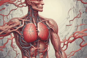
Blood Flow Overview
- Deoxygenated blood from the body flows to the right atrium, then the right ventricle, before going to the lungs.
- Oxygenated blood returns from the lungs to the left atrium, then the left ventricle, and is pumped out to the body.
Overview of Components
- Atria are the upper chambers; receive blood.
- Ventricles are the lower chambers that send blood out.
- The septum divides the heart into left and right sides. The right side contains deoxygenated blood, and the left side contains oxygenated blood.
- Valves ensure unidirectional blood flow facilitated by passive movement due to blood pressure.
Valves
- Atrioventricular (AV) valves are thin tissue flaps between the atria and ventricles prevent backflow into the atria and are anchored by connective tendons with Bicuspid/mitral and tricuspid valves.
- Semilunar valves are crescent-shaped flaps between the ventricles and arteries and prevent backflow facilitated by cup-like leaflets that snap shut when filled with back-flowing blood: pulmonary and aortic valves.
- Blood vessels include: veins that carry blood to the heart and arteries that carry blood away from the heart.
Blood Flow
- Deoxygenated blood enters the heart via the superior and inferior vena cavae into the right atrium, and is sent to the right ventricle.
- The tricuspid valve (AV valve) prevents backflow from the right ventricle into the right atrium.
- The right ventricle sends blood to the lungs.
- The semilunar pulmonary valve prevents backflow from the pulmonary arteries to the right ventricle.
- Via the pulmonary arteries, deoxygenated blood is carried to the lungs.
- Oxygenated blood travels via pulmonary veins back to the heart where it enters the left atrium, then is sent to the left ventricle.
- The bicuspid (mitral AV) valve prevents backflow from left ventricle into left atrium.
- From the left ventricle, blood is sent to the system via the aorta.
- The aortic valve (semilunar) prevents backflow from the aorta to the left ventricle. The aorta (artery) carries oxygenated blood to the systemic blood stream.
- Blood is squeezed from the atria into the ventricles; contraction proceeds from the bottom up with ventricles depolarizing from the bottom up.
Intrinsic Cardiac Conduction System
- The Intrinsic Conduction System is a system of autorhythmic nodes and conductive pathways, producing the heartbeat.
It includes:
- Sinoatrial Node
- Atrioventricular Node
- Conductive System: pathways of conducting autorhythmic cells connected via gap junctions.
- The conductive system functions to spread depolarization but doesn't set heart rate.
- Intermodal Pathway: branched conduction pathway from SA to AV node.
- Ventricular conduction system: branched conduction pathway in ventricles
- AV Bundle (aka Bundle of His)
- Purkinje fibers
- Electrical Conduction: Action potentials at autorhythmic cells in the intrinsic conduction system trigger action potentials in adjacent contractile cells.
Cardiac Cycle Overview
- The Cardiac Cycle is one contraction-relaxation cycle in the heart from the end of one heartbeat to the end of the next.
- Mechanical events lag slightly behind electrical signals with diastole (cardiac muscle relaxes) and systole (cardiac muscle contracts).
Sequencing the Cardiac Cycle
- Atrial and ventricular diastole occurs when all chambers are at rest, atria fill with blood from veins, ventricles have just completed contraction.
- Ventricles relaxing opens AV valves, enabling blood flow passively from atria to ventricles.
- Depolarization at SA node occurs and electrical activity travels rapidly to AV node via intermodal pathways
- Atrial Systole: atrial contraction finishes moving blood from atria to ventricles: Triggered by depolarization at atria and atrial contractile atrial cells contract leading to blood finishes flowing from atria to ventricles to end-diastolic volume.
- AV Node Delay: Conduction/depolarization slows across the AV node because of slower signal conduction through nodal cells, which allows the atria to complete contraction before ventricular contraction.
- Depolarization travels rapidly through ventricular conduction system to apex. Apex contractile cells contract approximately simultaneously during early ventricular systole where ventricles contract & push blood up, pushing the AV valves closed resulting in a “lub” heart sound. Semilunar valves are closed, isovolumic ventricular contraction, blood volume in the ventricle doesn't change.
- Depolarization spreads upward from apex through ventricles.
- Ventricular ejection: ventricular contraction forces open semilunar valves & blood pushes into arteries → End-systole.
- Ventricular diastole: ventricles repolarize and relax where ventricular pressure is less than arterial pressure, blood flows backward from arteries, semilunar valves close. Ventricular pressure is greater than atrial pressure (still). → AV valves remain closed→ Isovolumic ventricular relaxation.
- End of cardiac cycle → Atrial and ventricular diastole.
Complete Block
- In a complete block (third-degree AV block), conduction from atria to ventricles is disrupted.
- The SA node fires, but the signal isn't conducted to ventricles. Ventricles coordinate firing with ventricular autorhythmic cells 35 bpm.
- Contraction may be too slow to maintain blood flow: Treatment: mechanical pacemaker stimulates heart at a predetermined rate.
Electrocardiogram
- Electrocardiogram (ECG or EKG): recording of the summed electrical events of cardiac cycle.
- Einthoven's Triangle relates to Leads that record heart activity with electrodes stuck to the skin to record electrical activity of contractile fibers.
Spread of Electrical Events
- Occurs through depolarization and repolarization of cells
Differential Recording of Depolarization
- Involves the relative number of positive to negative charges at a point
- Depolarization is fast and repolarization is slow
Reading an EKG
- P wave: atrial depolarization, phase 0 in atrial contractile fibers, width less than 0.1s - If P wave it too wide it suggests atrial disease or use of Na+ channel blockers
- QRS complex: ventricular depolarization, phase 0 in ventricular contractile fibers. Shape of QRS complex varies, and its width is less than 0.09s. If too wide it implies conduction system problem, abnormal pacemaker in ventricles, ventricular disease, or to much Na.
- Atrial repolarization (Tp wave):
- Phase 3 in atrial contractile fibers that is typically masked by QRS complex
- T-wave: ventricular repolarization: Phase 3 in ventricular contractile fibers
Intervals
- Contain waves and segments, but do not contain waves.
RR Interval
- R wave to R wave indicates the period/time between contractions and indicates HR.
- Heart Rate (beats/minute) = 60 (sec/min)/RR (sec/beat).
PR Interval
- P wave to R wave representing time for atrial depolarization + AVN plus and indication of dromotropic state.
Segments
- Do not contain waves.
S-T Segment
- All ventricular fibers in Phase 2 (Ca2+ plateau): and usually returns to baseline STEMI elevation indicates heart attack.
T-P Segment
- The end of 1 heartbeat to starting of next ones.
EKG Analysis
- Heart Rate: 1 beat = beginning of one P wave to the beginning of the next P wave. a normal sinus rhythm beats around 60-100 bpm, bradycardia = sow HR, tachycardia = rapid HR.
- Cardiac Arrhythmia: irregular heartbeat, a benign extra heat that involves a “quivering”, where cells do not contact together. In atrial fibrillation: irregular heartbeat due to failure of the SA node, resulting in atrial contraction and poor ventricular filling.
- Ventricular fibrillation: irregular heartbeat due to irregular ventricular contraction, resulting in low blood pressure and cardiac output. The treatment is defibrillator shock that depolarizes all cells simultaneously. Action potentials in all cells being triggered simultaneously and coordinated contraction.
Calculating Blood Flow
- Follows Ohm's Law: Q = Δ Pressure/Resistance, Q (volume of blood), Resistance (resistance to blood flow), Δ pressure (pressure difference between chambers/vessel)
Wigger's Diagram
- Graph of electrical and mechanical events of 1 cardiac cycle, including an ECG, pressure, and left ventricular volume.
Atrial Systole
- EKG: P wave precedes atrial systole
- Left atrial pressure: first increases then decreases
- The diastolic blood pressure occurs because of pressure in the arteries when the heart rests between beats.
- It increases to EDV
Isovolumetric Ventricular Contraction
- EKG The QRS complex precedes ventricular systole
- Pressure The Bicuspid valve closes The ventricular pressure increases
- The LV Volume does no change
Ventricular Ejection
- EKG S-T segment, which occurs when the ventricles are in the Ca2+ plateau phase
- Pressure Ventricular pressure is greater than aortic pressure The aortic valve opens The aortic pressure increases (systolic blood pressure) The ventricular pressure first increases then decreases
- LV Volume Ejection decreases volume toward ESV
Early Ventricular Diastole
- EKG: T-wave precedes diastole
- Pressure: the aortic valve closes(B)
- Dicrotic notch
- Ventricular pressure decreases
- The LV Volume in isovolumic diastole
Late Ventricular Diastole
- EKG: T-P segment
- Pressure: ventricular pressure < atrial pressure while AV valves open.
- The LV Volume increases, and the volume during passive filling of the ventricles.
Cardiac Output
- Cardiac Output (CO): the volume of blood pumped by one ventricle in a period of time measured in mL/minute.
- Increases with exertion and O2 requirements.
- CO = HR x SV
- Average CO = 5040 mL/min. ((72 bpm * 70 mL/beat))
Stroke Volume
- Stroke Volume (SV): the amount of blood pumped by one ventricle per contraction, measured in mL per beat.
- Used to measure the strength of ventricular contraction. Increases with exertion and O2 requirements
- SV = EDV - ESV
Regulation of Stroke Volume
- Comparing factors determining SV:
- Preload: Degree of myocardial stretch before contraction that beings and is determined by end-diastolic volume (EDV). A high EDV leads to a stretch on the myocardial with many cross bridges, creating greater tension and contractile force.
- Frank-Starling Law of the Heart: within physiological limits, the heart will pump all of the blood that returns to it.
- Venous return: the amount of blood entering the heart from venous circulation, is determined by respiration and sympathetic control. which also determines EDV
- Starling Curve: EDV is related to SV
- Contractility: the intrinsic ability of contractile fibers to contract at any length (independent of preload).
- The increased cytosolic leads to greater number of crossbridges generating stronger contraction
- Inotropic Agents: substances that affect contractility
- Positive inotropic effect (increased force of contraction) occurs via catecholamines.
- Negative inotropic effect: (decreased force of contraction) the effect occurs via beta-blockers.
Factors determining Contractility
- NT include: catecholamines. Cells that are targeted include contractile cells, adrenergic receptors
- Cellular mechanism of action utilizes GPCR-AC,Gs protein
- Activation of L-type Ca2+ channels a. increased Ca2+ permeability
- The speeds up and is deactivation of phospholamban: regulatory protein that inhibits Ca2+-ATPase activity in SR increases Ca2+ increase in SR: More Ca2+ available for CICR (increased force of contraction). speeds removal of Ca2+ from cytoplasm to shorten the muscle twitch and match the contritction (HR).
- Activation of RyR2: a. Rapid Ca2+ release during CICR with increased fore of contraction.
Calculating Blood Pressure
- Blood flow follows Ohm's Law: Q = Δ Pressure / Resistance Where, Q = cardiac output (CO), Resistance = resistance to blood flow, Δ pressure = pressure difference between beginning and end of tube for systemic circulation
- Mean Arterial Pressure (MAP): average aortic and arterial pressure throughout one cardiac cycle. Δ Ρ = MAP = Paorta - Pvena cava
Pulsatile changes during cardiac cycles (systole and diastole)
- Dependent on blood viscosity, and length/radius of blood vessels. Radius can be affected with vasoconstriction/dilation (small vasoconstriction or dilation).
- Total Peripheral Resistance: (TPR) total resistance throughout the systemic circulation, determined by arteriolar tone and blood viscosity
- Calculating Pulse Pressure Is Q = ΔPressure/Resistance, also related to Blood pressure.
- Pulsatile changes to blood pressure.
- The mean Arteriole Pressure is dependent on systolic (SBP) and diastolic (DBP) blood pressure:
Kidney Functions
- The kidney provides waste excretion, homeostatis, and blood pH regulation.
- Waste Excretion: includes both metabolic (urea, uric acid, creatinine, urobilinogen) and foreign (drugs, toxins) substances.
- Blood pH regulation: accomplished by reducing acidity and alkalinity by reducing/conserving H+ and HCO3-.
Fluid Homeostatis
- Regulated by the concentration of solutes in plasma.
- Osmolarity: Is proportional to osmatically active particles a liter that may be ions (like intact particles).
- Osmotic Movement: Is affected by concentration rate solute/volume.
- Isosmotic -Hyperosmotic , Hypersmostic
- If a membrane is only permeable for H2o there will high water to the hyper-osmotic soultion.
Nephron Structure and Function Overview
- Nephron is the functional unit of the kidney and is and exchange in solutes and fluids.
Tubular System
- It is the struture that has 1 layer of epitheilal cells between the cell lumen.
- Tight cell-to-cell
- Microvilli Increase surface area
- Basolateral surface intersitial fluids.
Renal System
- Renal Vein
- Renal Artery
- Lymph Vessels
- Nerves
- Urethra all located at the Concave Surface of each body.
Filtration
- Blood vessels
- Tubles
Excretion
- Remaining
- Tubular System
- Urethala
Filtration @renal Corpuscle
- The blood enters Nephron Via renal System. Affernt carrying bood to Gloumes.
GFR (Glomerulous Filtration)
- Hight Pressure in Glomular Capillares push Fluids between the membrane
Filtration
- H20
- Glucose
- Salt
- Urea
- Few Proteins
- Small pepetides
In BOWman CAPSULES
- ISOSMotiv -Plasma (Minerals Plasma protiends
- Blood - EFFERNT arterolies - Paratubular
Calcination of GFR
Gfr= ALL Nephron in a Unit
- Avergae is 125 Ml/min
FIltration pressure -
###Filtration Coefficient
Reabsorbtion
-
Solite / FLuids
-
Medullary
2 -Types of Nephrons
- Short Loops of Henles
- Juxtamedullary Nephrons
Vasa Recta
Functions of Reabsorbtion and Counter-Current Exchange
- Loop of Henle
- Filtrated high -High Prmeability
ProxIal tubule
Descending loops
ascending loops
Mechanisms of Reabsorbtion
- Paracellular Transport: passive transport through semi-permeable cell-cell junctions between adjacent cells
- Substances can go through Epithetical via water, NA,k CA and A
Sodium Absorbtion.
- FILTRATE leaving Bowman capsule
- Transepithelial
Potassiusum Absorbtion
SODIUM Linked Absorbtion
- SGLT -apcial membrane and facilitated proteins that transport glucos.
Plasma protiens.
Osmosis
REABosorption solutes.
- Diffusion + ISF
Capilarries movement
- Pressure graditents
- pc< Prsf fluids moved from isf peritubular capiaries
4 summery
Secretion
- Selective removal of
- Proixmal tubkle. .tubule lumen
What gets Secreted
- H
- K
- FOregin SUBSTANTCE
- Orgaanince Comounds
- Metobilics
sequence mechanisms
- Active transported
- Anion transported
- OAT: tranports salts + benzoate a
PT II RHE FIltratiobns
1 - gfr - relativly consistant
- Blood flow 8-180 . major effects on gdr (Compensary- vasollidation)
i- systemic pressure
- smooth muscle (Arterreals will contratc)
2 REduce system
- Smooth muscle- Arterial- relaxes
Autoregulations
- Loacal - Mntains normale GPR Mygogic- responsive intrinsic ability
Tube
- GFR altering
1 BP
.increases . arterial walls strech .stretch sensirtivee . channels Muscles depolarize .vascular compract
Tubular Feedback
. increased blood flow- in Maccullate - denase cells. . Arent Arteries or Granular cells- speclaied - AFFERERNT AEROLIE - vasconstriction
- Granule
RAAS System
-
Low blood volume
-
Renning release
-
Angoetin
-
Enzyumes- (ACE)
-
Ang 11 Binds the Bowmenes capsules -
-
Ang 11 binds the arterilies
ANS System
-
Vasoconstriction
-
Prostagidins
-
Nephenine at
-
DEydration/
-
Blood pressure
-
Nep released.
Studying That Suits You
Use AI to generate personalized quizzes and flashcards to suit your learning preferences.