Podcast
Questions and Answers
Which of the following muscles of mastication does not protrude the mandible?
Which of the following muscles of mastication does not protrude the mandible?
- Medial Pterygoid
- Lateral Pterygoid
- Temporalis (correct)
- Masseter
Which nerve supplies the muscles of mastication?
Which nerve supplies the muscles of mastication?
- Trigeminal Nerve
- Mandibular Nerve (correct)
- Olfactory Nerve
- Maxillary Nerve
What is the origin of the Temporalis muscle?
What is the origin of the Temporalis muscle?
- Infatemporal Fossa
- Mandibular Ramus
- Temporal Fascia (correct)
- Zygomatic Bone
Which of the following muscles of mastication depresses the mandible?
Which of the following muscles of mastication depresses the mandible?
Which branch of the Trigeminal Nerve is associated with the Pterygo-palatine Ganglion?
Which branch of the Trigeminal Nerve is associated with the Pterygo-palatine Ganglion?
Which muscle is responsible for depression of the mandible?
Which muscle is responsible for depression of the mandible?
Which nerve supplies the muscles of mastication?
Which nerve supplies the muscles of mastication?
What is the main function of the medial pterygoid muscle?
What is the main function of the medial pterygoid muscle?
Which muscle is responsible for protraction of the mandible?
Which muscle is responsible for protraction of the mandible?
Which nerve supplies the Temporalis Muscle?
Which nerve supplies the Temporalis Muscle?
Which branch of the trigeminal nerve is responsible for supplying the lateral pterygoid muscle?
Which branch of the trigeminal nerve is responsible for supplying the lateral pterygoid muscle?
What is the result of anterior dislocation of the mandible?
What is the result of anterior dislocation of the mandible?
What is the action of the Lateral Pterygoid Muscle?
What is the action of the Lateral Pterygoid Muscle?
Which branch of the Trigeminal Nerve supplies the Masseter Muscle?
Which branch of the Trigeminal Nerve supplies the Masseter Muscle?
Which muscle is responsible for side to side movement of the mandible?
Which muscle is responsible for side to side movement of the mandible?
Where does the Medial Pterygoid Muscle originate?
Where does the Medial Pterygoid Muscle originate?
Which ligament is responsible for stabilizing the TMJ?
Which ligament is responsible for stabilizing the TMJ?
What is the insertion of the Temporalis Muscle?
What is the insertion of the Temporalis Muscle?
Which muscle of mastication is responsible for elevation of the mandible?
Which muscle of mastication is responsible for elevation of the mandible?
Where does the Lateral Pterygoid Muscle insert?
Where does the Lateral Pterygoid Muscle insert?
What is the function of the Deep Temporal Nerves?
What is the function of the Deep Temporal Nerves?
Flashcards are hidden until you start studying
Study Notes
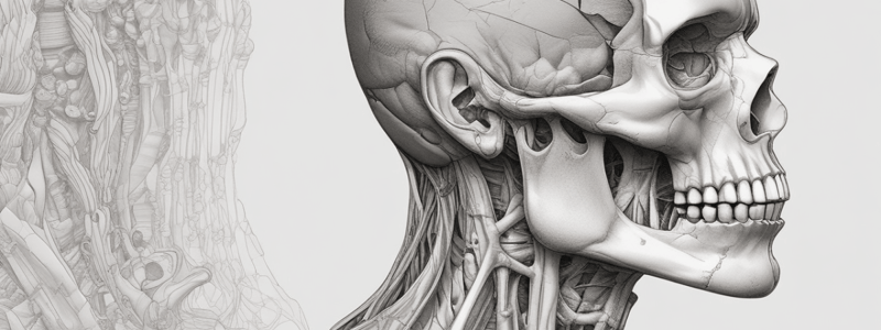
Temporal and Infratemporal Regions
- Muscles of mastication: 4 muscles that arise from the 1st branchial arch, inserted into the mandible, and take nerve supply from the mandibular nerve
- The 4 muscles of mastication are:
- Temporalis
- Masseter
- Lateral Pterygoid
- Medial Pterygoid
Temporalis Muscle
- Origin: temporal fascia, inserted into the tip, anterior border, and medial surface of the coronoid process
- Nerve supply: deep temporal nerves
- Action:
- Elevation of the mandible
- Retraction of the mandible
- Relations:
- Superficial relations: superficial temporal vessels, auriculo-temporal nerve, zygomatic arch, and masseter muscle
- Deep relations: maxillary artery and pterygoid plexus of veins, buccal nerve and artery, deep temporal vessels and nerve, and temporal fossa
Temporomandibular Joint (TMJ)
- Composed of:
- Synovial membrane
- Articular cartilage
- Articular disc (an oval plate of fibrous tissue that divides the joint into upper and lower compartments)
- Capsule (lined by synovial membrane)
- Accessory ligaments:
- Lateral temporomandibular ligament
- Sphenomandibular ligament
- Stylomandibular ligament
- Pterygomandibular ligament
- Blood supply: superficial temporal artery and maxillary artery
- Nerve supply: auriculotemporal nerve
Movements of the Mandible
- Position at rest:
- Slight interval between upper and lower teeth
- Lower teeth lie slightly behind the level of the upper teeth
- Movements:
- Protraction of the mandible by lateral and medial pterygoid muscles and masseter
- Retraction of the mandible by posterior fibers of temporalis, digastric, and geniohyoid muscles
- Depression of the mandible (opening of the mouth) by lateral pterygoid, platysma, digastric, mylohyoid, and geniohyoid muscles
- Elevation of the mandible (closure of the mouth) by temporalis, masseter, and medial pterygoid muscles
- Side to side movement of the mandible (grinding) by lateral and medial pterygoid muscles
Dislocation of the Mandible
- Anterior dislocation: mandible displaced in front of the eminence, causing compression of the blood vessels
- Dislocation is very dangerous and can be treated by using 2 fingers to depress the mandible downward and then backward
Masseter Muscle
- Origin: lower border and inner surface of zygomatic arch
- Insertion: lateral surface of ramus of mandible
- Action:
- Elevation of the mandible
- Protraction of the mandible
- Nerve supply: nerve to masseter
Lateral Pterygoid Muscle
- Origin: by 2 heads, upper head from infratemporal crest of greater wing of the sphenoid, and lower head from lateral surface of lateral pterygoid plate
- Insertion: fovea in front of the head of the mandible
- Action:
- Protraction of the mandible
- Depression of the mandible
- Side to side movement
- Nerve supply: anterior division of the mandibular nerve
Medial Pterygoid Muscle
- Origin: by 2 heads, superficial head from maxillary tuberosity, and deep head from medial surface of lateral pterygoid plate
- Insertion: medial surface of the ramus of mandible
- Action:
- Elevation of the mandible
- Protraction of the mandible
- Side to side movement
- Nerve supply: medial pterygoid nerve
Studying That Suits You
Use AI to generate personalized quizzes and flashcards to suit your learning preferences.


