Podcast
Questions and Answers
Which of the following joints is NOT found in digits 2-4 of the hand?
Which of the following joints is NOT found in digits 2-4 of the hand?
- Distal interphalangeal (DIP) joint
- Proximal interphalangeal (PIP) joint
- Metacarpophalangeal (MCP) joint
- Carpometacarpal (CMC) joint (correct)
The thumb (1st digit) differs from digits 2-5 in which key aspect regarding interphalangeal joints?
The thumb (1st digit) differs from digits 2-5 in which key aspect regarding interphalangeal joints?
- The thumb typically has both a PIP and DIP joint, while digits 2-5 only have a single IP joint.
- The thumb possesses a distal interphalangeal (DIP) joint, which is absent in other digits.
- The thumb lacks an interphalangeal (IP) joint, relying entirely on the MCP joint for flexion and extension.
- The thumb has only one interphalangeal (IP) joint, whereas digits 2-5 each have both a PIP and DIP joint (correct)
Which statement accurately describes the relative lengths of the metacarpals?
Which statement accurately describes the relative lengths of the metacarpals?
- The 1st metacarpal is the longest, with the remaining metacarpals progressively decreasing in length from radial to ulnar.
- The 5th metacarpal is the longest, allowing for greater mobility on the ulnar side of the hand.
- The 2nd metacarpal is typically the longest, while the 1st metacarpal is the shortest and stoutest. (correct)
- All metacarpals are approximately the same length to facilitate uniform grasping.
Posterior tubercles on the metacarpals provide attachment sites for which structures?
Posterior tubercles on the metacarpals provide attachment sites for which structures?
The unique orientation and rotation of the 1st metacarpal is critical for:
The unique orientation and rotation of the 1st metacarpal is critical for:
Which of the following statements accurately describes the arches of the hand?
Which of the following statements accurately describes the arches of the hand?
Which feature is characteristic of the proximal transverse arch of the hand?
Which feature is characteristic of the proximal transverse arch of the hand?
In the distal transverse arch, which metacarpals are the most mobile, allowing the hand to adapt to different shapes?
In the distal transverse arch, which metacarpals are the most mobile, allowing the hand to adapt to different shapes?
The longitudinal arch of the hand is primarily stabilized by:
The longitudinal arch of the hand is primarily stabilized by:
Which of the following describes the open-packed position of the carpometacarpal (CMC) joints?
Which of the following describes the open-packed position of the carpometacarpal (CMC) joints?
What structural elements contribute to the stability of the CMC joints?
What structural elements contribute to the stability of the CMC joints?
Mobility at the 4th and 5th CMC joints is crucial for:
Mobility at the 4th and 5th CMC joints is crucial for:
What type of joint is the 1st carpometacarpal (CMC) joint?
What type of joint is the 1st carpometacarpal (CMC) joint?
Which ligament is the prime stabilizer of the opposed 1st CMC joint?
Which ligament is the prime stabilizer of the opposed 1st CMC joint?
During flexion of the 1st CMC joint, arthrokinematics involve which of the following?
During flexion of the 1st CMC joint, arthrokinematics involve which of the following?
What is a key characteristic of the metacarpophalangeal (MCP) joints in terms of joint capsule?
What is a key characteristic of the metacarpophalangeal (MCP) joints in terms of joint capsule?
The open-packed position of the MCP joints is:
The open-packed position of the MCP joints is:
How do the collateral ligaments of the MCP joints behave during flexion?
How do the collateral ligaments of the MCP joints behave during flexion?
What is the primary function of the volar (palmar) plates in the MCP joints?
What is the primary function of the volar (palmar) plates in the MCP joints?
During MCP joint flexion, the proximal phalanx rolls and slides in what direction relative to the metacarpal?
During MCP joint flexion, the proximal phalanx rolls and slides in what direction relative to the metacarpal?
Which statement is most accurate regarding the arthrokinematics of the 1st MCP and IP joints?
Which statement is most accurate regarding the arthrokinematics of the 1st MCP and IP joints?
Which of the following is characteristic of the interphalangeal (IP) joints?
Which of the following is characteristic of the interphalangeal (IP) joints?
At what degree of finger flexion are collateral ligaments of the PIP joints maximally taut?
At what degree of finger flexion are collateral ligaments of the PIP joints maximally taut?
Which statement is true regarding the DIP joints, compared to the PIP joints?
Which statement is true regarding the DIP joints, compared to the PIP joints?
What is the primary function of the volar (palmar) plate at the IP joints?
What is the primary function of the volar (palmar) plate at the IP joints?
Which feature distinguishes DIP joints from PIP joints?
Which feature distinguishes DIP joints from PIP joints?
What best describes the relationship between flexion of MCP, PIP, and DIP joints?
What best describes the relationship between flexion of MCP, PIP, and DIP joints?
Why does passive tension in the dorsal capsule help guide arthrokinematics at the PIP and DIP joints?
Why does passive tension in the dorsal capsule help guide arthrokinematics at the PIP and DIP joints?
What is the key distinction between intrinsic and extrinsic muscles of the hand?
What is the key distinction between intrinsic and extrinsic muscles of the hand?
The flexor digitorum superficialis (FDS) primarily flexes which joints?
The flexor digitorum superficialis (FDS) primarily flexes which joints?
What is the primary action of the flexor digitorum profundus (FDP)?
What is the primary action of the flexor digitorum profundus (FDP)?
Which tendon travels through the split in the flexor digitorum superficialis (FDS) tendon?
Which tendon travels through the split in the flexor digitorum superficialis (FDS) tendon?
What structures enclose the extrinsic flexor tendons of the digits?
What structures enclose the extrinsic flexor tendons of the digits?
What is the function of the digital synovial sheaths?
What is the function of the digital synovial sheaths?
What is the primary function of the flexor pulleys in the fingers?
What is the primary function of the flexor pulleys in the fingers?
Severing or overstretching which pulleys would alter the mechanics of finger flexion?
Severing or overstretching which pulleys would alter the mechanics of finger flexion?
To isolate function at PIP, synergistically contracts to stabilize the MCP jt & wrist
To isolate function at PIP, synergistically contracts to stabilize the MCP jt & wrist
The extensor mechanism is the primary site of attachment for which muscle groups?
The extensor mechanism is the primary site of attachment for which muscle groups?
Which of the following describes the role of oblique retinacular ligaments?
Which of the following describes the role of oblique retinacular ligaments?
Lumbricals produce
Lumbricals produce
What occurs with isolated contraction of the extensor digitorum (ED)?
What occurs with isolated contraction of the extensor digitorum (ED)?
The optimal length-tension relationship for finger flexors during a power grip is achieved with the wrist in:
The optimal length-tension relationship for finger flexors during a power grip is achieved with the wrist in:
Which carpal bone serves as the keystone for the proximal transverse arch of the hand?
Which carpal bone serves as the keystone for the proximal transverse arch of the hand?
During thumb flexion at the 1st CMC joint, which arthrokinematic motion occurs?
During thumb flexion at the 1st CMC joint, which arthrokinematic motion occurs?
Which statement is most accurate regarding the position of the volar plate during MCP joint extension?
Which statement is most accurate regarding the position of the volar plate during MCP joint extension?
What best describes the resting position of the carpometacarpal joints 2-5?
What best describes the resting position of the carpometacarpal joints 2-5?
Which statement accurately relates to the roles of the palmar interossei muscles?
Which statement accurately relates to the roles of the palmar interossei muscles?
Which of the following best describes the primary role of the digital synovial sheaths?
Which of the following best describes the primary role of the digital synovial sheaths?
What is the functional consequence of the oblique retinacular ligament's unique orientation?
What is the functional consequence of the oblique retinacular ligament's unique orientation?
Where does the Flexor Digitorum Profundus (FDP) tendon attach distally?
Where does the Flexor Digitorum Profundus (FDP) tendon attach distally?
Which statement best describes the contribution of the wrist position to optimal finger flexor function during a power grip?
Which statement best describes the contribution of the wrist position to optimal finger flexor function during a power grip?
What is the primary arthrokinematic motion occurring during MCP joint abduction?
What is the primary arthrokinematic motion occurring during MCP joint abduction?
In the context of the extensor mechanism, the transverse fibers of the dorsal hood have what important function?
In the context of the extensor mechanism, the transverse fibers of the dorsal hood have what important function?
What is the functional effect of tension in the dorsal capsule of the PIP and DIP joints during motion?
What is the functional effect of tension in the dorsal capsule of the PIP and DIP joints during motion?
In what plane is the 1st metacarpal oriented differently compared to the other metacarpals?
In what plane is the 1st metacarpal oriented differently compared to the other metacarpals?
What describes the interaction between the extrinsic and intrinsic muscles of the hand?
What describes the interaction between the extrinsic and intrinsic muscles of the hand?
Why are annular pulleys, specifically A2 and A4, critical for finger flexion?
Why are annular pulleys, specifically A2 and A4, critical for finger flexion?
Which muscles are responsible for positioning the first metacarpal in an abducted, flexed, and rotated posture?
Which muscles are responsible for positioning the first metacarpal in an abducted, flexed, and rotated posture?
What is the primary function of check-rein ligaments at the PIP joints?
What is the primary function of check-rein ligaments at the PIP joints?
Which kinematic description accurately represents the motion at the PIP joint during finger flexion?
Which kinematic description accurately represents the motion at the PIP joint during finger flexion?
Which event will occur with isolated ED contraction?
Which event will occur with isolated ED contraction?
Why does arthrokinematics at the DIP and PIP joints have a similar pattern?
Why does arthrokinematics at the DIP and PIP joints have a similar pattern?
During thumb abduction at the 1st CMC joint, which arthrokinematic motion occurs?
During thumb abduction at the 1st CMC joint, which arthrokinematic motion occurs?
The stability of the MCP joints primarily comes from:
The stability of the MCP joints primarily comes from:
What is the effect of the thumb moving towards the 4th and 5th digits during firm opposition of the thumb?
What is the effect of the thumb moving towards the 4th and 5th digits during firm opposition of the thumb?
Which structure acts as the primary distal attachment for the extensor digitorum, extensor indicis, and extensor digiti minimi muscles?
Which structure acts as the primary distal attachment for the extensor digitorum, extensor indicis, and extensor digiti minimi muscles?
What occurs to the volar plate attachments with MCP joint flexion?
What occurs to the volar plate attachments with MCP joint flexion?
Which description accurately represents how the extrinsic finger flexor tendons are anchored distally?
Which description accurately represents how the extrinsic finger flexor tendons are anchored distally?
Which structure acts as the keystone at the distal transverse arch?
Which structure acts as the keystone at the distal transverse arch?
What component is responsible for limiting abduction and adduction at the PIP joint?
What component is responsible for limiting abduction and adduction at the PIP joint?
The flexor digitorum superficialis splits into two parts to allow passage of which of the following structures?
The flexor digitorum superficialis splits into two parts to allow passage of which of the following structures?
The extensor mechanism is the primary distal site for which muscle groups?
The extensor mechanism is the primary distal site for which muscle groups?
The second metacarpal primarily articulates with which carpal bone?
The second metacarpal primarily articulates with which carpal bone?
How do the insertions of the interossei muscles contribute to the function of the hand?
How do the insertions of the interossei muscles contribute to the function of the hand?
Which action would be limited after laceration of the flexor digitorum profundus tendon to the index finger?
Which action would be limited after laceration of the flexor digitorum profundus tendon to the index finger?
At what degree does the collateral ligaments become maximally taut at the Metacarpophalangeal joints?
At what degree does the collateral ligaments become maximally taut at the Metacarpophalangeal joints?
What defines an “intrinsic plus” hand position?
What defines an “intrinsic plus” hand position?
What is the functional significance of the palmar surface of the first metacarpal facing the midline of the hand?
What is the functional significance of the palmar surface of the first metacarpal facing the midline of the hand?
The interossei muscles are optimally positioned for maximum ABDuction and ADDuction when are extended, thus:
The interossei muscles are optimally positioned for maximum ABDuction and ADDuction when are extended, thus:
The oblique retinacular ligament is stretches with is joint action of the fingers?
The oblique retinacular ligament is stretches with is joint action of the fingers?
When considering the carpometacarpal (CMC) joints, which carpals articulate with the 3rd metacarpal?
When considering the carpometacarpal (CMC) joints, which carpals articulate with the 3rd metacarpal?
Which statement accurately describes the behavior of the volar plate at the MCP joints during extension?
Which statement accurately describes the behavior of the volar plate at the MCP joints during extension?
Which description accurately represents the attachment of the flexor digitorum profundus (FDP) tendon distally?
Which description accurately represents the attachment of the flexor digitorum profundus (FDP) tendon distally?
How does the interplay between extrinsic and intrinsic muscles influence finger movement?
How does the interplay between extrinsic and intrinsic muscles influence finger movement?
Why are the annular pulleys A2 and A4 critical for finger flexion?
Why are the annular pulleys A2 and A4 critical for finger flexion?
During isolated contraction of the extensor digitorum (ED), what movement is expected to occur?
During isolated contraction of the extensor digitorum (ED), what movement is expected to occur?
In the extensor mechanism, what is the function of the transverse fibers of the dorsal hood?
In the extensor mechanism, what is the function of the transverse fibers of the dorsal hood?
Which carpal serves as the keystone of the proximal transverse arch?
Which carpal serves as the keystone of the proximal transverse arch?
What key feature allows the coordination of movement between PIP and DIP joints?
What key feature allows the coordination of movement between PIP and DIP joints?
Flashcards
CMC Joints
CMC Joints
Joints between distal carpals and metacarpal bases.
CMC joint movement
CMC joint movement
Movements permitted are only gliding at these joints.
1st CMC Joint
1st CMC Joint
The thumb's CMC joint allows opposition, flexion, extension, abduction, and adduction.
Open-packed position of CMC Joint
Open-packed position of CMC Joint
Signup and view all the flashcards
Close-packed position of CMC Joint
Close-packed position of CMC Joint
Signup and view all the flashcards
MCP Joints
MCP Joints
Signup and view all the flashcards
MCP Joint Movements
MCP Joint Movements
Signup and view all the flashcards
Open-packed position of MCP Joint
Open-packed position of MCP Joint
Signup and view all the flashcards
Close-packed position of MCP Joint
Close-packed position of MCP Joint
Signup and view all the flashcards
Interphalangeal (IP) Joints
Interphalangeal (IP) Joints
Signup and view all the flashcards
Close-packed position of IP Joint
Close-packed position of IP Joint
Signup and view all the flashcards
Open-packed position of IP Joint
Open-packed position of IP Joint
Signup and view all the flashcards
Flexor Digitorum Superficialis (FDS)
Flexor Digitorum Superficialis (FDS)
Signup and view all the flashcards
Flexor Digitorum Profundus (FDP)
Flexor Digitorum Profundus (FDP)
Signup and view all the flashcards
Flexor Pollicis Longus (FPL)
Flexor Pollicis Longus (FPL)
Signup and view all the flashcards
Flexor Pulleys
Flexor Pulleys
Signup and view all the flashcards
Central Band
Central Band
Signup and view all the flashcards
Lateral Bands
Lateral Bands
Signup and view all the flashcards
Dorsal Hood
Dorsal Hood
Signup and view all the flashcards
Oblique Retinacular Ligament
Oblique Retinacular Ligament
Signup and view all the flashcards
Lumbricals
Lumbricals
Signup and view all the flashcards
Power grip characteristics
Power grip characteristics
Signup and view all the flashcards
Precision Handling
Precision Handling
Signup and view all the flashcards
Study Notes
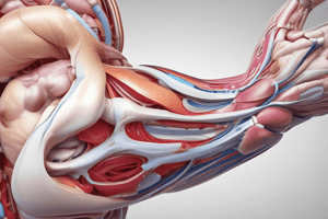
Hand Osteology and Joints
- The hand consists of five digits, each containing multiple joints
- The carpometacarpal (CMC), metacarpophalangeal (MCP), and interphalangeal (IP) joints are all present
- Fingers 2 through 4 each have a proximal (PIP) and distal (DIP) interphalangeal joint, while the thumb has only one IP joint
- The metacarpals are numbered 1 through 5, moving radially to ulnarly
- Metacarpal 1 (the thumb) is the shortest and stoutest, while metacarpal 2 is usually the longest
- The remaining metacarpals (3-5) decrease in length ulnarly
- Metacarpal bases are proximal and articulate with different numbers of carpal bones
- The heads of metacarpals 2-5 form the knuckles on the hand's dorsal side
- The distal end of all metacarpals have large convex heads
- Posterior tubercles on the metacarpals serve as attachment sites for the MCP joint's collateral ligaments
- The first metacarpal is in a different plane and rotated about 90 degrees medially, with the palmar surface facing the midline, enabling thumb opposition for grip and pinch
- Proximal and middle phalanges feature a concave base and convex head
- Palmar surfaces of middle and proximal phalanges have a slightly concave longitudinal profile
- Distal phalanges have a concave base and a distal tuberosity
Arches of the Hand
- Three arch systems support the concavity of the hand
- 2 transverse arches
- 1 longitudinal arch
- Concavity control allows for grasping, gripping, and object manipulation
- The proximal transverse arch comprises of the distal row of carpal bones
- It is a static, rigid arch that that forms a portion of the carpal tunnel
- The capitate bone serves as the keystone
- It gains reinforcement from bone contacts and intercarpal ligaments
- Distal transverse arch goes through the MCP joints
- Sides of the distal transverse arch are mobile
- Transverse flexibility enables the 1st, 4th, and 5th metacarpals to "fold" around the more stable 2nd and 3rd
- The keystone for the distal transverse arch gets formed by the MCP joints of the 2nd and 3rd metacarpals
- The longitudinal arch follows the general shape of the 2nd and 3rd rays
- The arch's proximal end is rigidly linked to the carpals via the CMC joints
- Provides longitudinal stability to the hand
- The arch's distal end is mobile
- Keystone of the arch can be found at the 2nd and 3rd MCP joints
Thumb Movements
- Thumb motions include flexion, extension, abduction, adduction, and opposition
CMC Joints
- The distal carpals articulate with the base of the metacarpals at these joints
- Gliding movements are only permitted
- Anterior (palmar), posterior (dorsal), and intermetacarpal ligaments provide stability
- The open-packed position is midway between flexion and extension
- The closed-packed position is fingers in full flexion
- The 2nd and 3rd metacarpals have relatively restricted movement
- The 1st, 4th, and 5th metacarpals are more mobile
- Mobility facilitates manipulation of diverse items
- The 4th and 5th CMC joints have greater mobility
- Observed by looking at the metacarpal heads while making a fist
- Jt capsules, dorsal CMC ligaments, palmar CMC ligaments, and intermetacarpal ligaments support the joints
- Dorsal ligaments are better developed and more comprehensive compared to palmar ligaments
- The 2nd CMC joint consists of the base of the 2nd metacarpal with the distal trapezoid, and exhibits smaller articulations with the capitate and trapezium.
- The base of the 3rd metacarpal has a joint with distal capitate
- The 4th CMC joint has a base of the 4th metacarpal with a distal hamate, displaying a smaller articulation with the capitate
- The 5th CMC joint's base of is linked with with the distal hamate
1st Carpometacarpal (CMC) Joint
- It's a saddle-shaped joint with reciprocally shaped joint surfaces
- The articulation exists between the 1st metacarpal base and the distal trapezium
- Flexion, extension, adduction, abduction, and opposition are allowed at the joint
- The joint capsule has a large and lax size
- Stability stems from muscular activity and ligamentous support
- The open-packed position is characterized by being midway between abduction and adduction, as well as midway between flexion and extension
- The closed-packed position involves full opposition
- The radial collateral ligament is strong and taut during opposition, flexion, and abduction
- It is the key stabilizer of the opposed CMC joint
- The ulnar collateral ligament remains taut during abduction and extension
- The anterior (palmar) oblique ligament, is thin and weak, and taut when in full extension
- The posterior oblique ligament works similarly to the radial collateral ligament
Arthrokinematics of the 1st CMC Joint
- Flexion involves a medial (ulnar) roll and glide of the 1st metacarpal on the trapezium
- Extension entails a lateral (radial) roll and glide of the 1st metacarpal on the trapezium
- Abduction is achieved through a palmar roll and dorsal slide of the 1st metacarpal on the trapezium
- Adduction includes a dorsal roll and palmar slide of the 1st metacarpal on the trapezium
MCP, PIP, & DIP Joints
- MCP = Metacarpophalangeal
- PIP = Proximal Interphalangeal
- DIP = Distal Interphalangeal
- MCP joints are formed between the convex heads of metacarpals and concave proximal surface of proximal phalanges
- 2nd though 5th metacarpals articulate at biaxial joints with their respective phalanges
- Widened proximal bases articulate with carpals
- Jts allow flexion, extension, abduction, and adduction
- From 1st to 5th MCP, flexion ROM increase
- Joint capsules are lax and stability gets derived from collateral ligaments
- Extension generally has general laxity
- Collateral ligaments become taut in 70–90° of flexion
- Open-packed position is slight flexion
- Closed-packed position is full opposition (thumb)/full flexion (fingers)
- Collateral ligaments cross joints in an oblique way
- The dorsal cord is thicker and stronger, while the accessory portion is thinner, fan-shaped, and attached to the palmar (volar) plate
- Dense fibrocartilage, called volar (palmar) plates are on the palmar side of jts
- Bases get attached to the proximal phalanx
- Necks of metacarpals are part of the attachment process
- Capsule blends with the volar part, which resists overextension and improves jt stability
- Flexible attachments allow the sliding of the plate proximally on the metacarpal head when the MCP joint flexes
- Deep transverse metacarpal ligaments comprised of three ligaments that interconnect and loosely bind the 2nd to 5th metacarpals into a wide, flat structure
- Proximal phalanx performs a palmar (anterior) roll and slide along metacarpal during flexion
- Activation of the flexor digitorum profundus (FPD) happens during flexion
- Dorsal capsule plus the radial collateral ligament creates tension
MCP Arthrokinematics During Extension
- The proximal phalanx rolls and slides dorsally on the metacarpal
- It results from coactivation of ED and finger's intrinsic muscles
- The palmar plate becomes taut, and the cord portion of the radial collateral ligament develops slack
- Abduction gets driven by activating the 1st dorsal interosseus muscle (DI1)
- The ulnar collateral ligament becomes taut, whereas the radial collateral ligament becomes slack
- Proximal phalanx performs rolls and slides in the equivalent direction to the digits
- The thumb's MCP joint has a hinge design that permits flexion and extension
- Both the bone structure supports and enhances stability
- Ligamentous support gets acquired through anterior (palmar) and collateral ligaments
- An existing convex head of the metacarpal plus a concave surface on the base of the phalanx facilitates stabilization
- Greater ROM is correlated to Volar plate increases articular surface stretch
1st MCP and IP Joint Arthrokinematics
- The phalanx slides and rolls in the same direction at both joints, which means that flexion generates a volar roll and slide, whereas extension produces a dorsal roll and slide
Interphalangeal (IP) Joints
- They're hinge joints, which enable extension and flexion
- Stability comes from the the form of the osseous surfaces
- PIP joints are quite stable in every position
- Capsules are composed of a volar plate, collateral ligaments, and extensor expansion
- The collateral ligaments become maximally taut when the finger is flexed at 25° with the PIP
- DIP joints are less stable than PIP joints
- Open-packed position happens with minimal flexion
- Full extension comprises the closed-packed position
- Proximal interphalangeal (PIP) Joint articulations exist between proximal phalanges and middle phalanges
- Heads of the proximal phalanx have two condyles separated by a shallow groove
- Middle phalanx has two shallow concave facets divided by a centre ridge
- The IP jt capsule gets reinforced by radial and ulnar ligamentous bands
- The collateral ligament's cord parts reduce adduction and abduction with with PIP bending
- Accessory parts blend with and strengthen the palmar plate
- Proximal-lateral areas along the palmar aspect that are responsible for stabilizing ligaments
- These structural areas will provide Check-rein strength.
- Assist in resisting hyperextension
- Mobile, thickened fibrocartilaginous structure
- Serves as a restraint to hyperextension
DIP Joints
- Articulations occur between the bases and heads of the middle phalanges
- DIP jt structure/tissues are comparable with the PIP structure
- No check-rein ligaments are present
- MCP, PIP, and DIP articulation in the ulnar digits happens better overall
- The PIP jts enable extremely limited hyperextension
- MCP and DIP joints enable some degree of hyperextension
- Virtually continuous quiescent stabilization exists due to collateral ligaments with IP joints at the target level for range of motion
- Arthrokinematics for DIP joints and PIP joints mirror
Musculature of Hand
- The muscles with distal and proximinal attachments to the hand
- Muscles of the hand with attachments proximal to the forearm/humerous
- Hand movements work best if movements coordinate between extrinsic muscles,intrinsic muscles,plus wrist musculature
Intrinsic Muscles of the Hand
- Thenar Eminence: abductor pollicis brevis, flexor pollicis brevis, and opponens pollicis
- Hypothenar Eminence: abductor digiti minimi, flexor digiti minimi, opponens digiti minimi, and palmaris brevis
- Other: adductor pollicis (2 heads), lumbricals (4), and interossei (4 palmar & 4 dorsal)
Extrinsic Muscles of the Hand
- Flexors of the digits: flexor digitorum superficialis, flexor digitorum profundus, and flexor pollicis longus
- Extensors of the fingers: extensor digitorum, extensor indicis, and extensor digiti minimi
- Extensors of the thumb: extensor pollicis longus, extensor pollicis brevis, and abductor pollicis longus
Extrinsic Flexors of the Digits
- Flexor Digitorum Superficialis (FDS): responsible for flexion of the PIP joints while also assisting in flexing every joint
- Flexor Digitorum Profundus (FDP): responsible for flexing at every crossing joint while performing primary functions by flexing the distal joint
- Flexor Pollicis Longus (FPL): responsible for flexion of the thumb IP joints.
- The muscle belly of the FDS lies deep to the 3 wrist flexors & pronator teres
- The FDS has four tendons that cross at the wrist and enter palmar side of the hand
- Splits near proximal phalanx allowing access and passage to the FDP as well
- After tendon splits it crosses the PIP joints and connects on either the end of the palmar at the middle phalanx
Flexor Digitorum Profundus (FDP)
- Muscles from within the belly reach the deepest areas for all the muscles at the forearm
- Each tendon is connected directly by the FDS allowing attachment on the palmar surface to the distal phalanx
- Sole source of flexion activity for flexor stabilization that is independent of joints that cross it
- Motion limited with interconnection on both tendons plus single fingers
Flexor Pollicis Longus (FPL)
- Located close and side by side from FDP at the surface muscles with the forearm
- Extracellular membrane passes through the wrist attaching distally towards lateral side on the distal joints on the thumb side
- Force of flexion with exertion with motion by torque (rotation) during MCP for thumb use
Flexor Structures of the Hand
- Surrounds FDS/FDP that extend to capral tunnel
- Ends inside primary hand area
- Tendons get found and connected at the 5th digit
- FPL tendons extend to tip
- Flex tendons get secured due to fibrous coatings
Flexor pulleys
- Flexor tendons get weaved across every sheath
- Tendons get coated deeply as well all the way up.
- Nutrients can supply more due to sheathing.
- Synovials also lubricate the tendons too.
Flex Pulleys:
- Every finger area has 5 annular pulleys numbered as A5 going across
- The more important, the A2 and A4 connect to where the bone is by each finger.
- The rest of the area has pulleys that attach directly inside these areas including C1-3 that secure in distinct areas and are thin/flexible
- For thumbs, ligaments give support for this as well.
Passive Flexor Elements Include
- Connects tendons at areas around the joint to hold together
- The A1 and A4 pulleys keep bowstringing from tendons going bad during tension
- The altered bio-mechanics of the finger flexion could then get severed or over time worn down resulting in elite climbers having finger issues related
Proximal Stabilization During Finger Flexion
- Extensor tendons help by joining and helping synergistic activity to single areas.
- ED contracts to stabilize MCP.
- Distal tension will get amplified more with FDS but needs stabilized with ECRL ED EDBC more than force
- The extrinsic digit extensors are the extensor digitorum (ED), extensor indicis (EI), and extensor digiti minimi (EDM), which sometimes has two tendons
Extensor Mechanism
- The attachment point exists in most muscles that are on the fingers and distal to wrist with the extensor assembly
- Small parts of the ED tendons will get combined with connecting muscles
- The lateral areas branch out for distal areas to attach
- Forces to the end parts could allow these extensions to do transfer for the best finger use
Extensor Mechanism Function
- The most identifiable aspect out is a hooded structure that has fiber support built in and connects transverse points allowing constant tension to the joint to keep in place
Parts of The active extension mechanism
- There are fibers connecting and stabilizing ED as well around it making every part connected with the transverse fibers
- Running all sides will allow these stabilizers to have best tension and support. Connective sides can allow good mobility and fusion to fibers
- Ret Lig prevent pull on joints and also enable muscle activation
Active Component of Extension
- Dorsal interossei, palmar interossei, and lumbricals participate
- Interossei muscles arise from side of MCP plus phalanxes attaching around it
- Fibers get formed into side band structures in a connecting form mostly that connects areas around them. Also attaching the lateral muscles all connected through side cords that enable every force and allow fibers to travel effectively to the finger as much as possible. Lats travel to joints too
- Every part works in support of the thumb too as much as possible allowing every kind of strength as best
Lumbricals
- Strong support for tendons for joints on the hand that will help connect and protect the rest of the finger that will have a lot of mobility and also support stabilization in each joint
- The lumbricals function with strong and synergistic effects that can give best performance of the thumb All these connections perform their peak action with synergy and control all different phases
Retinacular Ligaments
- Will run side by side and allow support with the joint as the major objective
- By assisting each muscle, it allows full control and motion of the hand preventing injury and giving support so muscles don’t take full force
Hyperextension of MCP
- Prevents force while preventing flexion while both parts need each ability and synergistic ability to maintain its posture
Synergistic Movement
- Tendons have active functions that allow each one to keep the synergistic motion in play along where most function is generated and most power will get delivered
Structure of thumb extension
- Extensors, like the EPL helps stabilize the thumb from losing joint support
- Synergistic work is made to happen to provide best performance and support between both
Extrinsic Musculature of the thumb
- Flexor Pollicis Longus (FPL) flexes at the thumbs IP JT for bending at thumbs
- The EPB (extensor Poll Brev), enables extension motion while taking from hand the support is required
- EPL, provides stability and extensions at the joint.
- Pull motions provided by these muscles help hand use
Active movements with thumbs
- ABD/AD can enable the thumb for peak Rom
- Thumb function is key to every hand motion such as grasping This allows stabilization also
Studying That Suits You
Use AI to generate personalized quizzes and flashcards to suit your learning preferences.