Podcast
Questions and Answers
What is the function of the bare parts of the fingers?
What is the function of the bare parts of the fingers?
- Assist in flexion of the fingers
- Act as windows for Lumbricals (correct)
- Protect underlying tissues
- Provide grip strength
Which structure is described as having separate synovial sheaths?
Which structure is described as having separate synovial sheaths?
- Other fingers' synovial sheaths (correct)
- Digital nerves
- Muscles of the hand
- Tendons of the flexor digitorum profundus
What do the synovial sheaths in the fingers primarily cover?
What do the synovial sheaths in the fingers primarily cover?
- The lumbrical muscles
- The flexor tendons in their resting position (correct)
- The bones of the fingers
- The extensor tendons
What anatomical feature do the Lumbricals support in the fingers?
What anatomical feature do the Lumbricals support in the fingers?
Which statement is true regarding the covered and uncovered portions of the fingers?
Which statement is true regarding the covered and uncovered portions of the fingers?
Which fingers possess complete sheaths according to the information provided?
Which fingers possess complete sheaths according to the information provided?
What type of sheath do the other fingers have?
What type of sheath do the other fingers have?
How many fingers possess complete sheaths?
How many fingers possess complete sheaths?
The presence of separate synovial sheaths means that:
The presence of separate synovial sheaths means that:
Which finger is NOT mentioned as having a complete sheath?
Which finger is NOT mentioned as having a complete sheath?
What is the main function of the synovial sheath in fingers?
What is the main function of the synovial sheath in fingers?
Which statement is true regarding the fingers with complete sheaths?
Which statement is true regarding the fingers with complete sheaths?
What characterizes the fibrous flexor sheath?
What characterizes the fibrous flexor sheath?
Which aspect of the fingers does the synovial sheath primarily affect?
Which aspect of the fingers does the synovial sheath primarily affect?
In terms of anatomical structure, the term 'complete sheath' refers to a sheath that:
In terms of anatomical structure, the term 'complete sheath' refers to a sheath that:
What are the main structures protected by the flexor retinaculum in the wrist?
What are the main structures protected by the flexor retinaculum in the wrist?
Which of the following bones is NOT part of the lateral attachment of the flexor retinaculum?
Which of the following bones is NOT part of the lateral attachment of the flexor retinaculum?
In which structure do the tendons of the superficialis and profundus group together?
In which structure do the tendons of the superficialis and profundus group together?
What unique feature does the flexor pollicis longus and flexor carpi radialis exhibit in relation to their sheaths?
What unique feature does the flexor pollicis longus and flexor carpi radialis exhibit in relation to their sheaths?
Where do the ulnar artery and nerve pass in relation to the flexor retinaculum?
Where do the ulnar artery and nerve pass in relation to the flexor retinaculum?
What type of structure is the common flexor sheath described as?
What type of structure is the common flexor sheath described as?
Flashcards
Lumbrical Windows
Lumbrical Windows
The part of a lumbrical muscle that is not covered by a synovial sheath.
Synovial Sheath
Synovial Sheath
A thin, fluid-filled sac that surrounds certain tendons, reducing friction during movement.
Fibrous Tendon Sheath
Fibrous Tendon Sheath
A fibrous connective tissue sheath that surrounds a tendon.
Lumbricals
Lumbricals
Signup and view all the flashcards
Lumbrical Windows and Synovial Sheaths
Lumbrical Windows and Synovial Sheaths
Signup and view all the flashcards
Flexor Retinaculum
Flexor Retinaculum
Signup and view all the flashcards
Carpal Tunnel
Carpal Tunnel
Signup and view all the flashcards
Medial Attachment of Flexor Retinaculum
Medial Attachment of Flexor Retinaculum
Signup and view all the flashcards
Lateral Attachment of Flexor Retinaculum
Lateral Attachment of Flexor Retinaculum
Signup and view all the flashcards
Common Flexor Sheath
Common Flexor Sheath
Signup and view all the flashcards
Separate Sheaths for Thumb and Radial Flexors
Separate Sheaths for Thumb and Radial Flexors
Signup and view all the flashcards
Fibrous Flexor Sheath
Fibrous Flexor Sheath
Signup and view all the flashcards
Complete Synovial Sheath
Complete Synovial Sheath
Signup and view all the flashcards
Special Synovial Sheaths in Thumb and Little Finger
Special Synovial Sheaths in Thumb and Little Finger
Signup and view all the flashcards
Separate Synovial Sheaths in Other Fingers
Separate Synovial Sheaths in Other Fingers
Signup and view all the flashcards
Greater Flexibility in Thumb and Little Finger
Greater Flexibility in Thumb and Little Finger
Signup and view all the flashcards
Benefits of Complete Synovial Sheaths
Benefits of Complete Synovial Sheaths
Signup and view all the flashcards
Flexibility and Support in Other Fingers
Flexibility and Support in Other Fingers
Signup and view all the flashcards
Collaborative Role of Sheaths
Collaborative Role of Sheaths
Signup and view all the flashcards
Importance of Understanding Sheaths
Importance of Understanding Sheaths
Signup and view all the flashcards
Study Notes
Hand Anatomy: Flexor Retinaculum and Carpal Tunnel
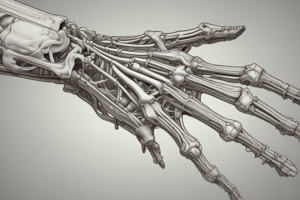
- The flexor retinaculum is a strong fibrous band covering structures passing through the carpal tunnel.
- The carpal tunnel is a narrow passageway in the wrist containing flexor tendons and nerves.
- Medial attachment of the flexor retinaculum: pisiform and hook of hamate
- Lateral attachment of the flexor retinaculum: scaphoid and trapezium
- Eight tendons of flexor superficialis and profundus share a common flexor sheath in two rows that open laterally.
- Flexor pollicis longus and flexor carpi radialis have separate sheaths.
- The ulnar artery and nerve pass above the flexor retinaculum in Guyon's canal.
Synovial Flexor Sheath
- Only the thumb and little finger have complete synovial sheaths.
- Other fingers have separate synovial sheaths lining the fibrous flexor sheath.
- Bare parts of the tendons are windows for lumbricals.
Digital Attachment of Long Tendons
- Branches of tendons insert into the dorsal digital expansion of the hand.
- Common digital artery
Extensor Expansion
- The extensor tendon's expansion across the metacarpophalangeal joint is called the extensor hood.
- This area provides insertion points for lumbricals, interossei, extensor indicis, and extensor digiti minimi muscles.
Thenar Muscles
- These muscles are located at the base of the thumb.
- Include abductor pollicis brevis, flexor pollicis brevis, opponens pollicis, and adductor pollicis
- The origin is the flexor retinaculum with scaphoid and trapezium.
- The insertion of these muscles is lateral to the base of the proximal phalanx.
- These muscles are innervated by the recurrent branch of the median nerve.
- Thenar muscles are used to abduct and flex the thumb.
Hypothenar Muscles
- These muscles are located at the base of the little finger.
- Include abductor digiti minimi, flexor digiti minimi brevis, and opponens digiti minimi
- Origin of these muscles are the flexor retinaculum and hook of hamate
- Insertion of these muscles is to the ulnar side of the 5th metacarpal
- These muscles are innervated by the deep branch of the ulnar nerve.
- Hypothenar muscles act on little finger–abduction, flexion, and opposition.
Palmaris Brevis Muscle
- Origin is the palmar aponeurosis.
- Insertion is on the skin of the ulnar border over the hypothenar base.
- Its action is to improve grip by steadying the skin on the ulnar side.
- Innervation by the superficial palmar branch of the ulnar nerve.
Interossei Muscles
- Palmar interossei muscles adduct the fingers towards the middle finger.
- Dorsal interossei abduct the fingers away from the middle finger.
- Both muscle groups originate from the metacarpals and insert into the extensor expansion and proximal phalanx.
- Both are innervated by the deep branch of the ulnar nerve
Lumbricals
- The origin of the lumbrical muscles is from the radial side of four profundus tendons.
- Their insertion is into the extensor expansion on the lateral side of the 2nd to 5th fingers.
- Innervation of these muscles is by median nerve and the deep branch of ulnar nerve.
- Lumbricals flex the metacarpophalangeal joints and extend the interphalangeal joints.
Studying That Suits You
Use AI to generate personalized quizzes and flashcards to suit your learning preferences.