Podcast
Questions and Answers
Which of the following muscles is NOT part of the Thenar Eminence?
Which of the following muscles is NOT part of the Thenar Eminence?
- Flexor pollicis brevis
- Opponens pollicis
- Abductor pollicis brevis
- Adductor pollicis (correct)
Which nerve is responsible for supplying the muscles of the Thenar Eminence?
Which nerve is responsible for supplying the muscles of the Thenar Eminence?
- Ulnar nerve
- Median nerve (correct)
- Radial nerve
- Palmar cutaneous branch of the median nerve
Which of the following structures does NOT pass deep to the Flexor Retinaculum?
Which of the following structures does NOT pass deep to the Flexor Retinaculum?
- Median nerve
- Flexor carpi radialis tendon
- Flexor digitorum profundus tendons
- Palmaris longus tendon (correct)
Which of the following bones is NOT part of the Distal row of the carpal bones?
Which of the following bones is NOT part of the Distal row of the carpal bones?
Which of the following muscles is NOT part of the Hypothenar Eminence?
Which of the following muscles is NOT part of the Hypothenar Eminence?
What are the two vessels that arise from the radial artery in the plane between the first dorsal interosseous and adductor pollicis?
What are the two vessels that arise from the radial artery in the plane between the first dorsal interosseous and adductor pollicis?
Which of the following nerves does not innervate any intrinsic muscles of the hand?
Which of the following nerves does not innervate any intrinsic muscles of the hand?
What is the origin of the Cephalic vein?
What is the origin of the Cephalic vein?
What is the function of the Extensor Retinaculum?
What is the function of the Extensor Retinaculum?
Which arteries arise from the dorsal carpal arch?
Which arteries arise from the dorsal carpal arch?
Which muscle is responsible for adduction of the thumb?
Which muscle is responsible for adduction of the thumb?
What is the main action of the Lumbrical Muscles?
What is the main action of the Lumbrical Muscles?
Which artery is responsible for supplying the thumb and lateral side of the index finger?
Which artery is responsible for supplying the thumb and lateral side of the index finger?
Which nerve supplies the medial two lumbrical muscles?
Which nerve supplies the medial two lumbrical muscles?
What is the name of the vascular arch formed by the radial artery on the lateral side of the palm?
What is the name of the vascular arch formed by the radial artery on the lateral side of the palm?
Flashcards are hidden until you start studying
Study Notes
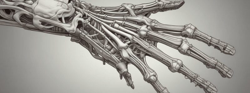
Hand Anatomy
- Carpal bones can be remembered using the mnemonic "She Looks Too Proud, Try To Chase Her"
- Proximal row of carpal bones: Scaphoid, Lunate, Triquetrum, Pisiform
- Distal row of carpal bones: Trapezium, Trapezoid, Capitate, Hamate
Flexor Retinaculum
- A thickening of deep fascia that holds long flexor tendons in position at the wrist
- Attaches medially to the pisiform bone and the hook of the hamate, and laterally to the tubercle of the scaphoid and the trapezium bones
Structures Passing Deep to Flexor Retinaculum
- Flexor digitorum superficialis tendons
- Median nerve
- Flexor pollicis longus tendon
- Flexor carpi radialis tendon
Structures Passing Superficial to Flexor Retinaculum
- Flexor carpi ulnaris tendon
- Ulnar nerve
- Ulnar artery
- Palmar cutaneous branch of the ulnar nerve
- Palmaris longus tendon
- Palmar cutaneous branch of the median nerve
Carpal Tunnel
- A fibro-osseous canal formed by the carpal bones and the flexor retinaculum
- Contents: tendons of FDS, FDP, and FPL, and the median nerve
Muscles of the Hand
- Divided into two groups: extrinsic and intrinsic
- Extrinsic group: muscles that originate outside the hand and insert into the hand bones
- Intrinsic group: arranged in five parts - thenar eminence, hypothenar eminence, adductor pollicis, interosseous muscles, and lumbrical muscles
Thenar Eminence
- Three muscles responsible for the prominent swelling on the lateral side of the palm at the base of the thumb
- Opponens pollicis, flexor pollicis brevis, and abductor pollicis brevis
- All supplied by the recurrent branch of the median nerve
Hypothenar Eminence
- Three muscles responsible for the swelling at the base of the little finger
- Branches: three palmar metacarpal arteries and three perforating branches
Dorsal Carpal Arch
- Formed by the radial artery before penetrating the back of the hand
- Gives rise to the dorsal carpal branch, which passes medially as the dorsal carpal arch, and subsequently divides to become small dorsal digital arteries
Veins of the Hand
- Deep veins follow the arteries
- Superficial veins drain into a dorsal venous network on the back of the hand over the metacarpal bones
- The Cephalic vein originates from the lateral side of the dorsal venous network and passes over the anatomical snuffbox into the forearm
- The Basilic vein originates from the medial side of the dorsal venous network and passes into the dorso-medial aspect of the forearm
Nerves of the Hand
- The hand is supplied by the ulnar, median, and radial nerves
- The ulnar nerve innervates all intrinsic muscles of the hand except for the three thenar muscles and the two lateral lumbricals, which are innervated by the median nerve
- The radial nerve only innervates skin on the dorsolateral side of the hand
Extensor Retinaculum
- A strong, fibrous band that extends obliquely across the back of the wrist
- Attached laterally to the anterior border of the radius, and medially to the triquetral and pisiform bones
Adductor Pollicis
- Originates as two heads: transverse head from the anterior aspect of the shaft of the 3rd metacarpal, and oblique head from the capitate and adjacent bases of the 2nd and 3rd metacarpals
- Inserts into the medial side of the base of the proximal phalanx of the thumb
- Supplied by the deep branch of the ulnar nerve
- Action: adduction of the thumb
Interosseous Muscles
- Divided into two groups: dorsal interossei and palmar interossei
- All are supplied by the deep branch of the ulnar nerve
- Action: PAD (palmar abduction of the fingers), DAB (dorsal abduction of the fingers)
Lumbrical Muscles
- Four worm-like muscles, each associated with one of the fingers
- Originate from the tendons of FDP in the palm
- The medial two lumbricals are innervated by the deep branch of the ulnar nerve; the lateral two lumbricals are innervated by digital branches of the median nerve
- Action: flex MPJ (metacarpophalangeal joint) and extend IPJ (interphalangeal joint)
Blood Vessels of the Hand
- Blood supply to the hand is by the radial and ulnar arteries
- These arteries form two interconnected vascular arches (superficial and deep) in the palm
- The radial artery contributes substantially to the supply of the thumb and the lateral side of the index finger
- The remaining digits and the medial side of the index finger are supplied mainly by the ulnar artery
Studying That Suits You
Use AI to generate personalized quizzes and flashcards to suit your learning preferences.


