Podcast
Questions and Answers
What is the primary function of the sensory areas in the cerebral cortex?
What is the primary function of the sensory areas in the cerebral cortex?
- Contribute to emotional responses
- Process sensory information and contribute to perception (correct)
- Integrate complex functions such as memory and reasoning
- Control voluntary movements
Which area of the cerebral cortex is primarily responsible for speech articulation?
Which area of the cerebral cortex is primarily responsible for speech articulation?
- Wernicke’s area
- Sensory association area
- Broca’s speech area (correct)
- Primary motor area
What characteristic of the primary motor area allows for specific muscle contractions?
What characteristic of the primary motor area allows for specific muscle contractions?
- It has a dedicated cortical region for each muscle group (correct)
- It solely activates the respiratory muscles
- It integrates sensory and motor functions
- It is located in the occipital lobe
In which hemisphere is Broca’s speech area predominantly located?
In which hemisphere is Broca’s speech area predominantly located?
What types of areas are included in Brodmann's classification of the cerebral cortex?
What types of areas are included in Brodmann's classification of the cerebral cortex?
What does the primary motor area primarily control?
What does the primary motor area primarily control?
Which of the following structures does not directly contribute to muscle activation for speech?
Which of the following structures does not directly contribute to muscle activation for speech?
Which statement best describes the relationship between cortical area size and muscle function?
Which statement best describes the relationship between cortical area size and muscle function?
What is the primary function of the precentral gyrus?
What is the primary function of the precentral gyrus?
Which of the following structures separates the frontal lobe from the temporal lobe?
Which of the following structures separates the frontal lobe from the temporal lobe?
What is the main role of commissural tracts in the brain?
What is the main role of commissural tracts in the brain?
Which tract is the largest commissural tract in the brain?
Which tract is the largest commissural tract in the brain?
The internal capsule is primarily composed of which type of tracts?
The internal capsule is primarily composed of which type of tracts?
Where is the insula located in relation to the other lobes of the cerebrum?
Where is the insula located in relation to the other lobes of the cerebrum?
Which of the following statements about association tracts is true?
Which of the following statements about association tracts is true?
How are projection tracts characterized?
How are projection tracts characterized?
Which structure contributes more cerebrospinal fluid (CSF) in the 4th ventricle?
Which structure contributes more cerebrospinal fluid (CSF) in the 4th ventricle?
What is the normal rate of reabsorption of CSF into the blood?
What is the normal rate of reabsorption of CSF into the blood?
Through which openings does CSF enter the subarachnoid space?
Through which openings does CSF enter the subarachnoid space?
What structure primarily reabsorbs CSF into the blood?
What structure primarily reabsorbs CSF into the blood?
Why is the pressure of CSF normally constant?
Why is the pressure of CSF normally constant?
What structure does the pia mater merge with at the level of the 2nd sacral vertebra?
What structure does the pia mater merge with at the level of the 2nd sacral vertebra?
Which of the following functions does cerebrospinal fluid (CSF) NOT perform?
Which of the following functions does cerebrospinal fluid (CSF) NOT perform?
Where do the lateral ventricles communicate with the third ventricle?
Where do the lateral ventricles communicate with the third ventricle?
Which structure lies between the right and left halves of the thalamus?
Which structure lies between the right and left halves of the thalamus?
What is the shape of the fourth ventricle in the brain?
What is the shape of the fourth ventricle in the brain?
How does the fourth ventricle communicate with the subarachnoid space?
How does the fourth ventricle communicate with the subarachnoid space?
What type of tissue primarily composes the pia mater?
What type of tissue primarily composes the pia mater?
Which part of the brain is situated above the lateral ventricles?
Which part of the brain is situated above the lateral ventricles?
What is the primary function of the premotor area in the brain?
What is the primary function of the premotor area in the brain?
Damage to which area would likely prevent a patient from recognizing familiar faces despite having a functioning visual field?
Damage to which area would likely prevent a patient from recognizing familiar faces despite having a functioning visual field?
Which of the following areas is directly involved in controlling the voluntary scanning movements of the eyes?
Which of the following areas is directly involved in controlling the voluntary scanning movements of the eyes?
Where do primary sensory areas primarily receive sensory information?
Where do primary sensory areas primarily receive sensory information?
What role do secondary sensory areas play in processing information?
What role do secondary sensory areas play in processing information?
What occurs if the primary somatosensory area is damaged?
What occurs if the primary somatosensory area is damaged?
Which sensory area is primarily associated with the sense of taste?
Which sensory area is primarily associated with the sense of taste?
Which brain structures communicate with the premotor area?
Which brain structures communicate with the premotor area?
Which substances easily cross the blood-brain barrier (BBB)?
Which substances easily cross the blood-brain barrier (BBB)?
What is the primary role of the hippocampus within the limbic system?
What is the primary role of the hippocampus within the limbic system?
Which structure is primarily involved in regulating autonomic functions like heart rate and blood pressure?
Which structure is primarily involved in regulating autonomic functions like heart rate and blood pressure?
The amygdala is primarily associated with which kind of stimuli?
The amygdala is primarily associated with which kind of stimuli?
Which of the following structures is considered part of the limbic system?
Which of the following structures is considered part of the limbic system?
Which area is primarily responsible for creating new memories and regulating happiness?
Which area is primarily responsible for creating new memories and regulating happiness?
Which structure is important for the formation of memory within the hypothalamus?
Which structure is important for the formation of memory within the hypothalamus?
What is the role of the parahippocampal gyrus?
What is the role of the parahippocampal gyrus?
Flashcards
Insula location
Insula location
The insula is a part of the cerebrum located deep within the lateral cerebral sulcus.
Central sulcus function
Central sulcus function
The central sulcus separates the frontal and parietal lobes of the cerebrum.
Cerebral white matter function
Cerebral white matter function
Cerebral white matter contains myelinated and unmyelinated axons that transmit impulses.
Corpus callosum
Corpus callosum
Signup and view all the flashcards
Commissural tracts
Commissural tracts
Signup and view all the flashcards
Association tracts
Association tracts
Signup and view all the flashcards
Projection tracts
Projection tracts
Signup and view all the flashcards
Frontal lobe location
Frontal lobe location
Signup and view all the flashcards
Primary Motor Area Location
Primary Motor Area Location
Signup and view all the flashcards
Primary Motor Area Function
Primary Motor Area Function
Signup and view all the flashcards
Broca's Area Location
Broca's Area Location
Signup and view all the flashcards
Broca's Area Function
Broca's Area Function
Signup and view all the flashcards
Motor Areas
Motor Areas
Signup and view all the flashcards
Sensory Areas
Sensory Areas
Signup and view all the flashcards
Association Areas
Association Areas
Signup and view all the flashcards
Brodmann's Areas
Brodmann's Areas
Signup and view all the flashcards
Premotor area function
Premotor area function
Signup and view all the flashcards
Frontal eye field location
Frontal eye field location
Signup and view all the flashcards
Frontal eye field function
Frontal eye field function
Signup and view all the flashcards
Primary sensory areas location
Primary sensory areas location
Signup and view all the flashcards
Primary somatosensory area location
Primary somatosensory area location
Signup and view all the flashcards
Primary visual area location
Primary visual area location
Signup and view all the flashcards
Primary auditory area location
Primary auditory area location
Signup and view all the flashcards
Secondary sensory areas function
Secondary sensory areas function
Signup and view all the flashcards
Dura mater
Dura mater
Signup and view all the flashcards
Arachnoid mater
Arachnoid mater
Signup and view all the flashcards
Pia mater
Pia mater
Signup and view all the flashcards
Ventricles
Ventricles
Signup and view all the flashcards
Lateral ventricles
Lateral ventricles
Signup and view all the flashcards
Third ventricle
Third ventricle
Signup and view all the flashcards
Fourth ventricle
Fourth ventricle
Signup and view all the flashcards
Cerebrospinal fluid (CSF)
Cerebrospinal fluid (CSF)
Signup and view all the flashcards
Cerebrospinal fluid flow
Cerebrospinal fluid flow
Signup and view all the flashcards
CSF production
CSF production
Signup and view all the flashcards
CSF reabsorption
CSF reabsorption
Signup and view all the flashcards
What are the openings in the fourth ventricle?
What are the openings in the fourth ventricle?
Signup and view all the flashcards
CSF pressure
CSF pressure
Signup and view all the flashcards
Blood Brain Barrier (BBB)
Blood Brain Barrier (BBB)
Signup and view all the flashcards
What substances can easily cross the BBB?
What substances can easily cross the BBB?
Signup and view all the flashcards
What substances cross the BBB slowly?
What substances cross the BBB slowly?
Signup and view all the flashcards
What substances cannot cross the BBB?
What substances cannot cross the BBB?
Signup and view all the flashcards
Limbic System
Limbic System
Signup and view all the flashcards
Cingulate Gyrus
Cingulate Gyrus
Signup and view all the flashcards
Hippocampus
Hippocampus
Signup and view all the flashcards
Amygdala
Amygdala
Signup and view all the flashcards
Study Notes
Overview of the Nervous System (Part 1)
- The nervous system is categorized into central and peripheral systems.
- The central nervous system (CNS) encompasses the brain and spinal cord.
- The peripheral nervous system (PNS) comprises peripheral nerves (cranial and spinal), ganglia, and receptors.
- The brain is composed of four major parts: brainstem, diencephalon, cerebrum, and cerebellum.
Brain Structures
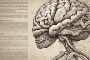
- Cerebrum: Seat of intelligence, responsible for reading, writing, speaking, and numerous other higher mental functions like making calculations, composing music, remembering the past, planning for the future, sensory perception, and initiating/coordinating skeletal muscle contractions.
- Divided into five lobes: frontal, parietal, temporal, occipital, and insula. Each lobe is named after the bone that covers it.
- Lobes and Functions
- Frontal: Voluntary motor functions, eye movements, planning, mood, smell, and social judgement.
- Parietal: Receives and integrates sensory information (touch, pain, temperature, etc.), interprets speech.
- Temporal: Areas for hearing and smell (auditory and olfactory perception). Involved in learning, memory, and emotional behaviours.
- Occipital: Visual center and visual perception.
- Insula: Deep within the lateral cerebral sulcus, not visible at the surface of the brain.
- Sulci and Fissures:
- Central sulcus: Separates frontal and parietal lobes
- Longitudinal fissure: Separates cerebrum into two hemispheres
- Lateral cerebral sulcus: Separates frontal and temporal lobes
- Parieto-occipital sulcus: Separates parietal and occipital lobes
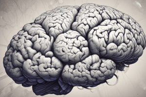
- Cerebral Cortex: Outer layer of the cerebrum (gray matter) composed of billions of neurons, including cell bodies. Embryonic development of the brain shows the gray matter enlarging faster than the white matter. This causes the cerebral cortex to fold which fits better in the cranial cavity.
- Gyri and Sulci: Gyri (folds) and sulci (shallow grooves) increase surface area.
- Hemispheres: Left and right hemispheres are connected by the corpus callosum. The Corpus Callosum is the largest fiber bundle in the brain with roughly 300 million nerve fibers.
- Internal Capsule: A v-shaped structure of white matter located between the lenticular nuclei (lateral) and thalamic/caudate nuclei (medial). Contains ascending and descending projection tracts.
- Genu: Part of the internal capsule, contains corticobulbar/corticonuclear tracts originating in the primary motor area for head and facial movement & terminating at brainstem cranial nuclei (III, IV, V, VI, VII, IX, X, XI, XII).
- Anterior Limb: Located between the lenticular nuclei and caudate nuclei. Contains major tracts for communication between the thalamus and frontal lobe (thalamocortical/thalamofrontal).
- Posterior Limb: Largest structure in the internal capsule, it contains ascending and descending tracts, Includes corticospinal, somatosensory, optic radiation, and auditory fibers.
Ventricles and Cerebrospinal Fluid (CSF)
- Four ventricles within the brain: two lateral, one third, and one fourth ventricle.
- CSF: Colorless liquid that protects the brain and spinal cord, providing shock absorption and nutrient delivery while removing waste.
- CSF composition: Glucose, proteins, lactic acid, urea, cations (Na+, K+, Ca2+, Mg2+), anions (Cl− and HCO3−), and some white blood cells.
- CSF circulation: Produced in choroid plexus. Flows from lateral to third and then fourth ventricles through the cerebral aqueduct and then into the subarachnoid space. CSF is reabsorbed into the venous circulation.
- Functions: Mechanical protection, chemical protection, and circulation (nutrients, waste). Involved in homeostasis.
Blood Brain Barrier (BBB)
- The BBB protects brain cells from harmful substances and pathogens by preventing their passage from blood into brain cells.
- It primarily consists of the endothelial cells in the choroid plexus.
- Tight junctions and astrocyte processes maintain the permeability characteristic.
- Some substances pass through by active transport (e.g., glucose). Others (e.g., creatinine, urea, & most ions) cross slowly.
Limbic System
- A complex system of structures in the cerebrum and diencephalon.
- Functions: Emotions, memory, olfaction (smell).
- Key structures include: cingulate gyrus, hippocampus, amygdala, fornix, septal nuclei, mammillary bodies, olfactory bulbs.
- Plays a role in spatial memory formation, long-term memories, and emotional responses like fear and aggression.
Basal Ganglia
- A group of nuclei deep within the cerebral hemispheres that play a role in regulating movement and posture. The basal ganglia include the caudate nucleus, putamen, globus pallidus, subthalamic nucleus, and substantia nigra
- Essential for motor control, procedural learning, and motor response selection.
Hemispheric Lateralization
- Functional specialization of brain hemispheres.
- Typically the left hemisphere is dominant for language, reasoning, and logical functions; the right hemisphere is dominant for spatial awareness, recognizing faces, and emotional content.
Learning Outcome Breakdown
- Describe the organization/function of the CNS, PNS, limbic system.
- Explain brain protection (meninges, CSF, BBB) and blood supply.
Studying That Suits You
Use AI to generate personalized quizzes and flashcards to suit your learning preferences.