Podcast
Questions and Answers
Where is the heart located?
Where is the heart located?
- Around the hips
- Above the diaphragm
- Between the second rib to the fifth (correct)
- Behind the lungs
Which direction does the base of the heart point towards?
Which direction does the base of the heart point towards?
- Left shoulder
- Neck
- Right shoulder (correct)
- Left hip
What is the mass of the heart approximately?
What is the mass of the heart approximately?
- ~ 500 grams
- ~ 1000 grams
- ~ 300 grams (correct)
- ~ 750 grams
Which layer of the heart wall is also known as the visceral pericardium?
Which layer of the heart wall is also known as the visceral pericardium?
In which cavity is the heart enclosed within a double-walled sac?
In which cavity is the heart enclosed within a double-walled sac?
Where does the apex of the heart point towards?
Where does the apex of the heart point towards?
What is the main component of the Myocardium?
What is the main component of the Myocardium?
What structures allow ions to flow from cell to cell in the cardiac muscle?
What structures allow ions to flow from cell to cell in the cardiac muscle?
Which chambers of the heart are primarily receiving chambers and not important in the pumping activity of the heart?
Which chambers of the heart are primarily receiving chambers and not important in the pumping activity of the heart?
Why are the walls of the left ventricle substantially thicker than those of the right ventricle?
Why are the walls of the left ventricle substantially thicker than those of the right ventricle?
Which vessel splits into right and left pulmonary arteries?
Which vessel splits into right and left pulmonary arteries?
Which vessels return blood to the heart from regions above and below the diaphragm?
Which vessels return blood to the heart from regions above and below the diaphragm?
What is the role of the semilunar valves during ventricles relaxation?
What is the role of the semilunar valves during ventricles relaxation?
What causes the AV valves to open?
What causes the AV valves to open?
What happens to the AV valves when the ventricles contract?
What happens to the AV valves when the ventricles contract?
What prevents semilunar valve flaps from everting into the atria?
What prevents semilunar valve flaps from everting into the atria?
When do the semilunar valves close?
When do the semilunar valves close?
What forces open the semilunar valves?
What forces open the semilunar valves?
What is the function of intercalated discs in cardiac muscle tissue?
What is the function of intercalated discs in cardiac muscle tissue?
Which protein complex is found on the thin filament in cardiac muscle cells?
Which protein complex is found on the thin filament in cardiac muscle cells?
What is the role of transverse tubules in cardiac muscle cells?
What is the role of transverse tubules in cardiac muscle cells?
What holds cardiac muscle fibers together at their ends?
What holds cardiac muscle fibers together at their ends?
Where are the Z discs located in cardiac muscle fibers?
Where are the Z discs located in cardiac muscle fibers?
What is the function of gap junctions in cardiac muscle tissue?
What is the function of gap junctions in cardiac muscle tissue?
What happens during the ventricular filling phase in the cardiac cycle?
What happens during the ventricular filling phase in the cardiac cycle?
What is the volume of blood in the ventricles at the end of atrial contraction?
What is the volume of blood in the ventricles at the end of atrial contraction?
What characterizes the ventricular systole phase in the cardiac cycle?
What characterizes the ventricular systole phase in the cardiac cycle?
What occurs during the isovolumetric contraction phase of ventricular systole?
What occurs during the isovolumetric contraction phase of ventricular systole?
How does atrial contraction affect the volume of blood in the ventricles?
How does atrial contraction affect the volume of blood in the ventricles?
What leads to the closing of the AV valves in the cardiac cycle?
What leads to the closing of the AV valves in the cardiac cycle?
Flashcards are hidden until you start studying
Study Notes
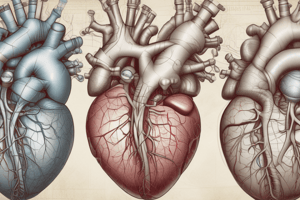
Heart Location and Structure
- The heart is located in the mediastinum, a region in the chest between the lungs.
- The base of the heart points towards the right shoulder, while the apex points towards the left hip.
- The heart's mass is approximately 250-350 grams in adults.
- The epicardium, also known as the visceral pericardium, is the outermost layer of the heart wall.
- The heart is enclosed within a double-walled sac called the pericardium, located within the thoracic cavity.
Cardiac Muscle Tissue
- The myocardium, the middle layer of the heart wall, mainly comprises cardiac muscle tissue.
- Intercalated discs, specialized junctions between cardiac muscle cells, allow ions to flow freely, enabling synchronized contractions.
- Gap junctions within intercalated discs facilitate the rapid spread of electrical impulses throughout the heart.
Heart Chambers and Valves
- The atria (right and left) are the receiving chambers of the heart, collecting blood returning from the body.
- The ventricles (right and left) are the pumping chambers, propelling blood to the lungs and the rest of the body.
- The left ventricle has thicker walls than the right ventricle to pump blood with greater force against higher resistance.
- The pulmonary trunk splits into the right and left pulmonary arteries, carrying deoxygenated blood to the lungs.
- The superior and inferior vena cava return deoxygenated blood to the right atrium from regions above and below the diaphragm, respectively.
- Semilunar valves prevent backflow of blood from the ventricles into the great arteries during ventricular relaxation.
- AV valves (tricuspid and bicuspid) open when ventricular pressure is lower than atrial pressure.
- When the ventricles contract, the AV valves close to prevent backflow of blood into the atria.
- Chordae tendineae anchor the AV valve flaps to papillary muscles, preventing them from everting into the atria.
- The semilunar valves close when ventricular pressure drops below arterial pressure during relaxation.
- The semilunar valves open when ventricular pressure exceeds arterial pressure.
Muscle Structure and Function
- Intercalated discs in cardiac muscle tissue facilitate communication and synchronous contractions, ensuring coordinated heartbeat.
- The troponin-tropomyosin complex is present on the thin filaments of cardiac muscle cells, regulating muscle contraction.
- Transverse tubules (T-tubules) in cardiac muscle cells help transmit action potentials, ensuring uniform contraction of the muscle fibers.
- Desmosomes, specialized junctions, hold cardiac muscle fibers together at their ends, providing structural integrity.
- Z discs, anchoring points for the thin filaments, are located at the ends of sarcomeres in cardiac muscle fibers.
Cardiac Cycle
- During ventricular filling, blood passively flows from the atria into the ventricles, followed by forceful atrial contraction to complete the filling
- End-diastolic volume (EDV) is the volume of blood in the ventricles at the end of atrial contraction.
- Ventricular systole is the period of ventricular contraction, marked by increased pressure within the ventricles.
- Isovolumetric contraction occurs at the beginning of ventricular systole when both AV and semilunar valves are closed, allowing for a brief increase in ventricular pressure without changing volume.
- Atrial contraction adds about 20% to the blood volume in the ventricles.
- Increased ventricular pressure leads to the closing of the AV valves.
Studying That Suits You
Use AI to generate personalized quizzes and flashcards to suit your learning preferences.