Podcast
Questions and Answers
Which organ is located on the left side of the upper abdomen?
Which organ is located on the left side of the upper abdomen?
- Liver
- Pancreas
- Spleen
- Stomach (correct)
What is the function of the pyloric sphincter?
What is the function of the pyloric sphincter?
- Allow food to pass from the stomach to the small intestine (correct)
- Digest food
- Secrete acid and enzymes
- Receive food from the esophagus
What is the purpose of rugae in the stomach?
What is the purpose of rugae in the stomach?
- Digest food
- Secrete acid and enzymes
- Receive food from the esophagus
- Allow expansion of the stomach (correct)
Which part of the alimentary canal receives food from the esophagus?
Which part of the alimentary canal receives food from the esophagus?
What is the most common malignancy of the GIT?
What is the most common malignancy of the GIT?
What is the term 'gut signature' used for in diagnosing bowel pathology?
What is the term 'gut signature' used for in diagnosing bowel pathology?
Which type of neoplasm commonly metastasizes to the gastrointestinal tract?
Which type of neoplasm commonly metastasizes to the gastrointestinal tract?
Which condition is characterized by thickening of the muscularis layer of the terminal ileum and increased vascularity?
Which condition is characterized by thickening of the muscularis layer of the terminal ileum and increased vascularity?
In which condition is tenderness in the inflamed area and increasing pain with cough commonly observed?
In which condition is tenderness in the inflamed area and increasing pain with cough commonly observed?
What is the best imaging plane for visualizing acute appendicitis?
What is the best imaging plane for visualizing acute appendicitis?
What is a characteristic feature of a normal appendix when visualized by ultrasound?
What is a characteristic feature of a normal appendix when visualized by ultrasound?
What is the recommended imaging modality for diagnosing obstruction of the intestines?
What is the recommended imaging modality for diagnosing obstruction of the intestines?
Which symptom is commonly associated with acute appendicitis?
Which symptom is commonly associated with acute appendicitis?
What is the term used for the worm-like extension of the caecum, which is commonly affected in acute appendicitis?
What is the term used for the worm-like extension of the caecum, which is commonly affected in acute appendicitis?
What is the peak incidence age for acute appendicitis?
What is the peak incidence age for acute appendicitis?
What is a characteristic feature associated with obstruction of the intestines?
What is a characteristic feature associated with obstruction of the intestines?
Which part of the small intestine absorbs most nutrients from food and drink?
Which part of the small intestine absorbs most nutrients from food and drink?
What is the role of the large intestine in the absorption process?
What is the role of the large intestine in the absorption process?
How many layers make up the walls of the gut?
How many layers make up the walls of the gut?
What is the purpose of patient preparation for gastrointestinal ultrasound?
What is the purpose of patient preparation for gastrointestinal ultrasound?
What does the 'Gut Signature' seen on ultrasound include?
What does the 'Gut Signature' seen on ultrasound include?
What role does ultrasound play in diagnosing gastrointestinal pathology?
What role does ultrasound play in diagnosing gastrointestinal pathology?
What are some pathological variations in the gut signature that should be considered suspicious?
What are some pathological variations in the gut signature that should be considered suspicious?
What are the ultrasound appearances of gastrointestinal neoplasms?
What are the ultrasound appearances of gastrointestinal neoplasms?
Which is the most common gastrointestinal malignancy?
Which is the most common gastrointestinal malignancy?
What are the ultrasound appearances of adenocarcinoma?
What are the ultrasound appearances of adenocarcinoma?
Where can specific ultrasound appearances of adenocarcinoma be found?
Where can specific ultrasound appearances of adenocarcinoma be found?
Flashcards are hidden until you start studying
Study Notes
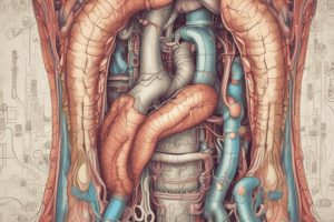
Anatomy and Pathology of the Gastrointestinal Tract
- The small intestine consists of the duodenum, jejunum, and ileum, and it absorbs most nutrients from food and drink.
- The large intestine includes the cecum, colon, rectum, and anal canal, and it absorbs water from wastes to create stool.
- The gut is a continuous hollow tube with 4-layer walls: mucosa, submucosa, muscularis propria, and serosa.
- Ultrasound equipment for imaging the gastrointestinal tract includes curvilinear and linear probes with specific frequencies for different scanning purposes.
- Patient preparation for gastrointestinal ultrasound involves fasting for 8-12 hours to remove excess gas from the stomach and bowel.
- The "Gut Signature" seen on ultrasound includes up to 5 layers, a uniform wall, and assessment of lumen content, diameter, peristalsis, and compressibility.
- Ultrasound is used to detect free fluid, masses, and surrounding tissue abnormalities in the gastrointestinal tract, and it plays a role in diagnosing neoplasms, inflammatory bowel disease, appendicitis, and obstruction.
- Pathological variations in the gut signature that should be considered suspicious include non-compressible gut, wall thickening, exophytic masses, dilated lumen, abnormal peristalsis, adjacent hyperechoic fat, and lymphadenopathy.
- Ultrasound appearances of gastrointestinal neoplasms include intra-mural lesions, exophytic lesions, and lesions that destroy the bowel wall.
- Adenocarcinoma is the most common gastrointestinal malignancy, affecting the stomach and large bowel more frequently than the small bowel.
- Ultrasound appearances of adenocarcinoma include large masses, thickened gut walls with symmetric or asymmetric patterns, and hypoechoic characteristics, along with target or pseudokidney signs.
- The role of ultrasound in diagnosing gastrointestinal pathology is described in the "Diagnostic Ultrasound" textbook, and specific ultrasound appearances of adenocarcinoma are detailed, including target and pseudokidney signs.
Studying That Suits You
Use AI to generate personalized quizzes and flashcards to suit your learning preferences.