Podcast
Questions and Answers
What is the primary tissue type that forms most of the gastrointestinal tract?
What is the primary tissue type that forms most of the gastrointestinal tract?
- Splanchnic mesoderm
- Endoderm (correct)
- Ectoderm
- Mesoderm
At what stage of development does the esophagus reach its final length?
At what stage of development does the esophagus reach its final length?
- 8 weeks
- 5 weeks
- 6 weeks
- 7 weeks (correct)
Which anatomical structure initially keeps the primordial gut closed at the cranial end?
Which anatomical structure initially keeps the primordial gut closed at the cranial end?
- Cloacal membrane
- Tracheoesophageal septum
- Esophageal membrane
- Oropharyngeal membrane (correct)
Which artery primarily supplies most of the foregut structures?
Which artery primarily supplies most of the foregut structures?
What separates the esophagus from the trachea during development?
What separates the esophagus from the trachea during development?
Where do the epithelium and glands of the esophagus originate from?
Where do the epithelium and glands of the esophagus originate from?
Which part of the muscularis externa of the esophagus is derived from striated muscle?
Which part of the muscularis externa of the esophagus is derived from striated muscle?
What occurs to the lumen of the esophagus during its development?
What occurs to the lumen of the esophagus during its development?
What initial shape does the distal part of the foregut take during stomach development?
What initial shape does the distal part of the foregut take during stomach development?
Which direction does the stomach rotate during its development?
Which direction does the stomach rotate during its development?
What is the role of the dorsal mesentery during stomach development?
What is the role of the dorsal mesentery during stomach development?
What happens to the original left side of the stomach during its rotation?
What happens to the original left side of the stomach during its rotation?
What forms as the clefts in mesenchyme combine during stomach development?
What forms as the clefts in mesenchyme combine during stomach development?
Which structure is carried to the left during the rotation of the stomach?
Which structure is carried to the left during the rotation of the stomach?
Which part of the omental bursa persists medial to the base of the right lung?
Which part of the omental bursa persists medial to the base of the right lung?
What does the greater curvature of the stomach correspond to after rotation?
What does the greater curvature of the stomach correspond to after rotation?
At what week does bile formation begin in the liver?
At what week does bile formation begin in the liver?
Which part of the diverticulum develops into the gallbladder?
Which part of the diverticulum develops into the gallbladder?
Which structure extends from the liver to the ventral abdominal wall?
Which structure extends from the liver to the ventral abdominal wall?
What initiates haematopoiesis in the liver?
What initiates haematopoiesis in the liver?
Which pancreatic bud appears first and contributes to most of the pancreas?
Which pancreatic bud appears first and contributes to most of the pancreas?
Where does the proximal part of the duct of the dorsal bud open, if it persists?
Where does the proximal part of the duct of the dorsal bud open, if it persists?
What developmental transformation occurs for the pancreas as the duodenum rotates?
What developmental transformation occurs for the pancreas as the duodenum rotates?
Which structure does NOT derive its development from the foregut?
Which structure does NOT derive its development from the foregut?
What structure does the omental bursa communicate with?
What structure does the omental bursa communicate with?
Which structures develop from the hepatic diverticulum?
Which structures develop from the hepatic diverticulum?
During which week does the duodenum begin to develop?
During which week does the duodenum begin to develop?
What happens to the lumen of the duodenum during the 5th and 6th weeks of development?
What happens to the lumen of the duodenum during the 5th and 6th weeks of development?
What is the primary artery supplying the duodenum?
What is the primary artery supplying the duodenum?
What occurs during recanalization of the duodenum?
What occurs during recanalization of the duodenum?
How does the omental bursa change as the greater omentum layers fuse?
How does the omental bursa change as the greater omentum layers fuse?
What is the main reason for the shrinking of the duodenal lumen during development?
What is the main reason for the shrinking of the duodenal lumen during development?
Which structure is formed from the cloacal membrane?
Which structure is formed from the cloacal membrane?
What happens to the mesentery of the descending colon?
What happens to the mesentery of the descending colon?
What differentiates the inferior one-third of the anal canal?
What differentiates the inferior one-third of the anal canal?
What does the urorectal septum contribute to during development?
What does the urorectal septum contribute to during development?
Which part of the colon retains its mesentery?
Which part of the colon retains its mesentery?
Which artery supplies all derivatives of the hindgut?
Which artery supplies all derivatives of the hindgut?
How does the appendix develop from the caecal swelling?
How does the appendix develop from the caecal swelling?
What is indicated by the pectinate line in the anal canal?
What is indicated by the pectinate line in the anal canal?
Flashcards are hidden until you start studying
Study Notes
Foregut Development
- The foregut is responsible for the development of vital organs like the esophagus, stomach, duodenum, liver, gallbladder, and pancreas.
- The esophagus forms from the caudal part of the pharynx.
- The esophagus rapidly elongates due to the growth and relocation of the heart and lungs.
- The esophagus is initially short, but reaches its full length by the 7th week.
- Muscle development in the esophagus involves both striated and smooth muscle fibers.
- Striated muscle (superior 3rd) develops from mesenchyme in the 4th and 6th pharyngeal arches.
- Smooth muscle (inferior 3rd) develops from surrounding splanchnic mesenchyme.
- Both types of muscle are innervated by the vagus nerve.
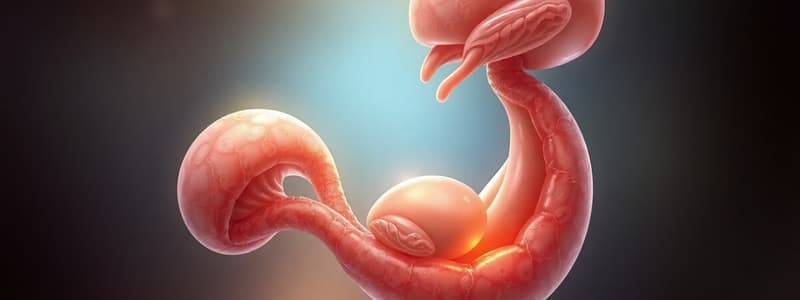
- The stomach forms from a slight dilation of the distal part of the foregut.
- This dilation occurs during the 4th week.
- The stomach undergoes a 90-degree clockwise rotation along its longitudinal axis as it enlarges.
- This rotation impacts the position of the stomach's borders.
Stomach Rotation and Omental Bursa Formation
- The stomach rotation repositions the ventral border (lesser curvature) to the right and the dorsal border (greater curvature) to the left.
- It flips the stomach’s original left side to become the ventral surface and the right side to become the dorsal surface.
- The stomach's cranial region shifts left and slightly inferiorly, while the caudal region moves right and superiorly.
- The stomach's long axis becomes almost transverse to the body's long axis.
- The stomach is suspended from the dorsal wall by the dorsal mesentery, also known as the primordial dorsal mesogastrium.
- This mesentery carries the spleen and coeliac artery.
- The primordial ventral mesogastrium connects the stomach, duodenum, liver, and ventral abdominal wall.
- Cleft formation in the dorsal mesogastrium leads to the development of the omental bursa, an isolated cavity.
- Stomach rotation pulls the dorsal mesogastrium leftwards, expanding the bursa.
- This expansion allows for efficient movement of the stomach within the abdominal cavity.
Omental Bursa and Duodenum Development
- The omental bursa develops into two regions: the infracardiac bursa (superior) and the superior recess of the omental bursa (inferior).
- The greater omentum is formed from two elongated layers of dorsal mesogastrium, covering the developing intestines.
- The omental bursa communicates with the peritoneal cavity through the omental foramen.
- The duodenum develops from the caudal part of the foregut, the cranial part of the midgut, and associated splanchnic mesenchyme.
- The junction of these parts is marked by the bile duct's origin.
- The duodenum grows rapidly, forming a C-shaped loop that projects ventrally.
- The loop rotates to the right with the stomach.
- It then becomes retroperitoneal by being pressed against the posterior abdominal wall.
- The duodenum is supplied by both the coeliac trunk and the superior mesenteric artery.
- The lumen temporarily obliterates due to epithelial cell proliferation during the 5th and 6th weeks.
- Recanalization occurs through vaculolation, restoring the lumen.
- The liver, gallbladder, and biliary duct system originate from a ventral endodermal outgrowth called the hepatic diverticulum.
- This diverticulum originates from the foregut's distal part during the 4th week.
- The hepatic diverticulum invades the septum transversum, which forms the ventral mesogastrium.
- During growth, the hepatic diverticulum divides into two layers of the ventral mesogastrium.
Liver and Biliary Development
- The larger part of the hepatic diverticulum becomes the primordium of the liver.
- The smaller caudal part forms the primordium of the gallbladder.
- Endodermal cells contribute to the epithelial lining of the intrahepatic biliary apparatus.
- Anastomosis between hepatic cords and endothelium-lined spaces forms the hepatic sinusoids.
- The liver grows rapidly during the 5th to 10th weeks, filling a significant part of the upper abdominal cavity.
- Initially, the left and right lobes of the liver are equal, but the right lobe eventually becomes larger.
- Haematopoiesis commences in the liver after the 6th week, and bile formation begins at the 12th week.
Pancreas Development
- The pancreas develops between layers of the mesentery.
- Two endodermal buds - dorsal and ventral pancreatic buds - arise from the caudal part of the foregut.
- The dorsal pancreatic bud (appears first) contributes the majority of the pancreas.
- It forms slightly cranial to the ventral bud.
- The smaller ventral pancreatic bud arises near the common bile duct's entry into the duodenum.
- It grows between layers of the ventral mesentery.
- As the duodenum rotates to the right, the ventral bud is carried dorsally, eventually lying posterior to the dorsal bud.
- The ventral pancreatic duct produces the uncinate process and head of the pancreas after fusion with the dorsal pancreatic bud.
- The pancreas becomes retroperitoneal along with the stomach, duodenum, and pancreas rotation.
- The ducts of the pancreatic buds anastomose, with the pancreatic duct forming from the ventral bud's duct and the distal dorsal bud duct.
- The proximal part of the dorsal bud's duct may persist as an accessory pancreatic duct, opening into the minor duodenal papilla.
Spleen Development
- Spleen development starts during the 5th week.
- It originates from mesenchyme in the dorsal mesogastrium.
- The mesentery associated with the spleen eventually fuses with parietal peritoneum and disappears.
Midgut Development
- The caecal swelling appears in the sixth week on the caudal limb of the midgut.
- Its apex, which forms the appendix, grows slowly, making the appendix variable in position.
- The appendix may be retrocaecal, retrocolic, or run over the pelvic brim.
Hindgut Development
- Distal third of the transverse colon, descending and sigmoid colons, rectum, the superior part of the anal canal, the urinary bladder, and most of the urethra are derived from the hindgut.
- These derivatives are supplied by the inferior mesenteric artery.
- The descending colon becomes retroperitoneal as its mesentery fuses with parietal peritoneum and disappears.
- The sigmoid colon retains its mesentery, remaining intraperitoneal.
Cloaca Development
- The cloaca is an endoderm-lined chamber in contact with surface ectoderm at the cloacal membrane.
- This membrane is composed of cloacal endoderm and anal pit ectoderm.
- The cloaca receives the allantois ventrally.
- The cloaca is partitioned into dorsal and ventral parts by the urorectal septum.
- The septum grows and produces extensions, causing infolding of the lateral cloacal walls.
- These folds fuse to form a partition, dividing the cloaca into the rectum, the cranial part of the anal canal, and the urogenital sinus.
- The anal pit is formed by anorectal canal recanalization through apoptosis of the epithelial anal plug, which temporarily closes the anorectal lumen.
Anal Canal Development
- The superior 2/3 of the anal canal originates from the hindgut and is supplied by the superior rectal artery.
- The inferior 1/3 develops from the anal pit and is supplied by the inferior rectal artery.
- The pectinate line marks the junction of epithelium from the anal pit ectoderm and hindgut endoderm.
Studying That Suits You
Use AI to generate personalized quizzes and flashcards to suit your learning preferences.


