Podcast
Questions and Answers
Which ligament is primarily injured in a low ankle sprain?
Which ligament is primarily injured in a low ankle sprain?
- Calcaneofibular Ligament
- Posterior Talofibular Ligament
- Syndesmotic Ligament
- Anterior Talofibular Ligament (correct)
What is a typical recovery time for a high ankle sprain?
What is a typical recovery time for a high ankle sprain?
- 15 days
- 1 week
- 6 weeks
- 2-7 weeks (correct)
Which test is used to assess the integrity of the ATFL?
Which test is used to assess the integrity of the ATFL?
- Lateral Hop for Distance Test
- Talar Tilt Test
- Anterior Drawer Test (correct)
- Royal London Hospital Test
What is the primary intervention recommended during the acute/protected motion phase of an ankle sprain?
What is the primary intervention recommended during the acute/protected motion phase of an ankle sprain?
What characteristics define Chronic Ankle Instability (CAI)?
What characteristics define Chronic Ankle Instability (CAI)?
What does the Bernese Ankle Rules focus on regarding functional tests?
What does the Bernese Ankle Rules focus on regarding functional tests?
Which of the following is NOT a sign of Achilles tendinopathy?
Which of the following is NOT a sign of Achilles tendinopathy?
Which functional test is NOT included in the assessment for ankle injuries?
Which functional test is NOT included in the assessment for ankle injuries?
Which ankle sprain mechanism of injury (MOI) is typical for a low ankle sprain?
Which ankle sprain mechanism of injury (MOI) is typical for a low ankle sprain?
Which ligament is known to provide primary restraint to the inversion moment when the ankle is in a plantar-flexed position?
Which ligament is known to provide primary restraint to the inversion moment when the ankle is in a plantar-flexed position?
What is the main distinguishing characteristic of reactive tendinopathy compared to degenerative tendinopathy?
What is the main distinguishing characteristic of reactive tendinopathy compared to degenerative tendinopathy?
Which intrinsic risk factor is associated with Achilles tendinopathy?
Which intrinsic risk factor is associated with Achilles tendinopathy?
What is the most common mechanism of injury leading to Achilles tendon ruptures?
What is the most common mechanism of injury leading to Achilles tendon ruptures?
Which symptom is typically associated with midportion Achilles tendinopathy?
Which symptom is typically associated with midportion Achilles tendinopathy?
Which physical performance measure is not typically used to assess Achilles tendinopathy?
Which physical performance measure is not typically used to assess Achilles tendinopathy?
What is the recommended approach to performing the eccentric phase of rehabilitation for Achilles tendinopathy?
What is the recommended approach to performing the eccentric phase of rehabilitation for Achilles tendinopathy?
Which test is commonly used to diagnose plantar fasciitis?
Which test is commonly used to diagnose plantar fasciitis?
What does the presence of a thickened nodule in the Achilles tendon suggest?
What does the presence of a thickened nodule in the Achilles tendon suggest?
Which anatomical range of motion (ROM) is needed for a successful lateral step down?
Which anatomical range of motion (ROM) is needed for a successful lateral step down?
What should be avoided during the rehabilitation of insertional Achilles tendinopathy?
What should be avoided during the rehabilitation of insertional Achilles tendinopathy?
What is a characteristic of Plantar Fasciitis pain throughout the day?
What is a characteristic of Plantar Fasciitis pain throughout the day?
In the progressive resistive eccentric exercise program for Achilles tendon rehabilitation, what is the initial loading weight for the eccentric phase?
In the progressive resistive eccentric exercise program for Achilles tendon rehabilitation, what is the initial loading weight for the eccentric phase?
Which biomechanical deficit is likely to exacerbate conditions like Achilles tendinopathy?
Which biomechanical deficit is likely to exacerbate conditions like Achilles tendinopathy?
Which of the following is NOT a typical presentation of rheumatoid arthritis?
Which of the following is NOT a typical presentation of rheumatoid arthritis?
What is a common pathological factor in Femoral Acetabular Impingement Syndrome?
What is a common pathological factor in Femoral Acetabular Impingement Syndrome?
Which rehabilitation intervention is considered beneficial for Gluteal Tendinopathy?
Which rehabilitation intervention is considered beneficial for Gluteal Tendinopathy?
Which statement about labral lesions is accurate?
Which statement about labral lesions is accurate?
What is a common symptom of Greater Trochanteric Pain Syndrome?
What is a common symptom of Greater Trochanteric Pain Syndrome?
What type of morphology is associated with CAM deformity in Femoral Acetabular Impingement Syndrome?
What type of morphology is associated with CAM deformity in Femoral Acetabular Impingement Syndrome?
Which test specifically indicates a posterior labral tear?
Which test specifically indicates a posterior labral tear?
Which type of exercise is emphasized for patients with Iliotibial Band Syndrome?
Which type of exercise is emphasized for patients with Iliotibial Band Syndrome?
What is the goal of the conservative treatment for labral lesions?
What is the goal of the conservative treatment for labral lesions?
Which is a recognized risk factor for Athletic Pubalgia or Core Muscle Injury?
Which is a recognized risk factor for Athletic Pubalgia or Core Muscle Injury?
Which treatment is most suited for Ischemic Necrosis of the Femoral Head?
Which treatment is most suited for Ischemic Necrosis of the Femoral Head?
What is the key muscle involved in the pathology of Gluteal Syndrome?
What is the key muscle involved in the pathology of Gluteal Syndrome?
Physical therapy for labral tears should primarily focus on which of the following?
Physical therapy for labral tears should primarily focus on which of the following?
Flashcards
Low Ankle Sprain
Low Ankle Sprain
An injury to the ligaments on the outside of the ankle, often caused by inverting the foot while plantar flexing. Usually involves the ATFL, PTFL, and CFL, and heals quicker than high ankle sprains.
High Ankle Sprain
High Ankle Sprain
Injury to the syndesmotic ligaments connecting the tibia and fibula above the ankle. Often caused by twisting or rolling the ankle. Takes longer to heal than a low ankle sprain.
Anterior Talofibular Ligament (ATFL)
Anterior Talofibular Ligament (ATFL)
A key ligament on the outside of the ankle, primarily resisting inversion when the foot is plantar flexed. Often injured in low ankle sprains.
Chronic Ankle Instability (CAI)
Chronic Ankle Instability (CAI)
Signup and view all the flashcards
Calcaneofibular Ligament (CFL)
Calcaneofibular Ligament (CFL)
Signup and view all the flashcards
Anterior Drawer Test
Anterior Drawer Test
Signup and view all the flashcards
Ottawa Ankle Rules
Ottawa Ankle Rules
Signup and view all the flashcards
Achilles Tendinopathy
Achilles Tendinopathy
Signup and view all the flashcards
MICE
MICE
Signup and view all the flashcards
Chronic Ankle Instability (CAI) ICF Impairments
Chronic Ankle Instability (CAI) ICF Impairments
Signup and view all the flashcards
Achilles Tendinopathy Midportion
Achilles Tendinopathy Midportion
Signup and view all the flashcards
Achilles Tendinopathy Insertional
Achilles Tendinopathy Insertional
Signup and view all the flashcards
Plantar Fasciitis
Plantar Fasciitis
Signup and view all the flashcards
Achilles Rupture MOI
Achilles Rupture MOI
Signup and view all the flashcards
Midportion Achilles Tenderness Sign
Midportion Achilles Tenderness Sign
Signup and view all the flashcards
Achilles Tendinopathy - Warm-up Phenomenon
Achilles Tendinopathy - Warm-up Phenomenon
Signup and view all the flashcards
Achilles Tendon Palpation Test
Achilles Tendon Palpation Test
Signup and view all the flashcards
Plantar Fasciitis - Risk Factors
Plantar Fasciitis - Risk Factors
Signup and view all the flashcards
Achilles Tendon - Physical Performance
Achilles Tendon - Physical Performance
Signup and view all the flashcards
Achilles Intervention Strength
Achilles Intervention Strength
Signup and view all the flashcards
Hip Dysplasia
Hip Dysplasia
Signup and view all the flashcards
Legg-Calve'-Perthes Disease
Legg-Calve'-Perthes Disease
Signup and view all the flashcards
Slipped Capital Femoral Epiphysis
Slipped Capital Femoral Epiphysis
Signup and view all the flashcards
Osteoarthritis
Osteoarthritis
Signup and view all the flashcards
What is the pathology of FAIS?
What is the pathology of FAIS?
Signup and view all the flashcards
What are the three types of FAIS?
What are the three types of FAIS?
Signup and view all the flashcards
What is Pincer Deformity?
What is Pincer Deformity?
Signup and view all the flashcards
What are some common interventions for FAIS?
What are some common interventions for FAIS?
Signup and view all the flashcards
What is the pathology of Labral Lesions?
What is the pathology of Labral Lesions?
Signup and view all the flashcards
What are the functions of the labrum?
What are the functions of the labrum?
Signup and view all the flashcards
What are common symptoms of a labral lesion?
What are common symptoms of a labral lesion?
Signup and view all the flashcards
What are common assessment tools for Labral Lesions?
What are common assessment tools for Labral Lesions?
Signup and view all the flashcards
What are common interventions for Labral Lesions?
What are common interventions for Labral Lesions?
Signup and view all the flashcards
What is the pathology of Osteoarthritis?
What is the pathology of Osteoarthritis?
Signup and view all the flashcards
What are common symptoms of Osteoarthritis?
What are common symptoms of Osteoarthritis?
Signup and view all the flashcards
What are common impairments with Osteoarthritis?
What are common impairments with Osteoarthritis?
Signup and view all the flashcards
What are common assessment tools for Osteoarthritis?
What are common assessment tools for Osteoarthritis?
Signup and view all the flashcards
What are common interventions for Osteoarthritis?
What are common interventions for Osteoarthritis?
Signup and view all the flashcards
Study Notes
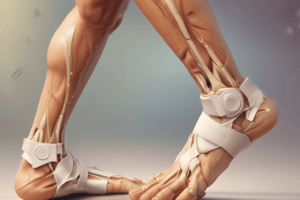
Foot and Ankle Sprains
- Low ankle sprains are more common than high ankle sprains.
- The most common mechanism of a low ankle sprain is inversion and plantar flexion.
- Structures involved in low ankle sprains include the anterior talofibular ligament (ATFL), calcaneofibular ligament (CFL), and posterior talofibular ligament (PTFL).
- Prognosis for low ankle sprains is shorter than high ankle sprains (approximately 1 week).
- High ankle sprains involve the syndesmotic ligaments.
- The typical mechanism of a high ankle sprain is external rotation of the tibia and dorsiflexion of the ankle.
- The prognosis for high ankle sprains is longer than low ankle sprains, approximately 2-7 weeks.
Chronic Ankle Instability (CAI)
- Chronic pain and reoccurring sensation of instability are common symptoms of CAI.
- Some cases of low ankle sprains can develop into chronic ankle instability.
- 40% of individuals develop persisting symptoms of CAI following an ankle sprain.
Lateral Ankle Sprain Grades
- Grade I: minimal loss of function, negative anterior drawer and talar tilt tests, little to no hemorrhaging, and minimal swelling.
- Grade II: some loss of function, positive anterior drawer test, negative talar tilt test, moderate hemorrhaging, moderate swelling, and moderate pain.
- Grade III: near total loss of function, positive anterior drawer and talar tilt tests, severe hemorrhaging, severe swelling, and severe pain.
Ottawa Ankle Rules
- Used to determine if ankle X-rays are needed.
- X-ray needed if pain in the malleolar or midfoot zone and bone tenderness at particular points, and/or inability to bear weight both immediately and in the emergency department.
Bernese Ankle Rules
- Procedure involves 3 stress tests: Indirect Fibular Stress, Direct Medial Malleolar Stress, and Mid- & Hind-Foot Compression Stress.
- Radiograph needed if symptoms reproduced from any 1 of the 3 above.
Hip Examination
- Hip dysplasia is a shallow acetabulum/decreased coverage of the femoral head.
- Hip instability is the increased stress on anterior capsul.
Developmental Dysplasia of the Hip (DDH)
- The typical presentation of an adult with DDH is an insidious onset of moderate to severe groin or lateral hip pain.
- Activity restriction, limping, and positive impingement signs are common indicators.
Slipped Capital Femoral Epiphysis (SCFE)
- Slippage of the femoral head relative to the femoral neck, often occurring in adolescents.
- Clinically, there may be gradual or sudden pain in the hip, groin, thigh, or knee.
- The pain is typically worse with activity but can occur at rest.
Legg-Calvé-Perthes Disease
- Idiopathic avascular necrosis of the femoral head, typically affecting children aged 4-10.
- It's more common in boys.
- The presence of pain in the groin, thigh, or medial knee along with an antalgic gait and reduced hip abduction/muscle spasms is indicative.
Osteoarthritis
- Results from the degradation of the articular cartilage with subsequent osteophyte formation, narrowing of the joint space, and sclerosis of bone.
- Clinically, there may be moderate to severe anterolateral hip pain during weight-bearing, morning stiffness lasting under an hour, and limited hip IR below 24º.
Rheumatoid Arthritis
- A systemic autoimmune inflammatory disease affecting the synovial lining, cartilage, and bone.
- Common presentation includes symmetrical involvement of multiple joints, pain, swelling, redness, and stiffness.
- Morning stiffness typically exceeding 1 hour, decreased range of motion, and presence of rheumatoid factor support diagnoses.
Greater Trochanteric Pain Syndrome (GTPS)
- A cluster of symptoms including trochanteric bursitis, abductor tendinopathy, and abnormal mechanical loads.
- Clinically, there may be insidious, chronic, and intermittent pain in the proximal lateral hip radiating to the distal thigh.
- Positive 30-second single-leg stance test (pain due to, and not necessarily weakness).
Labral Lesions
- Labral function includes shock absorption, pressure distribution, joint stabilization, and proprioception.
- Presentation features include anterior hip/groin pain, clicking, catching, and difficulty with range-of-motion, particularly rotation.
Iliotibial Band Syndrome
- Lateral hip or knee pain, weaker hip abductor strength, and a relationship to increased hip adduction and knee internal rotation during stance.
Avulsion Fractures
- common in 14-17 years old boys.
- Typically involve sudden onset pain.
- Limited active range of motion, weakness, and pain.
Ischemic Necrosis of the Femoral Head (AVN)
- Avascular necrosis of the femoral head characterized by interruption of blood supply.
- Pain in the groin (often worsened by weightbearing and relieved by rest), and limitation in range of motion in the non-capsular pattern.
Coxa Saltans/Snapping Hip
- Snapping hip is when various structures snap over the superior portion of the hip joint.
- Extra-articular snapping hip is more common and is often linked to GTPS.
Gluteal Syndrome
- Cluster of pain that may be caused by disorders of the piriformis or hamstrings.
- Common in those who sit a lot and have buttock pain.
- Pain often worsens with prolonged sitting or activity; referral to the knee may be common.
Athletic Pubalgia/Core Muscle Injury (CMI)
- Chronic pain in the abdominal/groin area radiating into the perineum or proximal adductors.
- Exacerbation of pain occurs with kicking, cutting, or sprinting.
- Imbalance between adductor and abductor muscle strength, and decreased ROM are often present.
Studying That Suits You
Use AI to generate personalized quizzes and flashcards to suit your learning preferences.