Podcast
Questions and Answers
What is the primary purpose of leaving gaps between slices during imaging?
What is the primary purpose of leaving gaps between slices during imaging?
- To prevent cross-talk between adjacent slices (correct)
- To optimize the phase gradient
- To enhance the signal-to-noise ratio
- To increase the image resolution
What happens to the nuclear precession frequency after the phase gradient pulse ends?
What happens to the nuclear precession frequency after the phase gradient pulse ends?
- It gradually increases without any external influence
- It returns to normal, but the phase difference remains (correct)
- It stabilizes entirely without phase difference
- It continues to change indefinitely
Which of the following equations represents the frequency encoding for the x-direction?
Which of the following equations represents the frequency encoding for the x-direction?
- ω=γ(B0+Gx) (correct)
- ϕ=γ(B0-Gy)ty
- ϕ=γ(B0+Gy)ty
- ω=γ(B0-Gx)
In the context of spatial encoding, what do the three directions refer to?
In the context of spatial encoding, what do the three directions refer to?
What is the result of performing an inverse Fourier transform on data collected in k-space?
What is the result of performing an inverse Fourier transform on data collected in k-space?
What does the BOLD signal in fMRI primarily reflect?
What does the BOLD signal in fMRI primarily reflect?
What is required for accurate detection of BOLD signal changes in fMRI?
What is required for accurate detection of BOLD signal changes in fMRI?
In spatial encoding for a 2D image, which of the following is NOT one of the three orthogonal axes?
In spatial encoding for a 2D image, which of the following is NOT one of the three orthogonal axes?
When a slice encoding gradient pulse is applied, how does it affect the Larmor frequencies of nuclei?
When a slice encoding gradient pulse is applied, how does it affect the Larmor frequencies of nuclei?
What is the purpose of using SPM (Statistical Parametric Mapping) software in fMRI?
What is the purpose of using SPM (Statistical Parametric Mapping) software in fMRI?
What limitation is associated with slice thickness in fMRI imaging?
What limitation is associated with slice thickness in fMRI imaging?
Why is the assumption of a slice being made in the z-direction considered convenient?
Why is the assumption of a slice being made in the z-direction considered convenient?
What problem arises from the imperfect slice profile during fMRI imaging?
What problem arises from the imperfect slice profile during fMRI imaging?
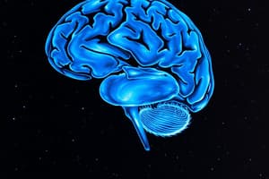
What characteristic of MRI allows it to detect variations in tissues?
What characteristic of MRI allows it to detect variations in tissues?
Which component of the MRI system is responsible for adjusting the static field's uniformity?
Which component of the MRI system is responsible for adjusting the static field's uniformity?
What happens to nuclei in an MRI when they are exposed to a magnetic field?
What happens to nuclei in an MRI when they are exposed to a magnetic field?
How does the strength of the magnet in medical MRI systems correlate with the subject size?
How does the strength of the magnet in medical MRI systems correlate with the subject size?
Which part of the MRI system maintains the low temperature necessary for superconducting magnets?
Which part of the MRI system maintains the low temperature necessary for superconducting magnets?
Which of the following best describes the principle behind generating an electromotive force (EMF) in MRI?
Which of the following best describes the principle behind generating an electromotive force (EMF) in MRI?
What is the role of gradient coils in the MRI process?
What is the role of gradient coils in the MRI process?
What is notably different about the world's strongest magnet compared to clinical MRI magnets?
What is notably different about the world's strongest magnet compared to clinical MRI magnets?
What is the main factor that affects signal intensity in MRI?
What is the main factor that affects signal intensity in MRI?
Which type of image is best suited for viewing anatomy on a clinical scanner?
Which type of image is best suited for viewing anatomy on a clinical scanner?
What is the purpose of using contrast agents in MRI?
What is the purpose of using contrast agents in MRI?
Which of the following can increase the Specific Absorption Rate (SAR) during an MRI?
Which of the following can increase the Specific Absorption Rate (SAR) during an MRI?
What is a potential side effect of rapidly changing gradient fields during an MRI scan?
What is a potential side effect of rapidly changing gradient fields during an MRI scan?
What type of blood has diamagnetic properties and repels magnetic fields?
What type of blood has diamagnetic properties and repels magnetic fields?
What happens to a magnet in the absence of adequate liquid helium or nitrogen?
What happens to a magnet in the absence of adequate liquid helium or nitrogen?
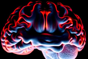
What is functional Magnetic Resonance Imaging (fMRI) primarily used for?
What is functional Magnetic Resonance Imaging (fMRI) primarily used for?
Which factor does NOT influence signal intensity in MRI?
Which factor does NOT influence signal intensity in MRI?
Why is magnetic field uniformity essential in MRI?
Why is magnetic field uniformity essential in MRI?
How do shimming coils adjust the magnetic field?
How do shimming coils adjust the magnetic field?
What can high magnetic fields disrupt?
What can high magnetic fields disrupt?
Which of the following is not a parameter conveyed in RF signals?
Which of the following is not a parameter conveyed in RF signals?
What do Fourier Transforms do in the context of MRI?
What do Fourier Transforms do in the context of MRI?
What is the role of gradients in MRI?
What is the role of gradients in MRI?
What is measured in T/m or mT/m?
What is measured in T/m or mT/m?
What is the primary purpose of adding a gradient in the z-direction during MR imaging?
What is the primary purpose of adding a gradient in the z-direction during MR imaging?
What causes the loud 'machine-gun' sound during MR imaging?
What causes the loud 'machine-gun' sound during MR imaging?
Which term best describes the shape of the RF pulse needed to excite a specific slice of tissue?
Which term best describes the shape of the RF pulse needed to excite a specific slice of tissue?
What effect does the strength and duration of the RF pulse have on magnetization?
What effect does the strength and duration of the RF pulse have on magnetization?
What happens when a gradient in the x or y direction is applied?
What happens when a gradient in the x or y direction is applied?
How does the enclosed MRI scanner amplify noise for the patient?
How does the enclosed MRI scanner amplify noise for the patient?
What is the role of the phase gradient in 3D imaging during MR?
What is the role of the phase gradient in 3D imaging during MR?
What is the outcome of a perfect sinc pulse in MR imaging?
What is the outcome of a perfect sinc pulse in MR imaging?
Flashcards
Cross-Talk
Cross-Talk
The phenomenon where neighboring slices are excited before nuclei return to equilibrium, causing signal interference.
Phase Encoding Gradient
Phase Encoding Gradient
A type of magnetic field gradient applied briefly during MRI data collection to encode spatial information along the y-axis (usually perpendicular to the image's length).
Frequency Encoding Gradient
Frequency Encoding Gradient
A type of magnetic field gradient applied continuously during MRI data collection to encode spatial information along the x-axis (usually parallel to the image's length).
K-space
K-space
Signup and view all the flashcards
2D Data Collection in MRI
2D Data Collection in MRI
Signup and view all the flashcards
What is the importance of a uniform magnetic field in MRI?
What is the importance of a uniform magnetic field in MRI?
Signup and view all the flashcards
What is Quenching?
What is Quenching?
Signup and view all the flashcards
What is Gradient Strength?
What is Gradient Strength?
Signup and view all the flashcards
What are Gradient Directions?
What are Gradient Directions?
Signup and view all the flashcards
What is the z-direction in MRI?
What is the z-direction in MRI?
Signup and view all the flashcards
What is the y-direction in MRI?
What is the y-direction in MRI?
Signup and view all the flashcards
How is NMR signal frequency related to magnetic field strength?
How is NMR signal frequency related to magnetic field strength?
Signup and view all the flashcards
What is the purpose of adding a gradient to the magnetic field in MRI?
What is the purpose of adding a gradient to the magnetic field in MRI?
Signup and view all the flashcards
BOLD Signal
BOLD Signal
Signup and view all the flashcards
fMRI (Functional Magnetic Resonance Imaging)
fMRI (Functional Magnetic Resonance Imaging)
Signup and view all the flashcards
SPM (Statistical Parametric Mapping)
SPM (Statistical Parametric Mapping)
Signup and view all the flashcards
Slice Axis
Slice Axis
Signup and view all the flashcards
Slice Encoding Gradient
Slice Encoding Gradient
Signup and view all the flashcards
Spatial Encoding
Spatial Encoding
Signup and view all the flashcards
Slice Thickness Limitation
Slice Thickness Limitation
Signup and view all the flashcards
Cross-talk between slices
Cross-talk between slices
Signup and view all the flashcards
Signal Intensity in MRI
Signal Intensity in MRI
Signup and view all the flashcards
T1 and T2 Relaxation Times
T1 and T2 Relaxation Times
Signup and view all the flashcards
Weighted MRI Images
Weighted MRI Images
Signup and view all the flashcards
Contrast Agents in MRI
Contrast Agents in MRI
Signup and view all the flashcards
SAR in MRI
SAR in MRI
Signup and view all the flashcards
Functional MRI (fMRI)
Functional MRI (fMRI)
Signup and view all the flashcards
Oxyhemoglobin and Deoxyhemoglobin in fMRI
Oxyhemoglobin and Deoxyhemoglobin in fMRI
Signup and view all the flashcards
Applications of fMRI
Applications of fMRI
Signup and view all the flashcards
Magnetic Field Gradient
Magnetic Field Gradient
Signup and view all the flashcards
Larmor Equation
Larmor Equation
Signup and view all the flashcards
Slice Selection Gradient
Slice Selection Gradient
Signup and view all the flashcards
Gradient Directions
Gradient Directions
Signup and view all the flashcards
MRI Noise
MRI Noise
Signup and view all the flashcards
RF Pulse
RF Pulse
Signup and view all the flashcards
Sinc Pulse
Sinc Pulse
Signup and view all the flashcards
Phase Gradient
Phase Gradient
Signup and view all the flashcards
Chemical Differences in MRI
Chemical Differences in MRI
Signup and view all the flashcards
Blood Flow Imaging in MRI
Blood Flow Imaging in MRI
Signup and view all the flashcards
Various Views in MRI
Various Views in MRI
Signup and view all the flashcards
Long Slices in MRI
Long Slices in MRI
Signup and view all the flashcards
MRI Process: Nuclear Magnetic Resonance
MRI Process: Nuclear Magnetic Resonance
Signup and view all the flashcards
Magnetic Fields in MRI
Magnetic Fields in MRI
Signup and view all the flashcards
MRI Magnet Strength
MRI Magnet Strength
Signup and view all the flashcards
Superconductors in MRI
Superconductors in MRI
Signup and view all the flashcards
Study Notes
MRI Capabilities
- Displays tissue variations in grayscale intensities, like tumors.
- Captures high-intensity vessel images in thin slices or 3D.
- Provides axial, coronal, sagittal, oblique, or 3D views.
- Useful for sagittal views, especially for the spine.
Magnets and Electricity
- Changing a magnetic field creates a current (electromotive force).
- A current in a loop produces a magnetic field.
Nuclear Spin
- Each nucleus has an intrinsic magnetic moment.
- Nuclei rotate in the magnetic field.
MRI Process
- Applies known gradients to the main magnetic field (B0).
- Detects electromotive force (EMF) from precessing nuclei.
- Uses discrete Fourier transform (DFT) to reconstruct images.
MRI Components
- Main Magnet: Provides the static magnetic field.
- Cryostats: Keep the superconductor magnet cold.
- Shim Coils: Adjust the static field for uniformity.
- Gradient Coils: Deliver changing magnetic fields.
- RF Coils: Deliver RF pulses to excite nuclei and record their precession frequencies.
- Patient Table: Supports the patient.
- Electronics: Process nuclear magnetic resonance (NMR) signals.
- Computer: Processes and reconstructs images.
Magnet Strength
- Magnet strength depends on the object size being studied.
- Clinical magnets:
- Previously 1.5T magnets,
- Now commonly 3T magnets.
- Smaller subjects require stronger magnets.
MRI Coils
- Used to deliver and receive radiofrequency (RF) energy pulses.
- Active shielding coils are used to reduce interference.
Main Magnet - Magnet Types
- Used in MRIs with 1.5T and above magnetic fields.
- Operate at extremely cold temperatures (4 Kelvin, -269°C).
- Surrounded by liquid helium in cryostats.
- Sometimes surrounded by liquid nitrogen (reduces helium boil-off).
- Materials must be maintained in good condition to ensure MRI functionality.
- Magnet quenching can cause significant damage.
Safety Measures
- Maintain a marked 0.5mT contour line (5 Gauss) for 3T, 1.5T and 0.7T magnets.
- High magnetic fields can disrupt pacemakers and affect electronics.
Image Data (Simplified)
- MRI uses radiofrequency (RF) electromagnetic waves to form images.
- RF signals carry information about frequency, amplitude, and phase.
- Fourier transforms process the raw data to create images.
Magnetic Field Gradients (Simplified)
- NMR signal frequency is proportional to the magnetic field strength.
- Shimming ensures uniform magnetic fields.
- Adding gradients (G) creates variations in the magnetic field.
- Gradients are necessary for NMR signal localization.
Noise During MR Imaging
- Loud "machine-gun" sound due to gradient field switching.
- Gradient coil expansion and movement cause the noise.
The RF Pulse
- Used to excite a specific slice of tissue.
- Sinc pulses create sharp rectangular slices.
- RF pulse characteristics determine flip angle of magnetization.
3-D Imaging (Simplified)
- A third gradient is added to create 3-D images by collecting one phase at a time.
- Slice encoding gradients can enhance 3-D data.
Image Contrast (Simplified)
- Signal intensity in MRI depends on nuclear density and parameters like T1 and T2 relaxation times.
- Different sequences emphasize different parameters.
- Contrast agents alter relaxation times for better image clarity.
Harmful Effects of MRI
- Specific Absorption Rate (SAR) measures energy absorption by RF fields in tissue.
- Higher field strength and larger flip angles increase SAR.
Functional MRI (fMRI)
- fMRI maps brain activity during specific tasks.
- Changes in blood oxygenation can indicate brain activity.
Source of Signal in fMRI
- Oxyhemoglobin (oxygenated blood) is diamagnetic and deoxyhemoglobin is paramagnetic.
- Concentration changes with oxygen levels reflect brain activity.
Visual Stimulation in fMRI
- Subjects view pictures at certain intervals.
- Data is analyzed using software like SPM (Statistical Parametric Mapping).
Slice Axis
- Slices are commonly assumed to be made in the z-direction, though this can vary.
- Changing the direction changes the equations.
Slice Encoding Gradient
- A slice encoding gradient is applied in the z-direction.
- This creates different nuclear frequencies in the z-direction.
- This allows for creating specific slices to be excited.
Spatial Encoding
- 2-D images use three orthogonal axes.
- There are slice, frequency and phase encoding.
2D Data Collection Process
- Data is collected encoding in 3 directions:
- Slice
- Phase
- Frequency.
Studying That Suits You
Use AI to generate personalized quizzes and flashcards to suit your learning preferences.