Podcast
Questions and Answers
What property of protons is utilized in MRI technology?
What property of protons is utilized in MRI technology?
- Their spinning alignment (correct)
- Their mass
- Their size
- Their charge
What is the main physiological basis for the signals measured in MRI?
What is the main physiological basis for the signals measured in MRI?
- Blood flow changes
- Diffusion of oxygen
- Rate of neuron firing
- Proton alignment in a magnetic field (correct)
What does diffusion tensor imaging (DTI) primarily measure in the brain?
What does diffusion tensor imaging (DTI) primarily measure in the brain?
- Diffusion of water molecules (correct)
- Electrical activity of neurons
- Rate of blood flow
- Structure of cell bodies
Which of the following statements about MRI scanners is true?
Which of the following statements about MRI scanners is true?
What aspect of brain anatomy is primarily captured using MRI techniques?
What aspect of brain anatomy is primarily captured using MRI techniques?
What is the primary function of PET imaging in the human brain?
What is the primary function of PET imaging in the human brain?
What notable finding was observed in the taxi driver study regarding brain anatomy?
What notable finding was observed in the taxi driver study regarding brain anatomy?
What does DTI primarily measure in brain imaging?
What does DTI primarily measure in brain imaging?
Which compound is used in PET imaging to bind to beta-amyloid in Alzheimer's disease?
Which compound is used in PET imaging to bind to beta-amyloid in Alzheimer's disease?
What aspect of fMRI imaging allows it to surpass the capabilities of PET?
What aspect of fMRI imaging allows it to surpass the capabilities of PET?
What is the primary problem associated with the introduction of radioisotopes in PET?
What is the primary problem associated with the introduction of radioisotopes in PET?
What did the first human fMRI study by Kwong et al. (1992) establish?
What did the first human fMRI study by Kwong et al. (1992) establish?
Which of the following processes is involved in the change of image intensity in fMRI?
Which of the following processes is involved in the change of image intensity in fMRI?
What design is primarily used in fMRI to account for the slow BOLD signal?
What design is primarily used in fMRI to account for the slow BOLD signal?
Which area of the brain is specifically responsive to moving stimuli according to the content?
Which area of the brain is specifically responsive to moving stimuli according to the content?
What specific area of the brain responds to motion perception?
What specific area of the brain responds to motion perception?
Which area of the brain is selectively responsive to color?
Which area of the brain is selectively responsive to color?
What happens in the case of prosopagnosia?
What happens in the case of prosopagnosia?
What is the advantage of using event-related designs in fMRI studies?
What is the advantage of using event-related designs in fMRI studies?
Which of the following correctly describes the resting state fMRI technique?
Which of the following correctly describes the resting state fMRI technique?
In multiverse analyses, what is primarily being compared?
In multiverse analyses, what is primarily being compared?
When flickering checks are presented, which area shows an increase in activity?
When flickering checks are presented, which area shows an increase in activity?
How do the results of fMRI correlate with neuropsychological conditions like achromatopsia?
How do the results of fMRI correlate with neuropsychological conditions like achromatopsia?
What is a common feature of moving stimuli in brain response studies?
What is a common feature of moving stimuli in brain response studies?
What could limit the effectiveness of measuring brief events in fMRI studies?
What could limit the effectiveness of measuring brief events in fMRI studies?
Flashcards
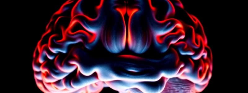
What is MRI?
What is MRI?
MRI uses strong magnetic fields to align the protons in water molecules within the brain. The difference in alignment between parallel and anti-parallel protons generates signals that are measured to create detailed images of the brain.
What is fMRI?
What is fMRI?
fMRI is a technique that measures brain activity by detecting changes in blood flow. When a brain area is more active, it requires more oxygenated blood. fMRI detects these changes in blood flow creating images of brain activity.
What is DTI?
What is DTI?
Diffusion Tensor Imaging (DTI) is a technique that uses MRI to visualize water diffusion in the brain's white matter. Water diffuses more easily along axons than across them, providing information about the direction and integrity of neuronal connections.
Why are high-field MRI scanners used?
Why are high-field MRI scanners used?
Signup and view all the flashcards
How does brain anatomy help scientists?
How does brain anatomy help scientists?
Signup and view all the flashcards
Area MT (V5)
Area MT (V5)
Signup and view all the flashcards
Area V4
Area V4
Signup and view all the flashcards
Face Recognition
Face Recognition
Signup and view all the flashcards
Prosopagnosia
Prosopagnosia
Signup and view all the flashcards
Fusiform Face Area (FFA)
Fusiform Face Area (FFA)
Signup and view all the flashcards
Event-Related Design
Event-Related Design
Signup and view all the flashcards
Multivariate Analyses
Multivariate Analyses
Signup and view all the flashcards
Resting State fMRI
Resting State fMRI
Signup and view all the flashcards
Mind Reading (with fMRI)
Mind Reading (with fMRI)
Signup and view all the flashcards
Brain Similarity Measures
Brain Similarity Measures
Signup and view all the flashcards
How do atlases aid functional measurement?
How do atlases aid functional measurement?
Signup and view all the flashcards
What is the purpose of comparing anatomy between groups?
What is the purpose of comparing anatomy between groups?
Signup and view all the flashcards
What brain structure is larger in taxi drivers?
What brain structure is larger in taxi drivers?
Signup and view all the flashcards
How does DTI reveal prosopagnosia?
How does DTI reveal prosopagnosia?
Signup and view all the flashcards
What does PET measure in the brain?
What does PET measure in the brain?
Signup and view all the flashcards
How does the most common PET tracer work?
How does the most common PET tracer work?
Signup and view all the flashcards
How is PET used for visual mapping?
How is PET used for visual mapping?
Signup and view all the flashcards
What does PiB bind to in Alzheimer's disease?
What does PiB bind to in Alzheimer's disease?
Signup and view all the flashcards
What does fMRI measure?
What does fMRI measure?
Signup and view all the flashcards
Why are block designs used in fMRI studies?
Why are block designs used in fMRI studies?
Signup and view all the flashcards
Study Notes
MRI and fMRI: Brain Imaging Techniques
- MRI and fMRI provide valuable insights into brain function, allowing researchers to observe and measure neuron responses in specific brain regions.
- MRI, originally known as Nuclear Magnetic Resonance (NMR), measures the magnetic properties of atomic nuclei (specifically protons).
- Protons spin on an axis, creating tiny magnetic fields. When placed in a strong magnetic field, these internal magnetic fields align either parallel or anti-parallel to the external field.
- The difference in the populations aligned parallel vs. anti-parallel provides the signal MRI measures.
- Stronger magnetic fields (e.g., 7T, 10T) yield more detailed information.
- Advanced MRI techniques like Diffusion Tensor Imaging (DTI) map the diffusion of water in white matter tracts. This reveals the direction of neural fiber pathways, which is crucial for understanding connectivity.
- Brain atlases aid in identifying anatomical regions in individual participants, allowing researchers to correlate structure with function and to compare groups or conditions.
- Studying groups with different experiences (e.g., taxi drivers vs. controls) reveals potential structural differences in the hippocampus.
- DTI can also assess the integrity of white matter tracts. Damage to the ventral visual pathway (as seen in prosopagnosia) is associated with decreased tract integrity.
PET: Positron Emission Tomography
- PET measures metabolic activity in the brain by detecting radioactive emissions.
- A common PET method involves injecting radioactive oxygen-labeled water into the participant.
- The unstable isotope releases positrons; their collision with electrons generates gamma rays.
- PET scanners detect these photons' paths, allowing for calculation of their origin, therefore the locations of active brain regions.
- PET enables the visualization of blood flow and metabolic activity in the brain. Activity is measured by subtracting baseline measurements from measurements taken with stimulation; regions active during the stimulation have higher counts.
- PET can track the binding of specific molecules (e.g., to amyloid plaques in Alzheimer's disease by injecting patients with a specific PET dye).
fMRI: Functional Magnetic Resonance Imaging
- Functional magnetic resonance imaging revolutionized cognitive neuroscience.
- fMRI measures changes in blood oxygenation (BOLD signal) associated with neural activity.
- Neural activity consumes oxygen, which triggers an increase in blood flow to supply more oxygen, hence the BOLD signal.
- Blood oxygenation level-dependent (BOLD) signal provides a measure of neural activity.
- fMRI experiments often employ block designs.
- Block designs present one condition followed by another for a certain time (e.g., 40 seconds), enabling tracking of activity changes across different conditions.
Visual Mapping using Imaging Techniques
- The response patterns of different brain areas (e.g., primary visual cortex, area MT, and area V4) vary significantly depending on the stimulus (e.g., static vs. moving images, color).
- The V5/MT area in the brain responds only to moving stimuli
- Area V1 shows consistent responses during stimulation-periods, while area MT increases only during motion.
Face Recognition and Other Cognitive Functions
- The fusiform gyrus is a brain area particularly active during face recognition.
- Damage in this area (e.g., from stroke) can cause prosopagnosia (face blindness).
- fMRI results show good correlation with neuropsychological findings of face processing.
Event-Related Designs
- Event-related designs are used to study responses to brief events quickly, without lengthy scanning times.
- They vary the timing and order of events to optimize statistical power in detecting responses to individual events.
- Multivoxel pattern analyses (MVPA) of fMRI data can involve analyzing patterns of activation across many voxels in response to stimulus categories, helping to extract more detailed information.
Resting State fMRI
- Researchers use resting-state fMRI to monitor brain activity while participants are at rest.
- This method can identify brain networks and how they fluctuate in response to cognitive processes while they are not actively engaged with tasks.
Mind Reading with fMRI
- Researchers are investigating the possibility of interpreting thoughts and intentions based on observed patterns of brain activity. Tasks such as motor imagery can be distinguished from other tasks.
Studying That Suits You
Use AI to generate personalized quizzes and flashcards to suit your learning preferences.