Podcast
Questions and Answers
What is fluoroscopy?
What is fluoroscopy?
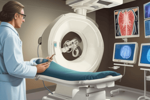
Fluoroscopy is a type of medical imaging that shows a continuous X-ray image on a monitor, much like an X-ray movie.
Which of the following are common applications of fluoroscopy? (Select all that apply)
Which of the following are common applications of fluoroscopy? (Select all that apply)
- Barium Swallows (correct)
- Radiation therapy
- Cardiac Angiography (correct)
- Orthopaedic - Arthrograms (correct)
Fixed systems are typically used for shorter or less complex interventions.
Fixed systems are typically used for shorter or less complex interventions.
False (B)
What is the main advantage of mobile systems in fluoroscopy?
What is the main advantage of mobile systems in fluoroscopy?
Which mode in fluoroscopy utilizes a higher dose and higher heat on the anode?
Which mode in fluoroscopy utilizes a higher dose and higher heat on the anode?
The input phosphor of the Image Intensifier emits _____ when X-rays pass through the patient.
The input phosphor of the Image Intensifier emits _____ when X-rays pass through the patient.
What does the photo emissive layer in the Image Intensifier do?
What does the photo emissive layer in the Image Intensifier do?
What can be a consequence of continuous high wear on the anode?
What can be a consequence of continuous high wear on the anode?
Study Notes
Fluoroscopy
- Fluoroscopy is a medical imaging technique that uses continuous x-ray images to provide a live view in real-time.
- It is similar to an x-ray movie.
- Fluoroscopy can be used for diagnosis and treatment.
- It can show anatomical and physiological information.
- Uses contrast media to make internal structures visible.
- Modern fluoroscopes use flat panel detectors, but are still often referred to as Image Intensifiers.
Uses of Fluoroscopy
- Orthopaedic procedures (eg, arthrograms and theatre procedures)
- Angiography (eg, cardiac, cerebral, peripheral)
- Gastrointestinal procedures (eg, barium swallows, meals and enemas)
- Endoscopic procedures (eg, ERCP and OGD)
- Urology procedures (eg, Ureteroscopy, nephrostomy insertions, PCNL)
- Interventional Radiology (eg, Line insertions, Embolisations, PTC)
- Hysterosalpingography (HSG)
- Stent insertions
- RIG insertions
- Proctogram
- Many more!
Fixed Fluoroscopy Systems
- Permanently installed in a dedicated room or suite.
- Used for longer screening cases.
- Different styles include floor-mounted, ceiling-mounted, and biplane (allows imaging from multiple positions simultaneously).
Mobile Fluoroscopy Systems
- Portable and can be moved between rooms.
- Often used for shorter interventions or theatre cases.
- Less reliant on dedicated x-ray rooms.
Advantages of Mobile Systems
- Does not require a custom room design.
- Can be easily moved between rooms.
Advantages of Fixed Systems
- Usually have more features and better built-in radiation protection.
- More reliable x-ray generators.
- Room is designed for unimpeded movement of the fluoroscopy system.
- Can be replaced during a procedure without moving the patient.
X-ray Generation in Fluoroscopy
- Mechanism is similar to conventional x-ray systems.
- Different modes are used for various imaging purposes:
- Continuous fluoroscopy : Continuous exposure, high dose, used for dynamic procedures.
- Pulsed fluoroscopy : Intermittent exposure, lower dose, used for procedures requiring less frequent images.
- High-Dose "Acquisitions": Higher dose, higher frame rate, provides a clearer and smoother image, useful for capturing specific events.
- Single high-dose image: Individual image with maximum clarity, used for specific procedures.
Anode Heat Dissipation
- Rotational anodes prevent excess heat buildup by rotating the anode target.
- Oil or water-cooled systems are used to further dissipate heat.
- Focal spot size can be adjusted to balance detail with anode heating.
- Smaller focal spots generate more heat, requiring increased dissipation measures.
- High x-ray output requires greater heat dissipation, leading to higher wear on the anode.
- Radiographers must adhere to radiation protection measures.
Image Intensifier System
- The image intensifier is a key component of the fluoroscopic system
- Consists of input phosphor, photo emissive layer (photocathode), and output phosphor.
- X-rays passing through the patient activate the input phosphor, which emits light.
- Photo cathode emits electrons that are accelerated to the output phosphor.
- The output phosphor produces an amplified light signal that is then converted to a video signal.
Studying That Suits You
Use AI to generate personalized quizzes and flashcards to suit your learning preferences.
Related Documents
Description
Explore the fascinating world of fluoroscopy, a real-time medical imaging technique using continuous x-ray images. This quiz covers the various uses of fluoroscopy in different medical fields, including orthopaedic, gastrointestinal, and interventional radiology procedures. Test your knowledge of fixed fluoroscopy systems and their applications.


