Podcast
Questions and Answers
What is the primary function of fluoroscopy?
What is the primary function of fluoroscopy?
- To produce static images
- To screen patients for diseases
- To guide surgeons during procedures (correct)
- To create 3D images
What is the main difference between fluoroscopy and general x-ray imaging?
What is the main difference between fluoroscopy and general x-ray imaging?
- Image display
- Exposure parameters
- Image receptor (correct)
- X-ray source
What is the benefit of using digital fluoroscopy over image intensified fluoroscopy?
What is the benefit of using digital fluoroscopy over image intensified fluoroscopy?
- Is not available in mobile units
- Provides lower intensity images
- Requires higher tube current
- Provides real-time dynamic imaging (correct)
What is the purpose of the scintillation layer in an image intensifier tube?
What is the purpose of the scintillation layer in an image intensifier tube?
Why did Thomas Edison stop researching fluoroscopy?
Why did Thomas Edison stop researching fluoroscopy?
What is the typical frame rate of fluoroscopy?
What is the typical frame rate of fluoroscopy?
What is the primary function of the scintillation layer in an image intensifier tube?
What is the primary function of the scintillation layer in an image intensifier tube?
What is the main advantage of using a flat panel detector in fluoroscopy?
What is the main advantage of using a flat panel detector in fluoroscopy?
What is the typical range of frame rates in fluoroscopy?
What is the typical range of frame rates in fluoroscopy?
What is the primary difference between mobile and fixed fluoroscopy systems?
What is the primary difference between mobile and fixed fluoroscopy systems?
What is the purpose of the image intensifier tube in fluoroscopy?
What is the purpose of the image intensifier tube in fluoroscopy?
What is the typical range of kVp used in fluoroscopy, depending on the body region?
What is the typical range of kVp used in fluoroscopy, depending on the body region?
Flashcards are hidden until you start studying
Study Notes
Fluoroscopy
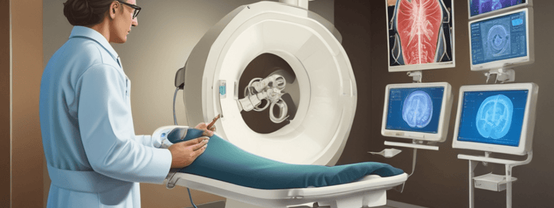
- Dynamic, real-time imaging technique used in operating theatres to guide surgeons in placement
- Provides continuous image of motion, with frame rate over 30 frames per second
- Capable of capturing static images (spot film) in 2D
Varieties of Fluoroscopy
- Mobile source and fixed set-ups, including interventional suite areas
- Image receptor can be placed over or under the table
History of Fluoroscopy
- Created by Thomas Edison, but he stopped research after his assistant died from metastatic skin cancer due to radiation exposure
Benefits of Fluoroscopy
- Provides images of continuous nature, enabling real-time dynamic imaging and screening
Physical Principles of Operation
- X-ray source: same as general x-ray tube
- Image receptor: different from x-ray, can be flat-panel detector or image intensifier tube
- Image intensifier tube:
- Complex electronic device
- Converts image to higher intensity and brightness
- Creates light of higher intensity and brightness
- Encased in glass with a scintillation layer
Exposure Parameters
- kVp (x-ray energy) depends on body region
- Tube current is typically low
Studying That Suits You
Use AI to generate personalized quizzes and flashcards to suit your learning preferences.


