Podcast
Questions and Answers
What is the primary purpose of fluoroscopy in radiology?
What is the primary purpose of fluoroscopy in radiology?
- To measure radiation exposure in patients
- To develop personalized treatment plans for patients
- To guide diagnostic and therapeutic procedures in real-time (correct)
- To produce high-resolution images for diagnostic purposes
What is the main advantage of digital fluoroscopy over traditional fluoroscopy?
What is the main advantage of digital fluoroscopy over traditional fluoroscopy?
- Increased cost-effectiveness
- Increased radiation exposure
- Reduced procedure time
- Improved image quality and resolution (correct)
Which of the following is a limitation of fluoroscopy?
Which of the following is a limitation of fluoroscopy?
- Real-time imaging capability
- High image resolution
- Portability of equipment
- Higher radiation exposure compared to other modalities (correct)
What is the purpose of using shielding and protective equipment in fluoroscopy?
What is the purpose of using shielding and protective equipment in fluoroscopy?
Which procedure is NOT typically guided by fluoroscopy?
Which procedure is NOT typically guided by fluoroscopy?
What is the benefit of using the lowest possible X-ray beam intensity in fluoroscopy?
What is the benefit of using the lowest possible X-ray beam intensity in fluoroscopy?
What is the primary difference between traditional and digital fluoroscopy?
What is the primary difference between traditional and digital fluoroscopy?
Why is fluoroscopy particularly useful in orthopedic and musculoskeletal procedures?
Why is fluoroscopy particularly useful in orthopedic and musculoskeletal procedures?
Flashcards are hidden until you start studying
Study Notes
Fluoroscopy in Radiology
Definition
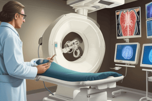
- Fluoroscopy is a type of medical imaging that uses continuous X-ray beams to guide diagnostic and therapeutic procedures.
Principle
- Fluoroscopy works by transmitting X-ray beams through the body, which are then detected by a digital image receptor or an image intensifier.
- The resulting images are displayed on a monitor, allowing the operator to visualize internal structures in real-time.
Applications
- Guiding interventional procedures:
- Angiography and angioplasty
- Biopsy and tumor treatment
- Pain management procedures
- Diagnostic imaging:
- Swallowing studies (e.g., barium swallows)
- Upper GI series
- Voiding cystourethrograms
- Orthopedic and musculoskeletal procedures:
- Guiding joint injections and aspirations
- Reduction of fractures and dislocations
Advantages
- Real-time imaging allows for precise guidance during procedures
- Enables dynamic assessment of anatomical structures and physiological processes
- Can reduce procedure time and radiation exposure
Limitations
- Higher radiation exposure compared to other imaging modalities
- Requires specialized equipment and trained operators
- Image quality may be affected by patient size, motion, and anatomical complexity
Radiation Safety
- Fluoroscopy operators must follow radiation safety guidelines to minimize exposure to patients, staff, and themselves.
- Techniques to reduce radiation exposure include:
- Using lowest possible X-ray beam intensity
- Limiting fluoroscopy time
- Using shielding and Protective Equipment
Digital Fluoroscopy
- Digital fluoroscopy systems use digital image receptors and computer algorithms to process and display images.
- Advantages over traditional fluoroscopy:
- Improved image quality and resolution
- Reduced radiation exposure
- Ability to store and review images electronically
Fluoroscopy in Radiology
Definition
- Uses continuous X-ray beams to guide diagnostic and therapeutic procedures
Principle
- Transmits X-ray beams through the body
- Detects X-ray beams using a digital image receptor or image intensifier
- Displays images on a monitor for real-time visualization
Applications
- Guides interventional procedures: angiography, angioplasty, biopsy, tumor treatment, pain management
- Used for diagnostic imaging: swallowing studies, upper GI series, voiding cystourethrograms
- Guides orthopedic and musculoskeletal procedures: joint injections, aspirations, fracture reduction, dislocation reduction
Advantages
- Real-time imaging for precise guidance
- Enables dynamic assessment of anatomical structures and physiological processes
- Reduces procedure time and radiation exposure
Limitations
- Higher radiation exposure compared to other imaging modalities
- Requires specialized equipment and trained operators
- Image quality affected by patient size, motion, and anatomical complexity
Radiation Safety
- Operators must follow radiation safety guidelines to minimize exposure
- Techniques to reduce radiation exposure: lowest X-ray beam intensity, limited fluoroscopy time, shielding, and protective equipment
Digital Fluoroscopy
- Uses digital image receptors and computer algorithms for image processing and display
- Advantages: improved image quality, reduced radiation exposure, electronic image storage and review
Studying That Suits You
Use AI to generate personalized quizzes and flashcards to suit your learning preferences.


