Podcast
Questions and Answers
Fluoroscopy is a medical imaging technique that captures single, high-resolution still images of the body's internal structures, rather than real-time video.
Fluoroscopy is a medical imaging technique that captures single, high-resolution still images of the body's internal structures, rather than real-time video.
False (B)
Fluoroscopy involves lower radiation doses compared to standard X-rays due to its real-time imaging capabilities.
Fluoroscopy involves lower radiation doses compared to standard X-rays due to its real-time imaging capabilities.
False (B)
Diagnostic fluoroscopy is only utilized for surgical procedures and is not employed for visualizing anatomy or dynamic biological processes.
Diagnostic fluoroscopy is only utilized for surgical procedures and is not employed for visualizing anatomy or dynamic biological processes.
False (B)
The image intensifier, introduced in the 1960s, decreases the light intensity of the X-ray image for better viewing in fluoroscopy.
The image intensifier, introduced in the 1960s, decreases the light intensity of the X-ray image for better viewing in fluoroscopy.
Before the introduction of image intensifiers, fluoroscopy screens were commonly made of zinc-cadmium sulfide.
Before the introduction of image intensifiers, fluoroscopy screens were commonly made of zinc-cadmium sulfide.
Interventional fluoroscopy imaging chains utilize a high voltage regulator to enhance image quality.
Interventional fluoroscopy imaging chains utilize a high voltage regulator to enhance image quality.
Fluoroscopy can only assist with orthopedic surgeries; it is not applicable for diagnosing heart conditions.
Fluoroscopy can only assist with orthopedic surgeries; it is not applicable for diagnosing heart conditions.
The flat panel detector in a fluoroscopy system directly emits the X-ray beam that passes through the patient.
The flat panel detector in a fluoroscopy system directly emits the X-ray beam that passes through the patient.
Image-intensifier tubes convert high intensity visible photons into low intensity X-ray photon fluence.
Image-intensifier tubes convert high intensity visible photons into low intensity X-ray photon fluence.
A fluoroscopy monitor displays static X-ray images only, not continuous, real-time images.
A fluoroscopy monitor displays static X-ray images only, not continuous, real-time images.
The glass or metal envelope surrounding the components of an image-intensifier tube primarily serves aesthetic purposes.
The glass or metal envelope surrounding the components of an image-intensifier tube primarily serves aesthetic purposes.
Fluoroscopy is limited to only guiding treatments involving implants.
Fluoroscopy is limited to only guiding treatments involving implants.
Decreasing the particle fluence in an Image Intensifier increases in the flux magnitude of visible photons.
Decreasing the particle fluence in an Image Intensifier increases in the flux magnitude of visible photons.
In an image intensifier, the electronic lens system primarily functions to decrease the energy of image-carrying electrons while magnifying the image onto the output screen.
In an image intensifier, the electronic lens system primarily functions to decrease the energy of image-carrying electrons while magnifying the image onto the output screen.
The brightness of the image at the output screen of an image intensifier is typically 50 to 100 times greater than the image emerging from the input screen.
The brightness of the image at the output screen of an image intensifier is typically 50 to 100 times greater than the image emerging from the input screen.
Cesium Iodide (CsI) crystals in the input phosphor are structured in a way that encourages lateral light spread to enhance image clarity.
Cesium Iodide (CsI) crystals in the input phosphor are structured in a way that encourages lateral light spread to enhance image clarity.
The photocathode is stimulated directly by X-rays to emit electrons in the image intensifier tube.
The photocathode is stimulated directly by X-rays to emit electrons in the image intensifier tube.
The anode voltage in an image intensifier is significantly lower than photoemission voltage.
The anode voltage in an image intensifier is significantly lower than photoemission voltage.
The photocathode is typically composed of lead and tin compounds.
The photocathode is typically composed of lead and tin compounds.
Photoemission is the process where electrons are emitted upon stimulation by ultraviolet light.
Photoemission is the process where electrons are emitted upon stimulation by ultraviolet light.
The primary purpose of electron optics is to diverge electrons generated in the active area of the input.
The primary purpose of electron optics is to diverge electrons generated in the active area of the input.
Electrostatic focusing lenses in an image intensifier tube expand the electron pattern from the large cathode end to fit the output phosphor.
Electrostatic focusing lenses in an image intensifier tube expand the electron pattern from the large cathode end to fit the output phosphor.
Flux gain is determined by dividing the number of x-rays at the input phosphor by the number of light photons at the output phosphor.
Flux gain is determined by dividing the number of x-rays at the input phosphor by the number of light photons at the output phosphor.
Minification gain arises from the increased quantity of light photons produced at the output phosphor as compared to the number of x-rays at the input phosphor.
Minification gain arises from the increased quantity of light photons produced at the output phosphor as compared to the number of x-rays at the input phosphor.
Brightness gain is the sum of minification gain and flux gain.
Brightness gain is the sum of minification gain and flux gain.
In image intensification, electronic gain refers to the kinetic energy gained by electrons through acceleration, typically around a factor of 50.
In image intensification, electronic gain refers to the kinetic energy gained by electrons through acceleration, typically around a factor of 50.
An image intensifier with an input phosphor diameter of 30 cm and an output phosphor diameter of 3 cm would have a minification gain of 100.
An image intensifier with an input phosphor diameter of 30 cm and an output phosphor diameter of 3 cm would have a minification gain of 100.
Veiling glare, which reduces contrast in image intensifiers, is primarily caused by external scatter radiation reaching the output phosphor.
Veiling glare, which reduces contrast in image intensifiers, is primarily caused by external scatter radiation reaching the output phosphor.
Flat-panel detectors increase x-ray attenuation and reduce DQE compared to image intensifiers due to their aluminum covers.
Flat-panel detectors increase x-ray attenuation and reduce DQE compared to image intensifiers due to their aluminum covers.
Flashcards
Fluoroscopy
Fluoroscopy
A medical imaging technique using X-rays to create real-time video inside the body.
Image Intensifier
Image Intensifier
A device that receives X-ray images, converts them to light, and enhances their brightness.
Diagnostic Fluoroscopy
Diagnostic Fluoroscopy
A modality that visualizes anatomy in real-time using radiation.
Interventional Fluoroscopy
Interventional Fluoroscopy
Signup and view all the flashcards
History of Fluoroscopy
History of Fluoroscopy
Signup and view all the flashcards
Evaluating health problems
Evaluating health problems
Signup and view all the flashcards
Imaging chain
Imaging chain
Signup and view all the flashcards
X-ray
X-ray
Signup and view all the flashcards
Flat panel detector
Flat panel detector
Signup and view all the flashcards
Monitor in imaging
Monitor in imaging
Signup and view all the flashcards
Image-intensifier tube
Image-intensifier tube
Signup and view all the flashcards
High voltage generator
High voltage generator
Signup and view all the flashcards
Fluence in imaging
Fluence in imaging
Signup and view all the flashcards
Input Phosphor
Input Phosphor
Signup and view all the flashcards
Photocathode
Photocathode
Signup and view all the flashcards
Photoemission
Photoemission
Signup and view all the flashcards
Electron Optics
Electron Optics
Signup and view all the flashcards
Electron Acceleration
Electron Acceleration
Signup and view all the flashcards
Visible Light Conversion
Visible Light Conversion
Signup and view all the flashcards
Brightness Amplification
Brightness Amplification
Signup and view all the flashcards
Electrostatic Focusing Lens
Electrostatic Focusing Lens
Signup and view all the flashcards
Flux Gain
Flux Gain
Signup and view all the flashcards
Minification Gain
Minification Gain
Signup and view all the flashcards
Brightness Gain
Brightness Gain
Signup and view all the flashcards
Veiling Glare
Veiling Glare
Signup and view all the flashcards
Output Phosphor
Output Phosphor
Signup and view all the flashcards
Optical System in XRII
Optical System in XRII
Signup and view all the flashcards
Study Notes
Overview of the Fluoroscopy Machine
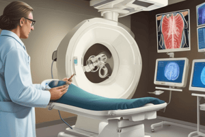
- Fluoroscopy is a medical imaging technique that uses X-rays to create a real-time video of the inside of the body.
- It is also referred to as "fluoro".
- The image intensifier is a complex device that receives the X-ray beam, converts it into light, and increases light intensity for better viewing.
History
- Thomas Edison invented the fluoroscope in 1896.
- It had a zinc-cadmium sulfide screen.
- Fluoroscopy has been a valuable tool in medical imaging.
- Image intensifiers became routine in the 1960s.
Overview
- Diagnostic fluoroscopy is a daily procedure performed in radiology suites across the country.
- This modality visualizes anatomy using radiation in real-time.
- Patient doses can be significant, increasing the risk of adverse reactions.
Interventional Fluoroscopy
- Interventional fluoroscopy guides minimally invasive medical procedures.
- It's used to evaluate dynamic biological processes, such as the movement of a body part, an instrument, or a contrast agent.
How it Works
- An X-ray beam passes through the body part being examined.
- X-ray images are transmitted to a monitor.
- Healthcare providers can see the body part in motion.
What it's Used For
- Healthcare providers use fluoroscopy to monitor and diagnose conditions.
- It guides treatments like implants and injections.
- It guides orthopedic surgery.
- It allows the evaluation of specific body areas like bones, muscles, joints, organs, and blood vessels.
Imaging Chain Components
- X-ray source: Emits an X-ray beam that passes through the patient's body.
- Flat panel detector (FPD) / Image-intensifier tube: A receptor that converts the X-ray signal into digital images.
- Monitor: Displays digital images as a continuous X-ray image.
Imaging Chain of Interventional Fluoroscopy
- The chain involves an X-ray source, a flat panel detector/image intensifier tube, and a monitor.
- The chain produces real-time X-ray images that guide medical procedures.
- Key components include: High Voltage Generator, X-Ray Tube, X-Ray Image Intensifier, and Video Camera/Monitor.
Image-Intensifier Tube
- It receives the image-forming X-ray beam and converts it into a high-intensity visible-light image.
- Tube components are contained within a glass or metal envelope that provides structural support and maintains the vacuum.
Input Phosphor
- X-rays that exit the patient and are incident on the image-intensifier tube are transmitted through the glass envelope and interact with the input phosphor.
- Energy is converted into visible light.
Input Phosphor (X-Rays to Light)
- The input phosphor converts X-rays into light.
- Cesium Iodide (CsI) crystals are commonly used, arranged in a dense needle-like structure (approximately 300 μm).
- It prevents lateral light spread.
Photocathode
- The photocathode is the next element in the image intensifier tube.
- It is bonded directly to the input phosphor.
- It is a thin metal layer, often composed of cesium and antimony compounds, responding to the input phosphor stimulation.
- Photocathode emits electrons through photoemission when illuminated by visible light from the input phosphor.
Photocathode converts Light to Electrons
- Photocathode converts light photons into electrons through the photoelectric effect.
- This occurs when photons strike a thin bi- or multi-alkali photocathode.
- Electrons are released and repelled from the photocathode.
Photoemission
- Photoemission is electron emission resulting from visible light stimulation.
- Numerous light photons are required to cause the emission of a single electron.
- The number of emitted electrons is directly proportional to the light intensity.
Electron Optics
- Proper electron travel is maintained.
- Electrons generated in the active area of the input are focused onto the fixed-size output screen.
- Electrostatic focusing lenses are responsible for reducing the electron pattern from the large cathode end of the tube to the small output phosphor.
Flux Gain
- Flux gain is the ratio of the number of output light photons to the number of input X-ray photons at the phosphor.
Minification and Brightness Gain
- Minification gain is the increased illumination of the image due to light photon multiplication at the output phosphor.
- Brightness gain is the ability of the image intensifier to increase illumination; it's the product of minification and flux gain.
Intensification
- Intensification has two mechanisms:
- Electronic (flux) gain - electrons gain kinetic energy from acceleration (~50 typically).
- Minification gain - reducing the large X-ray image at the input phosphor to a smaller diameter at the output phosphor (e.g., 40 cm to 2.5 cm).
Output Phosphor
- Accelerated electrons interact and produce visible light.
- Output phosphor converts electrons to light via focused electron beams onto a thin powder phosphor.
Optical System
- The optical system couples the image intensifier to the video camera.
- It includes a collimating lens to shape divergent light from the output phosphor,
- and an aperture to limit the light reaching the camera.
Flat-Panel Detector
- Replaces the image intensifier tube, optical system, and cameras.
- It directly converts X-rays into a digital form, improving image quality.
- Carbon fiber covers reduce X-ray attenuation and increase DQE (Detective Quantum Efficiency).
FPD (Indirect/Direct Systems)
- Indirect systems use a phosphor layer that converts X-rays to light, and a photodiode converts this light into a charge.
- Direct systems directly produce a charge from X-ray absorption in a semiconductor material.
Video Camera
- Video camera captures the XRII output image and converts it to an analogue electrical signal in a recognized video format (e.g., NTSC/PAL/SECAM).
- Older cameras used photoconductive targets scanned by electron beams. Modern cameras use Charge-Coupled Devices (CCDs).
Interventional Procedures and X-Ray Tube Positioning
- X-ray tubes are positioned beneath the patient for interventional procedures to minimize scattered radiation reaching medical staff. The majority of scattered radiation originates from the patient's entrance side.
Studying That Suits You
Use AI to generate personalized quizzes and flashcards to suit your learning preferences.