Podcast
Questions and Answers
What is the head of the femur?
What is the head of the femur?
- The area where the femur connects to the knee
- Part of the femur that fits into the hip socket (correct)
- The narrow region of the femur
- A projection on the femur for muscle attachment
What is the fovea capitis?
What is the fovea capitis?
A small depression in the head of the femur.
What is the neck of the femur?
What is the neck of the femur?
The region just below the head of the femur.
What is the greater trochanter?
What is the greater trochanter?
What is the lesser trochanter?
What is the lesser trochanter?
What is the linea aspera?
What is the linea aspera?
What is the patellar surface?
What is the patellar surface?
What is the medial epicondyle of the femur?
What is the medial epicondyle of the femur?
What is the lateral epicondyle of the femur?
What is the lateral epicondyle of the femur?
What is the gluteal tuberosity?
What is the gluteal tuberosity?
What is the medial condyle of the femur?
What is the medial condyle of the femur?
What is the lateral condyle of the femur?
What is the lateral condyle of the femur?
What is the pectineal line of the femur?
What is the pectineal line of the femur?
What is the adductor tubercle?
What is the adductor tubercle?
Flashcards are hidden until you start studying
Study Notes
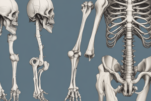
Femur Bone Anatomy
- Head of femur: The rounded uppermost part of the femur that fits into the acetabulum of the pelvis, forming the hip joint.
- Fovea capitis: A small depression located on the head of the femur, serves as an attachment point for the ligamentum teres, which stabilizes the hip joint.
- Neck of femur: The constricted region just below the head; it connects the head to the shaft and is a common site for fractures.
- Greater trochanter: A large protrusion on the lateral side of the femur, serving as an attachment point for muscles of the hip and thigh.
- Lesser trochanter: A smaller projection located on the medial side of the femur, also serving as a muscle attachment site, particularly for the iliopsoas muscle.
- Linea aspera: A prominent ridge along the posterior shaft of the femur that serves as an attachment for several muscles and helps stabilize the bone.
- Patellar surface: The smooth area on the distal end of the femur where it articulates with the patella (kneecap), facilitating knee movement.
- Medial epicondyle of femur: A bony prominence on the inner side of the distal femur, providing attachment for ligaments and muscles.
- Lateral epicondyle of femur: Similar to the medial epicondyle but located on the outer side, also acting as a site for muscle and ligament attachment.
- Gluteal tuberosity: A roughened area on the posterior surface of the femur that serves as an attachment point for the gluteus maximus muscle.
- Medial condyle of femur: The rounded bony end of the femur that articulates with the tibia on the inner side of the knee.
- Lateral condyle of femur: The rounded bony end that articulates with the tibia on the outer side of the knee, facilitating smooth knee joint movement.
- Pectineal line of femur: A faint ridge on the posterior surface of the femur, running from the lesser trochanter to the linea aspera, serving as an attachment for the pectineus muscle.
- Adductor tubercle: A small bony prominence located above the medial condyle, providing attachment for the adductor magnus muscle, important for thigh adduction.
Studying That Suits You
Use AI to generate personalized quizzes and flashcards to suit your learning preferences.