Podcast
Questions and Answers
What is the primary function of the head of the femur?
What is the primary function of the head of the femur?
- Forming the articular head of the hip joint (correct)
- Protecting the femoral artery
- Providing stability to the knee joint
- Connecting muscles in the thigh region
Which of the following best describes the location of the fovea of the head of the femur?
Which of the following best describes the location of the fovea of the head of the femur?
- Situated at the neck of the femur
- Located at the distal end of the femur
- Found at the proximal end of the femur
- On the articular surface of the head of the femur (correct)
What is the anatomical significance of the neck of the femur?
What is the anatomical significance of the neck of the femur?
- It provides attachment for major muscle groups
- It supports the body weight in standing position
- It serves as a primary attachment point for ligaments (correct)
- It is the site for the creation of new blood cells
Which region of the lower limb is categorized separately for descriptive purposes?
Which region of the lower limb is categorized separately for descriptive purposes?
What characteristic distinguishes the femur from other bones in the body?
What characteristic distinguishes the femur from other bones in the body?
Which muscle group is primarily discussed in relation to the gluteal region?
Which muscle group is primarily discussed in relation to the gluteal region?
Which of the following is NOT associated with the actions of the muscles of the gluteal region?
Which of the following is NOT associated with the actions of the muscles of the gluteal region?
Which of the following conditions can be associated with issues in the gluteal region?
Which of the following conditions can be associated with issues in the gluteal region?
What is the primary function of the gluteus minimus?
What is the primary function of the gluteus minimus?
Where is the most common site for intramuscular injection in adults?
Where is the most common site for intramuscular injection in adults?
Which muscle is considered the anatomical key to gluteal anatomy?
Which muscle is considered the anatomical key to gluteal anatomy?
Which function is NOT associated with the tensor fasciae latae?
Which function is NOT associated with the tensor fasciae latae?
Which of the following muscles is considered part of the pelvitrochanteric muscles?
Which of the following muscles is considered part of the pelvitrochanteric muscles?
Which muscle primarily inserts at the greater trochanter of the femur?
Which muscle primarily inserts at the greater trochanter of the femur?
What is the function of the intertrochanteric line?
What is the function of the intertrochanteric line?
Where does the pectineus muscle insert on the femur?
Where does the pectineus muscle insert on the femur?
Which of the following is NOT located on the shaft of the femur?
Which of the following is NOT located on the shaft of the femur?
What is the primary role of the lesser trochanter?
What is the primary role of the lesser trochanter?
What structure does the popliteal surface provide?
What structure does the popliteal surface provide?
Which part of the femur is attached to the tibial collateral ligament?
Which part of the femur is attached to the tibial collateral ligament?
The linea aspera serves as an attachment site for which of the following?
The linea aspera serves as an attachment site for which of the following?
Which muscle originates from the lateral supracondylar line of the femur?
Which muscle originates from the lateral supracondylar line of the femur?
Which of the following muscles insert at the trochanteric fossa?
Which of the following muscles insert at the trochanteric fossa?
Which muscle plays a role in the ventral flexion of the lumbar vertebral column?
Which muscle plays a role in the ventral flexion of the lumbar vertebral column?
What is the main function of iliacus and psoas major?
What is the main function of iliacus and psoas major?
Which group of muscles is primarily responsible for lateral stability of the trunk?
Which group of muscles is primarily responsible for lateral stability of the trunk?
Which structure does the gluteus maximus originate from?
Which structure does the gluteus maximus originate from?
What is the insertion point of the psoas minor?
What is the insertion point of the psoas minor?
Which of the following is NOT a function of the gluteus medius?
Which of the following is NOT a function of the gluteus medius?
Which nerve innervates the iliacus muscle?
Which nerve innervates the iliacus muscle?
Psoas major contributes to which movement when contracted unilaterally?
Psoas major contributes to which movement when contracted unilaterally?
Which muscle is primarily involved in maintaining the erect standing position?
Which muscle is primarily involved in maintaining the erect standing position?
The gluteus minimus primarily performs which action?
The gluteus minimus primarily performs which action?
What is the function of the lateral epicondyle of the femur?
What is the function of the lateral epicondyle of the femur?
At what age does fusion of the ilium, pubis, and ischium typically begin?
At what age does fusion of the ilium, pubis, and ischium typically begin?
Which angle is considered normal for the femoral neck in adults?
Which angle is considered normal for the femoral neck in adults?
What defines coxa vara?
What defines coxa vara?
What does the intercondylar fossa primarily serve as an attachment point for?
What does the intercondylar fossa primarily serve as an attachment point for?
Which condition is characterized by destruction and flattening of the head of the femur?
Which condition is characterized by destruction and flattening of the head of the femur?
What is the function of the patellar surface of the femur?
What is the function of the patellar surface of the femur?
Coxa magna is defined by which characteristic?
Coxa magna is defined by which characteristic?
What is the significance of the intercondylar line?
What is the significance of the intercondylar line?
A 75-year-old woman exhibits a femoral neck angle of 150 degrees. What condition is likely associated with this finding?
A 75-year-old woman exhibits a femoral neck angle of 150 degrees. What condition is likely associated with this finding?
Flashcards are hidden until you start studying
Study Notes
Femur
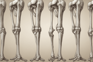
- The strongest and longest bone in the human body.
- Covered by a thick layer of muscle so only a small portion is palpable.
- Composed of three parts: upper end, shaft/body, and lower end.
- Head (caput femoris): forms the articular head of the hip joint.
- Fovea of head of femur (fovea capitis femoris): site of attachment of the ligament of the head of the femur.
- Neck (collum femoris): articular capsule of the hip joint attaches to the dorsal 2/3 of the neck.
- Shaft of the femur (corpus femoris): body of the femur.
- Greater trochanter (trochanter major): insertion of the gluteus medius, gluteus minimus, piriformis, obturator internus, gemellus superior and gemellus inferior.
- Trochanteric fossa (fossa trochanterica): insertion of the obturator externus.
- Lesser trochanter (trochanter minor): dorsomedial prominence, insertion of the iliopsoas.
- Intertrochanteric line (linea intertrochanterica): ventral line connecting both trochanters, attachment of the articular capsule of the hip joint.
- Intertrochanteric crest (crista intertrochanterica): dorsal crest connecting both trochanters.
- Pectineal line (linea pectinea): insertion of the pectineus, located below the lesser trochanter.
- Gluteal tuberosity (tuberositas glutea): insertion of the gluteus maximus, located below the greater trochanter.
- Linea aspera: attachment site for many muscles of the thigh.
- Lateral supracondylar line (linea supracondylaris lateralis): origin of the plantaris.
- Medial supracondylar line (linea supracondylaris medialis):
- Popliteal surface (facies poplitea): floor of the popliteal fossa.
- Condyles of femur (condyli femoris): distal end of the femur, articular surfaces that articulate with the tibia.
- Medial condyle (condylus medialis femoris):
- Medial epicondyle (epicondylus medialis): attachment of the tibial collateral ligament, origin of the medial head of the gastrocnemius
- Adductor tubercle (tuberculum adductorium): insertion of the extensor part of the adductor magnus.
- Lateral condyle (condylus lateralis femoris):
- Lateral epicondyle (epicondylus lateralis): attachment of the fibular collateral ligament, origin of the lateral head of the gastrocnemius.
- Intercondylar line (linea intercondylaris): attachment of the oblique popliteal ligament.
- Intercondylar fossa (fossa intercondylaris): attachment of the cruciate ligaments of the knee joint.
- Patellar surface (facies patellaris): ventral surface for articulation with the patella.
- Medial condyle (condylus medialis femoris):
Hip Bone (innominate bone)
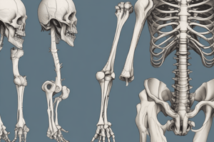
- Composed of ilium, pubis, and ischium.
- Prior to puberty, triradiate cartilage separates these parts, fusion begins at age 15-17.
- Together, ilium, pubis, and ischium form a cup-shaped socket called the acetabulum (Latin = 'vinegar cup').
- Head of the femur articulates with the acetabulum to form the hip joint.
Femoral Neck Abnormalities
- Caput-collum-diaphyseal angle (CCD angle): angle formed by the main axis of the femoral neck and the longitudinal axis of the femoral shaft.
- Normal angle:
- ~125° in adults
- ~150° in newborns
- Normal angle:
- Coxa vara: deformity of the proximal femur due to decreased femoral neck-shaft angle (< 120°), shortening and thickening of the femoral neck.
- Coxa valga: deformity of the femur due to an increased femoral neck-shaft angle (> 140°).
- Coxa magna: asymmetrical, circumferential enlargement and deformation of the femoral head and neck.
Legg-Calvé-Perthes Disease
- Also known as idiopathic avascular necrosis of the femoral head.
- Clinical condition characterized by destruction and flattening of the head of the femur with an increased joint space in the radiograph.
Muscles of the Hip Joint
- Anterior Group
- Iliacus (m.iliacus): origin: iliac fossa, insertion: femur – lesser trochanter.
- Functions: flexion and external rotation of the thigh.
- Psoas major (m.psoas major): origin: bodies of T12 and L1 (and intervertebral discs between), insertion: femur – lesser trochanter
- Functions: flexion and external rotation of the thigh, ventral flexion of the lumbar vertebral column, lateroflexion of the trunk to the side of the contracted muscle and rotation to the opposite side (unilateral contraction).
- Psoas minor (m.psoas minor): origin: bodies of T12 and L1, and the intervertebral discs between them, insertion: iliopubic ramus.
- Functions: ventral flexion of the lumbar vertebral column.
- Iliacus (m.iliacus): origin: iliac fossa, insertion: femur – lesser trochanter.
- Posterior Group (Superficial Layer)
- Gluteus maximus (m.gluteus maximus): origin: gluteal surface of the ilium – dorsal to the posterior gluteal line, iliac crest, thoracolumbar fascia – posterior layer, sacrum, coccyx, sacrotuberous ligament, gluteal aponeurosis. Insertion: femur – gluteal tuberosity, tibia – lateral condyle (via the iliotibial tract).
- Functions: abduction of the thigh (cranial fibers), extension, external rotation and adduction of the thigh (caudal fibers), maintains extension of the knee joint (by stretching the iliotibial tract), keeps the pelvis in retroversion, maintains the erect standing position, provides lateral stability to the trunk (most important function).
- Gluteus medius (m.gluteus medius): origin: gluteal surface of the ilium – between the posterior and anterior gluteal lines, iliac crest. Insertion: femur – greater trochanter.
- Functions: abduction of the thigh and tilting of the pelvis (middle fibers), flexion and internal rotation of the thigh (anterior fibers), extension and external rotation of the thigh (posterior fibers).
- Gluteus minimus (m.gluteus minimus): origin: gluteal surface of the ilium – between the anterior and inferior gluteal lines, iliac crest. Insertion: femur – greater trochanter.
- Functions: abduction of the thigh and tilting of the pelvis (middle fibers), flexion and internal rotation of the thigh (anterior fibers), extension and external rotation of the thigh (posterior fibers).
- Tensor fasciae latae / tensor of fascia lata (m.tensor fasciae latae): origin: ilium – anterior superior iliac spine. Insertion: tibia – tuberosity for the iliotibial (on the lateral condyle) tract of Gerdy (via the iliotibial tract).
- Functions: extension of the knee joint (“locking of the knee joint”), abduction, flexion and internal rotation of the thigh, stabilisation of both knee joints and the hip joint during walking.
- Gluteus maximus (m.gluteus maximus): origin: gluteal surface of the ilium – dorsal to the posterior gluteal line, iliac crest, thoracolumbar fascia – posterior layer, sacrum, coccyx, sacrotuberous ligament, gluteal aponeurosis. Insertion: femur – gluteal tuberosity, tibia – lateral condyle (via the iliotibial tract).
- Posterior Group (Deep Layer)
- Piriformis (m.piriformis): origin pelvic surface of the sacrum. Insertion: greater trochanter.
- Gemellus superior (m.gemellus superior):
- Obturator internus (m.obturatorius internus):
- Gemellus inferior (m.gemellus inferior):
- Quadratus femoris (m.quadratus femoris):
- Functions: maintain stability of the hip joint and have important postural functions.
Intra-Muscular Injection Site
- Muscles commonly used for intramuscular injection in the lower limb:
- Gluteus medius of the gluteal region in adults (most common).
- Vastus lateralis muscle of the thigh region in children.
- Gluteus medius:
- Posterior 1/3 is deep and covered by the gluteus maximus.
- Anterior 2/3 is superficial and not covered by the gluteus maximus.
- Intramuscular injection should ideally be given in the anterior 2/3.
Clinical Correlation
- Piriformis muscle: arises from the pelvic surface of the sacrum, passes through the greater sciatic notch, inserts at the greater trochanter.
- Considered the “anatomical key” to gluteal anatomy; the greater sciatic foramen is the “door.”
- Gluteus medius: lies posterior to the piriformis.
- Sciatic nerve emerges from the greater sciatic foramen, normally through the infrapiriformic space.
- Spine of the ischium: separates the greater and lesser sciatic foramina.
Studying That Suits You
Use AI to generate personalized quizzes and flashcards to suit your learning preferences.


