Podcast
Questions and Answers
Which structure of the eye is primarily responsible for refracting light?
Which structure of the eye is primarily responsible for refracting light?
- Cornea (correct)
- Iris
- Lens
- Pupil
A patient is diagnosed with a condition affecting central vision. Which part of the retina is most likely involved?
A patient is diagnosed with a condition affecting central vision. Which part of the retina is most likely involved?
- Choroid
- Macula (correct)
- Rods
- Fovea
Which of the following best describes the function of the tonometer?
Which of the following best describes the function of the tonometer?
- Evaluating the field of vision
- Testing distance visual acuity
- Examining the fundus and optic disc
- Measuring intraocular pressure (correct)
What does a 20/200 visual acuity measurement indicate?
What does a 20/200 visual acuity measurement indicate?
A patient reports difficulty seeing at night. Which retinal cells are likely affected?
A patient reports difficulty seeing at night. Which retinal cells are likely affected?
If a patient's Ishihara color plate test reveals a deficit, which condition is most likely?
If a patient's Ishihara color plate test reveals a deficit, which condition is most likely?
Which of the following best describes the condition known as 'emmetropia'?
Which of the following best describes the condition known as 'emmetropia'?
A patient presents with redness, flaking, and irritation of the eyelid margins. Which condition is most likely?
A patient presents with redness, flaking, and irritation of the eyelid margins. Which condition is most likely?
Which intervention is most appropriate for a patient experiencing bacterial conjunctivitis with crusting and drainage?
Which intervention is most appropriate for a patient experiencing bacterial conjunctivitis with crusting and drainage?
A patient reports a sudden onset of crooked or distorted lines. Which condition is most likely?
A patient reports a sudden onset of crooked or distorted lines. Which condition is most likely?
After cataract surgery, a patient is instructed to avoid activities that increase IOP. Which activity is LEAST LIKELY to be a concern?
After cataract surgery, a patient is instructed to avoid activities that increase IOP. Which activity is LEAST LIKELY to be a concern?
A patient with glaucoma is prescribed eye drops. What is the most important aspect of patient education?
A patient with glaucoma is prescribed eye drops. What is the most important aspect of patient education?
A patient reports seeing floating spots and flashing lights. Which eye condition is most likely?
A patient reports seeing floating spots and flashing lights. Which eye condition is most likely?
What is the primary purpose of a myringotomy?
What is the primary purpose of a myringotomy?
A patient reports a sensation that the room is spinning. Which condition is most likely?
A patient reports a sensation that the room is spinning. Which condition is most likely?
Flashcards
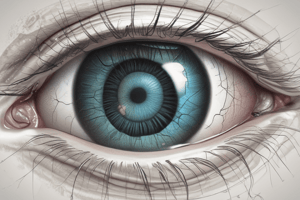
Cornea
Cornea
Transparent, avascular, dome-like structure refracting light.
Slit Lamp
Slit Lamp
A binocular microscope used to examine the eye.
Emmetropia
Emmetropia
Normal vision
Myopia
Myopia
Signup and view all the flashcards
Nystagmus
Nystagmus
Signup and view all the flashcards
Blepharitis
Blepharitis
Signup and view all the flashcards
Conjunctivitis
Conjunctivitis
Signup and view all the flashcards
Cataract
Cataract
Signup and view all the flashcards
Intracapsular extraction
Intracapsular extraction
Signup and view all the flashcards
Acute Glaucoma
Acute Glaucoma
Signup and view all the flashcards
Pupil in Acute Glaucoma
Pupil in Acute Glaucoma
Signup and view all the flashcards
Chronic Glaucoma
Chronic Glaucoma
Signup and view all the flashcards
Retinal Detachment
Retinal Detachment
Signup and view all the flashcards
Scleral Buckling
Scleral Buckling
Signup and view all the flashcards
Macular Degeneration
Macular Degeneration
Signup and view all the flashcards
Study Notes
Anatomy of the Eye
- The cornea is a transparent, avascular, dome-like structure
- It serves as the main refracting surface of the eye
- Aqueous humor is produced by the ciliary body and nourishes the cornea
- The uvea consists of the iris, ciliary body, and choroid
- The iris is the colored part of the eye
- The pupil is a space that dilates and constricts in response to light
- The lens is a colorless and biconvex structure
- Allows focusing for near vision and refocusing for distance vision (accommodation)
- A choroid is a vascular tissue
- It supplies blood to the portion of the sensory retina closest to it
- Vitreous humor occupies 2/3 of the eye's volume
- Helps maintain the shape of the eye
- The retina is composed of 10 microscopic layers
- It is a neural tissue and an extension of the optic nerve
- The macula is the part of the retina and is responsible for central vision
- Rods are responsible for night vision
- Cones are responsible for bright light and color vision
Testing vision
- The Snellen chart consists of progressively smaller rows of letters
- Used to test distance vision
- 20/20 vision is considered the standard of normal vision
- A person with 20/200 vision can see an object from 20 feet away that a person with 20/20 vision can see from 200 feet away
- An ophthalmoscope examines the fundus, optic cup, periphery of the retina, and macula
- A slit lamp is a binocular microscope
- It examines the eye with magnification of 10 to 40 times the real image
- A tonometer measures IOP
- It determines the pressure necessary to indent or flatten a small anterior area of the eye
- Normal IOP is 10-21 mmHg
- Perimetry evaluates the field of vision, the area, or the extent of physical space visible to an eye in a given position
- The ishihara color plate test identifies color vision deficits/color-blindness
- Dots of primary colors are integrated into a background of secondary colors, arranged in patterns like numbers or shapes
- An amsler grid tests patients with macular problems such as macular degeneration
- It consists of a geometric grid of identical squares with a central fixation point
- The patient stares at the central fixation spot
- Some squares may look faded, or the lines may be wavy for patients with macular problems
Eye-related Terms
- Aphakia is without a lens
- Astigmatism is the irregularity in the curve of the cornea
- Blindness visual acuity with best correction (BCVA) ranges from 20/400
- Legal blindness has a BCVA that does not exceed 20/200
- Diplopia is double vision
- Emmetropia is normal vision
- Hyperopia is far-sightedness
- Myopia is near-sightedness
- Hyperemia translates to red eye
- Nystagmus is the involuntary oscillation of the eyeball
- Proptosis is the downward displacement of the eyeball
- Ptosis is a drooping eyelid
- Papilledema is swelling of the optic disc
- Strabismus is a condition in which there is deviation from perfect ocular alignment
- An enucleation is the removal of the entire eye and the part of the optic nerve
- Evisceration involves the surgical removal of the intraocular contents
- This is achieved through an incision or opening in the cornea or sclera
- Exenteration is the removal of the eyelids, the eye, and various amounts of orbital contents
Conditions of the Eyelids and Conjunctiva
- Blepharitis is an inflammatory reaction of the eyelid margin, or seborrheic skin condition
- It is commonly caused by Staphylococcus aureus
- Manifestations include flaking, redness, irritation and recurrent styes
- If bacterium S. aureus is likely, use antibiotic ointment
- Scrub the margin of eyelid with cotton swab
- Apply ointment with cotton swab as directed
- Hordeolum (stye) refers to an inflammation/infection of glands and follicles of the eyelid margin
- External hordeolum impacts the hair follicles of the lashes
- Chalazion is a granulomatous, chronic infection of meibomian glands
- Etiological factors are the bacterium staphylococcus and seborrhea
- Manifestations include pain, redness, foreign body sensation and possible pustule
- Warm soaks, good handwashing, eyelid hygiene, and antibiotic ointment can assist
- Conjunctivitis is an inflammation or infection of the bulbar (covering the cornea and sclera) or palpebral (lining the eyelids) conjunctiva
- Pink eye is usually referring to infectious conjunctivitis
- Causes of bacterial conjunctivitis are Streptococcus pneumoniae, Haemophilus influenzae, and Staphylococcus aureus
- Adenovirus and herpes simplex virus cause viral conjunctivitis
- Allergic conjunctivitis is caused by hypersensitivity reaction in allergic rhinitis or environmental pollutants/allergens
- Manifestations are foreign body sensation, scratching or burning, itching, and light sensitivity (photophobia)
- Warm soaks are used with crusting/drainage, cold compress is used in viral cases of conjunctivitis
- Bacterial conjunctivitis needs topical antibiotics
- Allergic conjunctivitis needs topical or oral antihistamines, vasoconstrictors, and mast cell stabilizers
- Corneal abrasion/ulceration (Keratitis) happens when the layers of cornea are lost due to contact lens overwearing or trauma
- This may lead to corneal ulcer and secondary infection into cornea (keratitis) possibly resulting in blindness
- Manifestations are pain, redness, foreign body sensation, photophobia, increased tearing and difficulty opening the eye
- Fluorescein staining and woods lamp or slit lamp are diagnostic procedures
- Treatment is urgent
- Potent antibiotic eyedrops and eye patching should be done in first 24 hours
- Manage pain with cycloplegics
- See an ophthalmologist for ulceration
- Ensure the patch is secure
- Review safety practices (eye sheild use, hand washing)
- Iritis/Uveitis is inflammation of the intraocular structure
- This may involve the anterior uvea such as the iris (iritis) or the iris and ciliary body (iridocyclitis)
- It can also involve intermediate uveitis structures such as parts posterior to the lens (pars plantis or peripheral uveitis)
- The choroid (choroiditis), or the retina (retinitis), or vitreous near the optic nerve and macula of posterior uveitis can be involved
- Anterior uveitis is usually unilateral
- Posterior uveitis is often bilateral
- Etiologies are immune disorders or even idiopathic causes
- Can be from other conditions like ankylosing spondylitis, Crohn's, Reiter's syndrome, lupus or even trauma
- Acute onset with deep eye pain, light hypersensitivity, conjunctival redness and a pupil that does not respond briskly are manifestations
- Ophthalmology and intervention needed
- Corticosterioids and cycloplegic agents will treat inflammation
Disorders of the eyes
- Cataracts are clouding/opacity of the crystalline lens which impairs vision
- Etiologies are aging, congenital issues or trauma
- Risk factors are diabetes, UV light, high-dose radiation/corticosteroids, etc.
- Blurred central vision, glare from bright lights, gradual & painless vision loss, milky pupils are manifestations
- Diagnosed using slit-lamp examination, ophthalmoscopy, perimetry and visual acuity tests
- Surgical removal of the lens can be done, usually under local anesthetics
- Preoperative eyedrops lessen motor activity
- Postoperative oral medications drop intraocular pressure
- intraocular lens (IOL) implants replace the thick eye glasses with suboptimal refraction
- Otherwise, a contact lens/spectacles can correct refraction
- Two types of cataract extractions exist
- Intracapsular extraction requires removing lens and capsule via small cut
- Extracapsular extraction cuts the lens capsule and extracts the nucleus/cortex/anterior capsules
- The posterior capsule is usually kept in place and is the base where and IOL is implanted
- Cryosugery is a procedure where supercooled instrument freezes the exposed lens lifting the lens out
- In Phacoemulsification, a section of the anterior capsule is removed, and a device liquefies the nucleus and cortex
- After liquefaction, the nucleus and cortex is suctioned through a tube
- Orient and explain the procedure to drop anxiety
- Instruct avoiding touching eyes to decrease contamination
- Obtain conjunctival cultures, if requested, using aseptic technique
- Administer preoperative eye drops, antibiotic, mydriatic-cycloplegic and etc
- I.V. sedative, antiemetic, and opioids may be needed
- post-operative drugs mitigate pain and prevent nausea/vomiting
- Sudden pain coupled with with restlessness and increased pulse, and with fever is alarming
- Coughing and rapid movements can prevent increased IOP
- Prevent complications; sit upright with head elevated, ambulate but move gently
- Glasses and eye shields may b need at all times
- Wipe the postoperative eye with a tissue outwards
- Lens replacements include aphakic eyeglasses, contacts or IOL implants
- Aphakic causes objects to appear magnified
- Contacts allow almost normal vision
- IOL usual option for lens replacement
- IOLs can be single-focus/monofocal, multifocal or accomodative
- Acute (close angle) glaucoma is defined by an obstruction occurs at the meshwork and the Canal of Schlemm
- It is often progressive visual field loss, leading to eventual blindness
- Rapidly declining eyesight should be clinical indication of of Angle-Closure Glaucoma
- Expect intense periocular ache, conjunctival hyperemia/congestion, nausea/vomiting, bradycardia, peripheral visual loss
- Pupil that is vertically oval, may not react to light/accomodate
- Can be diagnosed with tonometry, ophthalmoscopy, gonioscopy and perimetry
- Should be medically treated with hyperosmotic drugs, hypotensives and through surgical procedure
- Patients should adhere to drug regimen and maintain appointments
- Remind health condition and remind when signs return
- Chronic open angle glaucoma:
- IOP rises from within.
- Loss field.
- Causes: Age, heredity, and ethnicity.
- Symptoms: Headache, blurred vision, halos, pain.
- Nursing intervention: Prescribed medications, exercise.
Retinal detachment
- Involves the sensory region detaching from the pigmented epithelium
- a break in the retina leads to the detachment
- Trauma and inflammation can lead to spontaneous detachment, which is when degenerative changes in the vitreous/retina lead to detachment
- The patient notes visual sensations where there are particles where the eyes look
- There is a sensation of a veil in the fields of eyesight
- Legal blindness occurs if holes are not filled and sealed
- Perform indirect ophthalmoscopy to confirm the detachment
- Perform a slit-lamp exam, as well as optical coherence tomograph
- Perform scleral buckling, where the retina is pulled to correct the detachment
- Inject silicone oil into the vitreous to push the retina
- Perform cryosurgery by using a freezing agent to cause scaring to adhere the retina
- To avoid complications, the bumping of the head must be avoided
- Prevent actions that can raise the eye pressure
- Encourage mobility as needed and ambulation
- Analgesics, sedatives and radio will help treatment
Macular Degeneration
- Common cause for vision loss in people over 60
- Slow breakdown of outer retina (dry form); abrupt onset with wet version
- Amsler grids should be used to monitor patient vision
- Injuries lead to tissue traumas
- Injuries that cause the soft tissue is from injury to the optic nerve
- Surgical action is needed right away
- Inspection causes: lid issues, hemorrhaging.
- Actions: Inspection, and thorough cleanings.
- Medical intervention: Antibiotics.
Other Information
- Muscles, fat and fascia in nerves may be caught
- Diagnose muscle entrapment with CT
- Operate on fractures; conduct surgical repair
- A metal shield can be used if bleeding is a concern
- Avoid injuries to the eyes
- Damage to the tympanic structure can impact the hearing
- Cerumen and foreign blockages can impact the ear
- Irrigation with the ear dislodges them
- Use suction or tools as a last resort
Studying That Suits You
Use AI to generate personalized quizzes and flashcards to suit your learning preferences.