Podcast
Questions and Answers
Which of the following best describes the primary function of the cornea?
Which of the following best describes the primary function of the cornea?
- Regulating the amount of light entering the eye by adjusting pupil size.
- Refracting and focusing light, while also providing a protective barrier. (correct)
- Detecting colors and enhancing vision in low-light conditions.
- Transmitting visual information from the retina to the brain.
How do the ciliary muscles contribute to vision?
How do the ciliary muscles contribute to vision?
- By controlling the shape of the lens to focus light. (correct)
- By detecting colors in bright light conditions.
- By regulating the amount of light that enters the eye.
- By transmitting visual signals from the retina to the optic nerve.
What is the primary function of the retina?
What is the primary function of the retina?
- To process light and convert it into electrical signals. (correct)
- To transmit visual information to the brain.
- To control the amount of light entering the eye.
- To focus light onto the lens.
What is the functional significance of the fovea within the retina?
What is the functional significance of the fovea within the retina?
What roles do rods and cones play in visual perception?
What roles do rods and cones play in visual perception?
Why does the optic disc create a blind spot in the visual field?
Why does the optic disc create a blind spot in the visual field?
What is unique about the optic nerve compared to other cranial nerves?
What is unique about the optic nerve compared to other cranial nerves?
What is the role of the optic chiasm in the visual pathway?
What is the role of the optic chiasm in the visual pathway?
What is the significance of the lateral geniculate body (LGB) in the visual pathway?
What is the significance of the lateral geniculate body (LGB) in the visual pathway?
What is the path taken by the optic radiations?
What is the path taken by the optic radiations?
Where does visual information get perceived and interpreted?
Where does visual information get perceived and interpreted?
A lesion in the right optic nerve will result in what visual field deficit?
A lesion in the right optic nerve will result in what visual field deficit?
A lesion at the optic chiasm impacting the crossing fibers typically leads to which visual field deficit?
A lesion at the optic chiasm impacting the crossing fibers typically leads to which visual field deficit?
What visual field deficit would you expect from a lesion in the right optic tract?
What visual field deficit would you expect from a lesion in the right optic tract?
What impact does damage to the occipital cortex have on vision?
What impact does damage to the occipital cortex have on vision?
An individual has a loss of vision in the left temporal region and the right nasal region. Which visual field defect is the correct diagnosis?
An individual has a loss of vision in the left temporal region and the right nasal region. Which visual field defect is the correct diagnosis?
If an individual has a right homonymous hemianopsia (loss of the right visual field in both eyes), which parts of the retina are impacted?
If an individual has a right homonymous hemianopsia (loss of the right visual field in both eyes), which parts of the retina are impacted?
What is unilateral spatial neglect?
What is unilateral spatial neglect?
Which functional consequence is associated with unilateral spatial neglect?
Which functional consequence is associated with unilateral spatial neglect?
If an individual presents neglect and a visual field cut like hemianopsia, what impact might that have?
If an individual presents neglect and a visual field cut like hemianopsia, what impact might that have?
Flashcards
Cornea
Cornea
The clear front surface of the eye, responsible for refracting and focusing light.
Iris
Iris
The colored part of the eye that regulates the amount of light entering.
Lens
Lens
Focuses light onto the retina and is posterior to the iris.
Retina
Retina
Signup and view all the flashcards
Fovea
Fovea
Signup and view all the flashcards
Rods
Rods
Signup and view all the flashcards
Cones
Cones
Signup and view all the flashcards
Optic Disc
Optic Disc
Signup and view all the flashcards
Optic Nerve
Optic Nerve
Signup and view all the flashcards
Optic Chiasm
Optic Chiasm
Signup and view all the flashcards
Optic Tracts
Optic Tracts
Signup and view all the flashcards
Lateral Geniculate Body (LGB)
Lateral Geniculate Body (LGB)
Signup and view all the flashcards
Optic Radiations
Optic Radiations
Signup and view all the flashcards
Occipital Lobe
Occipital Lobe
Signup and view all the flashcards
Optic Nerve Lesion
Optic Nerve Lesion
Signup and view all the flashcards
Optic Chiasm Lesion
Optic Chiasm Lesion
Signup and view all the flashcards
Homonymous Hemianopsia
Homonymous Hemianopsia
Signup and view all the flashcards
Unilateral Spatial Neglect
Unilateral Spatial Neglect
Signup and view all the flashcards
Study Notes
Key Structures and Layers of the Eye
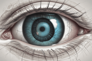
- The cornea, located at the front of the eye, refracts and focuses light, acts as a protective barrier, maintains transparency, and provides an immune privilege environment.
- The iris, the colored part of the eye, sits behind the cornea and functions similarly to a lens; its color depends on individual genetics.
- Posterior to the iris, the lens regulates incoming light with the help of the ciliary muscle, which bends or compresses the lens to prevent damage from excessive light.
- The retina, located at the back of the eye, performs initial lower-level processing of light.
- The fovea, a unique feature within the retina, is critical for high-resolution vision tasks like reading.
- Rods, located in the periphery of the retina, are highly sensitive to light, offer resolution in low light, and primarily detect monochromatic colors.
- Cones, located closer to the medial aspect of the retina, are helpful for distinguishing colors and are most active in daytime.
- The optic disc, located towards the optic nerve, is where retinal ganglion cell axons converge to form the optic nerve, contains no photoreceptors, resulting in a blind spot.
- The optic nerve, the second cranial nerve at the back of the eye, transmits visual information to the brain. The optic nerve is surrounded by dura mater, arachnoid, and pia mater.
- Vision occurs as light enters the eye, gets focused by the cornea and lens onto the retina, gets processed by photoreceptor cells, and then gets transmitted to the brain via the optic nerve.
Pathway from Retina to Occipital Cortex
- Light passes through the cornea, lens, and pupil to reach the retina where rods and cones perform initial lower-level processing of visual information, including contrast, color, and movement.
- Visual information is transmitted from the retina via the optic nerve (second cranial nerve); retinal ganglion cell axons converge at the optic disc to form the optic nerve.
- The optic nerve from each eye travels to the optic chiasm, a crucial point where nasal and temporal visual field information is segregated.
- At the optic chiasm, fibers from the nasal portion of each retina cross over to the contralateral side, while temporal portion fibers remain on the ipsilateral side.
- Reorganized fibers form the optic tracts after the optic chiasm; each optic tract contains information from the contralateral visual field, originating from the temporal retina of one eye and the nasal retina of the other eye.
- Optic tracts travel to the lateral geniculate body (LGB) in the thalamus, which relays optic tract fibers to the next set of neurons in the visual pathway; this is the only synaptic relay before the cortex.
- Optic radiations carry visual information from the LGB through the temporal and parietal lobes towards the back of the brain.
- Optic radiations reach the occipital lobe, specifically the primary visual cortex (V1), where visual information is perceived and interpreted, leading to conscious sight; the image projected onto the retina is flipped upside down, and the occipital cortex reconstructs it.
- The visual pathway ensures that information from the left visual field is processed in the right occipital cortex, and vice versa.
Localizing Lesions Based on Visual Field Deficits
- Optic Nerve Lesion (#1): Total vision loss occurs in the affected eye since visual information cannot reach the brain.
- Optic Chiasm Lesion: Bitemporal hemianopsia (loss of temporal visual fields in both eyes) results from disrupted nasal fibers (interpreting temporal visual fields) crossing at the chiasm.
- Optic Tract Lesion: A homonymous hemianopsia (loss of the visual field on the same side relative to the lesion) occurs after the optic chiasm.
- A right optic tract lesion causes a left homonymous hemianopsia, resulting in loss of the left temporal visual field (right eye) and the left nasal visual field (left eye).
- Conversely, a left optic tract lesion causes a right homonymous hemianopsia, with loss of the right visual field.
- Optic Radiation Lesion: Homonymous hemianopsia can be caused by effects to the optic radiation, depending on the affected portion of the optic radiation or visual cortex.
- Occipital Cortex Lesion: Contralateral homonymous hemianopsia can be caused by damage to the occipital cortex.
- Localizing lesions requires assessing lost visual field parts in each eye; the pattern of loss directly corresponds to the damage location along the visual pathway.
Visual Pathway & Lesions - Questions and Answers
- The visual pathway from the orbit to the visual cortex includes:
- Retina (rods and cones for light detection and initial processing)
- Optic Nerve (transmits visual information)
- Optic Chiasm (nasal fibers decussate, temporal fibers remain ipsilateral)
- Optic Tract (information from contralateral visual field)
- Lateral Geniculate Body (LGB) (synapse in the thalamus)
- Optic Radiations (project to the occipital cortex)
- Occipital Lobe/Primary Visual Cortex (perception and interpretation)
- Visual field deficits based on the lesion location:
- Optic nerve lesion: Total vision loss in the affected eye
- Optic chiasm lesion: Bitemporal hemianopsia (loss of temporal vision in both eyes)
- Optic tract lesion: Homonymous hemianopsia (loss of the visual field on the opposite side of the lesion)
- Optic radiation: Homonymous hemianopsia (contralateral to the lesion)
- Occipital cortex lesion: Contralateral homonymous hemianopsia
- Examples:
- Loss of vision in the left temporal and right nasal region: Left homonymous hemianopsia
- Right homonymous hemianopsia: Impacts the nasal portion of the left retina and the temporal portion of the right retina
- Unilateral spatial neglect results in failure to attend to stimuli on the opposite side of the body.
- Consequences:
- Ignoring objects/people on the neglected side
- Difficulties with tasks requiring attention to both sides
- Potential safety issues
- May worsen visual deficits where present, even if visual pathway is intact.
- Visual field cut is a loss of visual information; unilateral spatial neglect is an attentional deficits where individuals do not process information on one side.
Studying That Suits You
Use AI to generate personalized quizzes and flashcards to suit your learning preferences.