Podcast
Questions and Answers
ما هو الغرض الرئيسي للقرنية؟
ما هو الغرض الرئيسي للقرنية؟
- تكسير أشعة الضوء لمساعدة التركيز على الشبكية
- السماح بدخول الضوء إلى العين
- حماية العين من الغبار والأوساخ والعدوى
- جميع ما سبق (correct)
كم يبلغ سُمْك القرنية تقريبًا؟
كم يبلغ سُمْك القرنية تقريبًا؟
- 0.5 ملم
- 1 ملم (correct)
- 3 ملم
- 2 ملم
ما هي الطبقة الخارجية للقرنية؟
ما هي الطبقة الخارجية للقرنية؟
- طبقة بومان
- الطبقة الضامة
- الطبقة الظهارية (correct)
- غشاء ديسيميه
ما هي الطبقة الأكثر سُمْكًا في القرنية؟
ما هي الطبقة الأكثر سُمْكًا في القرنية؟
ما هي وظيفة الشبكية في العين؟
ما هي وظيفة الشبكية في العين؟
ما الوظيفة الأساسية للقرنية في الإدراك البصري؟
ما الوظيفة الأساسية للقرنية في الإدراك البصري؟
ما الطبقتان الرئيسيتان في الشبكية؟
ما الطبقتان الرئيسيتان في الشبكية؟
ما وظيفة الخلايا المخروطية في الشبكية؟
ما وظيفة الخلايا المخروطية في الشبكية؟
ما الطبقة المسؤولة عن تزويد الشبكية بالمغذيات؟
ما الطبقة المسؤولة عن تزويد الشبكية بالمغذيات؟
ما الوظيفة الرئيسية للشبكية؟
ما الوظيفة الرئيسية للشبكية؟
Flashcards are hidden until you start studying
Study Notes
Eye Anatomy: Understanding the Cornea and Retina
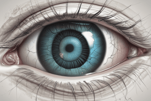
The human eye is a complex organ composed of several intricate components that work together to allow us to see the world around us. Two crucial parts of the eye that contribute significantly to our ability to perceive light and color are the cornea and the retina. Let's delve deeper into these fascinating structures and explore their roles within the context of eye anatomy.
Cornea

The cornea is a transparent dome-shaped structure that covers the front of the eye and serves as its protective barrier. It is approximately 1mm thick and accounts for about a third of the total refractive power of the eye. The cornea plays several vital roles in maintaining the clarity and health of the eye, including:
- Protecting the eye from dust, debris, and infection.
- Allowing light to enter the eye.
- Refracting light rays to help focus images on the retina.
This thin, curved surface is made up of five distinct layers:
- Epithelial layer: The outermost layer consisting of non-keratinized stratified squamous epithelial cells.
- Bowman's layer: A collagenous membrane beneath the epithelial layer.
- Stroma: The thickest layer of the cornea, composed mainly of collagen fibers and water, providing most of its refractive power.
- Descemet's membrane: A thin basement membrane lying beneath the stroma.
- Endothelium: The innermost layer of the cornea, made up of a single layer of hexagonal cells responsible for maintaining the overall transparency of the cornea through pumping out excess fluid.
The cornea's essential function in visual perception is emphasized by its healing capabilities. Injuries to the cornea often heal quickly due to the rapid regeneration of damaged cells, allowing the eye to maintain its visual integrity despite external damage.
Retina

The retina is a multilayered structure located at the back of the eye responsible for receiving light signals from the outside world. Light enters the eye through the cornea and lens and falls onto the retina, where it is transformed into electrical impulses that are then sent to the brain via the optic nerve. This process allows us to see various colors, shapes, and distances.
The retina consists of two main layers:
-
Photoreceptor layer: Contains specialized cells called rods and cones that detect light and transmit this information to other cells in the retina. Rods are primarily responsible for night vision and peripheral perception, while cones handle color vision and central focus.
-
Neural layer: Comprises a series of neurons called ganglion cells that receive signals from photoreceptors and send them along the optic nerve to the brain. Additionally, there are several supporting layers within the retina, such as the choriocapillaris, which provides nutrients to the retina, and the retinal pigment epithelium, responsible for maintaining the health and function of the retina by removing waste products and recycling components.
In summary, both cornea and retina play essential roles in our visual system. The cornea acts as a protective barrier while also contributing significantly to the eye's refractive power, ensuring clear and sharp images reach the retina. Meanwhile, the retina processes light signals and converts them into electrical impulses that travel to the brain, enabling us to perceive the world around us.
Studying That Suits You
Use AI to generate personalized quizzes and flashcards to suit your learning preferences.


