Podcast
Questions and Answers
Which part of the large intestine is referred to as the right ventral colon?
Which part of the large intestine is referred to as the right ventral colon?
- Right Ventral Colon (correct)
- Dorsal Diaphragmatic Flexure
- Left Ventral Colon
- Pelvic Flexure
What is the alternative name for the Ventral Diaphragmatic Flexure?
What is the alternative name for the Ventral Diaphragmatic Flexure?
- Sternal Flexure (correct)
- Caudal Flexure
- Pelvic Flexure
- Cervical Flexure
What differentiates the various compartments of the Rumen?
What differentiates the various compartments of the Rumen?
- Muscle contractions in the rumen
- Valves between the compartments
- Pillars on the luminal surface
- Grooves on the serosal surface (correct)
Which segments of the colon are referred to collectively as the 'large colon'?
Which segments of the colon are referred to collectively as the 'large colon'?
In regard to the equine gastrointestinal system, what does the term 'small colon' refer to?
In regard to the equine gastrointestinal system, what does the term 'small colon' refer to?
Which groove is associated with the cranial and caudal sections of the rumen?
Which groove is associated with the cranial and caudal sections of the rumen?
Which of the following is NOT a type of groove found in the Rumen?
Which of the following is NOT a type of groove found in the Rumen?
What anatomical structure follows the cecum in the flow of ingesta within the large intestine?
What anatomical structure follows the cecum in the flow of ingesta within the large intestine?
Where is the Insula ruminis located?
Where is the Insula ruminis located?
Which part of the rumen contains the caudoventral blind sac?
Which part of the rumen contains the caudoventral blind sac?
What stimulates the closure of the reticular groove to form a tube-like gastric groove?
What stimulates the closure of the reticular groove to form a tube-like gastric groove?
Which part of the gastric groove is found between the cranial and caudal lips?
Which part of the gastric groove is found between the cranial and caudal lips?
In what scenario would a thirsty adult bypass the rumen and send water directly to the abomasum?
In what scenario would a thirsty adult bypass the rumen and send water directly to the abomasum?
What is the primary function of the gastric groove?
What is the primary function of the gastric groove?
Which of the following parts follows the reticular groove in the sequence of flow when the reticular groove is closed?
Which of the following parts follows the reticular groove in the sequence of flow when the reticular groove is closed?
What is the primary function of the Tunica flava abdominis in horses?
What is the primary function of the Tunica flava abdominis in horses?
Which layer of the peritoneum lines the abdominal wall?
Which layer of the peritoneum lines the abdominal wall?
Which structure in the horse differentiates the non-glandular and glandular parts of the stomach?
Which structure in the horse differentiates the non-glandular and glandular parts of the stomach?
Where is the Tunica flava abdominis thickest in horses?
Where is the Tunica flava abdominis thickest in horses?
Which anatomical feature is associated with the inguinal region?
Which anatomical feature is associated with the inguinal region?
What characteristic of the lower esophageal sphincter is notable in horses?
What characteristic of the lower esophageal sphincter is notable in horses?
How do the mesenteries in dogs and horses compare?
How do the mesenteries in dogs and horses compare?
What is the purpose of careful suturing in the area of the Tunica flava abdominis?
What is the purpose of careful suturing in the area of the Tunica flava abdominis?
What is the function of the duodenal papillae in the ruminant small intestine?
What is the function of the duodenal papillae in the ruminant small intestine?
Which of the following structures is found at the ileocecocolic junction in ruminants?
Which of the following structures is found at the ileocecocolic junction in ruminants?
Where are jejunal lymph nodes primarily located in the ox as compared to the goat?
Where are jejunal lymph nodes primarily located in the ox as compared to the goat?
What is the main innervation source for the rumen, omasum, abomasum, and reticulum?
What is the main innervation source for the rumen, omasum, abomasum, and reticulum?
Which artery supplies blood to the greater curvature of the abomasum?
Which artery supplies blood to the greater curvature of the abomasum?
What distinguishes the jejunal lymph node location in goats and sheep from that in oxen?
What distinguishes the jejunal lymph node location in goats and sheep from that in oxen?
What is unique about the location of the jejunum compared to the ileum in ruminants?
What is unique about the location of the jejunum compared to the ileum in ruminants?
In ruminants, which part of the small intestine follows the duodenal flexures?
In ruminants, which part of the small intestine follows the duodenal flexures?
Which condition is characterized by a twisting of the intestine and can lead to obstructive issues?
Which condition is characterized by a twisting of the intestine and can lead to obstructive issues?
What is an expected finding during a physical examination if the small intestine is normal?
What is an expected finding during a physical examination if the small intestine is normal?
Which of the following diagnostic tools is NOT typically used for assessing abdominal conditions in equines?
Which of the following diagnostic tools is NOT typically used for assessing abdominal conditions in equines?
Which of the following could indicate an abnormal condition during a rectal examination?
Which of the following could indicate an abnormal condition during a rectal examination?
Which of these conditions does NOT belong in the differential diagnosis list for equine colic?
Which of these conditions does NOT belong in the differential diagnosis list for equine colic?
What is a common sign of abnormal small intestine during physical examination?
What is a common sign of abnormal small intestine during physical examination?
Which factor influences the decision to perform surgical exploration in cases of acute abdomen?
Which factor influences the decision to perform surgical exploration in cases of acute abdomen?
What does the '+' sign signify during gastrointestinal auscultation?
What does the '+' sign signify during gastrointestinal auscultation?
Flashcards
Right Ventral Colon (RVC)
Right Ventral Colon (RVC)
Part of the large intestine after the cecum.
Ventral Diaphragmatic Flexure
Ventral Diaphragmatic Flexure
A bend in the large intestine, also known as the sternal flexure.
Left Dorsal Colon (LDC)
Left Dorsal Colon (LDC)
Component of the large intestine, located on the left side.
Pelvic Flexure
Pelvic Flexure
Signup and view all the flashcards
Large vs Small Colon
Large vs Small Colon
Signup and view all the flashcards
Tunica flava abdominis
Tunica flava abdominis
Signup and view all the flashcards
External abdominal oblique muscle
External abdominal oblique muscle
Signup and view all the flashcards
Equine stomach
Equine stomach
Signup and view all the flashcards
Margo plicatus
Margo plicatus
Signup and view all the flashcards
Fundus (stomach)
Fundus (stomach)
Signup and view all the flashcards
Lower esophageal sphincter (horse)
Lower esophageal sphincter (horse)
Signup and view all the flashcards
Abdominal cavity vs. Peritoneal cavity
Abdominal cavity vs. Peritoneal cavity
Signup and view all the flashcards
Parietal peritoneum
Parietal peritoneum
Signup and view all the flashcards
Rumen Compartments
Rumen Compartments
Signup and view all the flashcards
Cranial and Caudal Grooves
Cranial and Caudal Grooves
Signup and view all the flashcards
Longitudinal Grooves
Longitudinal Grooves
Signup and view all the flashcards
Accessory Grooves
Accessory Grooves
Signup and view all the flashcards
Coronary Grooves
Coronary Grooves
Signup and view all the flashcards
Reticular Groove
Reticular Groove
Signup and view all the flashcards
Gastric Groove Function
Gastric Groove Function
Signup and view all the flashcards
Why Does the Gastric Groove Form?
Why Does the Gastric Groove Form?
Signup and view all the flashcards
What Stimulates Gastric Groove Formation?
What Stimulates Gastric Groove Formation?
Signup and view all the flashcards
Gastric Groove: Open vs. Closed
Gastric Groove: Open vs. Closed
Signup and view all the flashcards
Ruminant Stomach Innervation
Ruminant Stomach Innervation
Signup and view all the flashcards
Abomasal Blood Supply
Abomasal Blood Supply
Signup and view all the flashcards
Duodenal Papillae
Duodenal Papillae
Signup and view all the flashcards
Duodenocolic Fold
Duodenocolic Fold
Signup and view all the flashcards
Jejunal Lymph Nodes (Ox)
Jejunal Lymph Nodes (Ox)
Signup and view all the flashcards
Jejunal Lymph Nodes (Goat)
Jejunal Lymph Nodes (Goat)
Signup and view all the flashcards
Ileocecocolic Junction (Ruminants)
Ileocecocolic Junction (Ruminants)
Signup and view all the flashcards
Ileocolic & Cecocolic Orifice (Carnivores & Equine)
Ileocolic & Cecocolic Orifice (Carnivores & Equine)
Signup and view all the flashcards
Colic
Colic
Signup and view all the flashcards
GI Auscultation
GI Auscultation
Signup and view all the flashcards
Rectal Palpation
Rectal Palpation
Signup and view all the flashcards
Distended Intestines
Distended Intestines
Signup and view all the flashcards
Abdominocentesis
Abdominocentesis
Signup and view all the flashcards
Abdominal Ultrasound
Abdominal Ultrasound
Signup and view all the flashcards
Medical Management
Medical Management
Signup and view all the flashcards
Surgical Exploration
Surgical Exploration
Signup and view all the flashcards
Study Notes
Equine Gastrointestinal Tract Anatomy
- The equine gastrointestinal tract has a unique anatomy compared to other species.
- The abdominal wall is composed of skin, fascia, and muscles, including the tunica flava abdominis (a large animal only).
- The inguinal rings, inguinal canal, and vaginal ring are crucial anatomical features.
- The abdominal cavity is different from the peritoneal cavity, with various peritoneal parts including parietal and visceral peritoneum and mesenteries connecting different organs.
- The male and female vaginal tunic and vaginal process differ, as well as the vaginal ring.
- The tunica flava abdominis is elastic tissue found in horses and cattle, associated with the external abdominal oblique (EAO) muscle.
- It's thickest ventrally, where it merges with the EAO aponeurosis along the linea alba.
- Careful surgical suturing of this area is required.
Equine Stomach
- The equine stomach is a simple stomach with non-glandular and glandular mucosa.
- The Margo plicatus divides the non-glandular (fundus) from the glandular portion.
- The fundus is frequently called a "blind sac."
Equine Small Intestine
- The duodenum features a cranial flexure, descending, caudal flexure, ascending portions, and a duodenocolic fold.
- The jejunum is suspended by the root of the mesentery
- The ileum has an ileocecal fold, ileocecal orifice, and ileal papilla.
- The ileum in the equine is identified by the length of the ileocecal fold, as the antimesenteric ileal artery is absent.
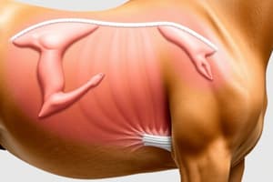
Equine Large Intestine
- The equine large intestine has taeniae coli (bands) and haustra (pouches), which vary in number.
- The cecum has a base, body, apex, and cecocolic fold.
- The ascending, transverse, and descending (small) sections of the colon have different numbers of bands.
Canine vs. Equine Gastrointestinal Differences
- The ascending colon of the canine is strikingly different from that of the equine, demonstrating different simplicity. The equine colon is a "hindgut fermenter".
Ruminant Gastrointestinal System
- The ruminant stomach has four compartments: rumen, reticulum, omasum, and abomasum.
- The first three (rumen, reticulum, omasum) are collectively termed the forestomach.
- The abomasum is similar to the simple stomach of monogastric animals. The left side of the abdominal cavity is dominated by the rumen, and the rumen does not contact the right abdominal wall.
- The rumen is laterally compressed and extends from the diaphragm to the pelvic inlet, covering one-half of the abdominal cavity.
Ruminant Stomach Anatomy
- Reticular groove, left longitudinal groove, and right longitudinal groove define the rumen's contours.
- Accessory grooves, cranial groove, coronary groove, and caudodorsal and caudoventral blind sacs also contribute to the rumen's complexity.
- The omasum is characterized by a variety of mucosal folds (laminae) of variable length.
- The abomasum is the equivalent of the simple stomach in monogastric animals.
Ruminant Stomach Additional Features
- The internal rumen aspect's papilla size depends on the diet and location.
- The rumen contents can vary in consistency depending on the location—gas dorsally to dense liquid ventrally.
- The reticulums' groove edges are able to contract to form a tube.
- The gastric groove helps the rumen to feed to the abomasum. An opening permits direct flow to the abomasum.
Ruminant Small Intestine
- The duodenum has cranial and caudal flexures, a duodenocolic fold, and duodenal papilla.
- The jejunum, mesoduodenum, and ileum (with ileocecal fold, ileocecocolic junction, and ileal papilla) are component parts of the ruminant small intestine.
- Jejunal lymph nodes are located in particular places in cattle vs. goats/sheep.
Ruminant Large Intestine
- The ruminant large intestine comprises the cecum and several colon segments, which vary in number and arrangements.
- The ascending colon's proximal loop, the spiral portion, and the distal loop are parts of its structure.
Clinical Application of Equine Gastrointestinal Anatomy
- Equine colic, or acute abdominal pain, has several possible causes, which are not all immediately apparent and/or discoverable based on the animal presentation.
- The correct identification of the source of the pain is often more significant in the success rate than in the speed of treatment
- Important diagnostic and physical assessment procedures that rely on location within the abdominal cavity are important.
- The locations of various anatomical structures within the abdominal cavity can help direct the practitioner in the identification of causes and possible solutions.
- Several surgical techniques can be employed to correct problems in the equine GI; some such procedures focus on fixation, or re-locating/re-directing compromised aspects of the GI tract.
Studying That Suits You
Use AI to generate personalized quizzes and flashcards to suit your learning preferences.