Podcast
Questions and Answers
Which organ is NOT derived from the foregut?
Which organ is NOT derived from the foregut?
- Proximal duodenum
- Stomach
- Large intestine (correct)
- Gallbladder
Which of the following structures is a derivative of the foregut?
Which of the following structures is a derivative of the foregut?
- Rectum
- Descending colon
- Pancreas (correct)
- Cecum
What is a function of the gallbladder?
What is a function of the gallbladder?
- Storing bile (correct)
- Secreting digestive enzymes
- Absorbing nutrients
- Producing insulin
Which of the following organs is developed from the foregut and plays a role in digestion?
Which of the following organs is developed from the foregut and plays a role in digestion?
Which structure connects the foregut derivatives to the rest of the digestive system?
Which structure connects the foregut derivatives to the rest of the digestive system?
What role does the mesenteric artery play in the structure described?
What role does the mesenteric artery play in the structure described?
Which direction does the arrow indicate in relation to the rotation of the primary intestinal loop?
Which direction does the arrow indicate in relation to the rotation of the primary intestinal loop?
What happens to the primary intestinal loop after 90° of rotation?
What happens to the primary intestinal loop after 90° of rotation?
In the primary intestinal loop's anatomy, what might be affected by the mesenteric artery's positioning?
In the primary intestinal loop's anatomy, what might be affected by the mesenteric artery's positioning?
What happens to the foregut, midgut, and hindgut by the end of the 5th week of development?
What happens to the foregut, midgut, and hindgut by the end of the 5th week of development?
What structure is responsible for maintaining the initial attachment of the gut tube?
What structure is responsible for maintaining the initial attachment of the gut tube?
What visualization can be expected when looking at the primary intestinal loop after the specified rotation?
What visualization can be expected when looking at the primary intestinal loop after the specified rotation?
Which of the following processes begins by the end of the 5th week in gut development?
Which of the following processes begins by the end of the 5th week in gut development?
What role does the dorsal mesentery play during the gut development process?
What role does the dorsal mesentery play during the gut development process?
At what stage does the attachment of the gut tube to the abdominal wall occur?
At what stage does the attachment of the gut tube to the abdominal wall occur?
What does the anorectal canal develop into?
What does the anorectal canal develop into?
Which structure is formed from the urogenital sinus?
Which structure is formed from the urogenital sinus?
What is the role of the urorectal septum in development?
What is the role of the urorectal septum in development?
Which of the following does NOT originate from the urorectal septum?
Which of the following does NOT originate from the urorectal septum?
What anatomical structures are formed from the division created by the urorectal septum?
What anatomical structures are formed from the division created by the urorectal septum?
What significant developmental change occurs in the seventh week regarding the cloacal membrane?
What significant developmental change occurs in the seventh week regarding the cloacal membrane?
Which two structures are specifically separated by the development that occurs in the seventh week?
Which two structures are specifically separated by the development that occurs in the seventh week?
What role does the cloacal membrane play during the seventh week of embryonic development?
What role does the cloacal membrane play during the seventh week of embryonic development?
In which week of development are the anal canal and urogenital sinus openings created?
In which week of development are the anal canal and urogenital sinus openings created?
What physiological outcome results from the separation of the openings in the seventh week?
What physiological outcome results from the separation of the openings in the seventh week?
Flashcards
Foregut
Foregut
The first section of the developing digestive tract, giving rise to several important organs.
Esophagus
Esophagus
A muscular tube that transports food from the mouth to the stomach.
Stomach
Stomach
A muscular organ responsible for churning food and mixing it with digestive juices.
Liver
Liver
Signup and view all the flashcards
Gallbladder
Gallbladder
Signup and view all the flashcards
Mesenteric artery
Mesenteric artery
Signup and view all the flashcards
Primary intestinal loop
Primary intestinal loop
Signup and view all the flashcards
Counterclockwise rotation
Counterclockwise rotation
Signup and view all the flashcards
90° counterclockwise rotation
90° counterclockwise rotation
Signup and view all the flashcards
Axis of the loop
Axis of the loop
Signup and view all the flashcards
Initial Gut Attachment
Initial Gut Attachment
Signup and view all the flashcards
Dorsal Mesentery
Dorsal Mesentery
Signup and view all the flashcards
5th Week Development
5th Week Development
Signup and view all the flashcards
Midgut
Midgut
Signup and view all the flashcards
StatPearls
StatPearls
Signup and view all the flashcards
Treasure Island (FL)
Treasure Island (FL)
Signup and view all the flashcards
DM 103
DM 103
Signup and view all the flashcards
THANK YOU FOR LISTENING!
THANK YOU FOR LISTENING!
Signup and view all the flashcards
Available from: https://www.ncbi.nlm.nih.gov/books/NBK553156/
Available from: https://www.ncbi.nlm.nih.gov/books/NBK553156/
Signup and view all the flashcards
Urorectal Septum
Urorectal Septum
Signup and view all the flashcards
Anorectal Canal
Anorectal Canal
Signup and view all the flashcards
Urogenital Sinus
Urogenital Sinus
Signup and view all the flashcards
Rectum
Rectum
Signup and view all the flashcards
Urethra
Urethra
Signup and view all the flashcards
Seventh Week of Development
Seventh Week of Development
Signup and view all the flashcards
Cloacal Membrane
Cloacal Membrane
Signup and view all the flashcards
Anal Canal
Anal Canal
Signup and view all the flashcards
Separation of Openings
Separation of Openings
Signup and view all the flashcards
Study Notes
Embryology of the Human Digestive System
- The digestive system develops from germ tissues during gastrulation.
- By week 2, the endoderm, mesoderm, and ectoderm are developed.
- The primitive gut tube is formed by week 3-4 by incorporating the yolk sac.
- Foregut: develops into the esophagus, stomach, liver, gallbladder, pancreas, and proximal duodenum.
- Midgut: develops into the distal duodenum, jejunum, ileum, cecum, appendix, ascending colon, and proximal 2/3 of the transverse colon.
- Hindgut: develops into the distal 1/3 of the transverse colon, descending colon, sigmoid colon, and upper anal canal.
Molecular Regulation of Gut Tube Development
- Retinoic acid (RA) gradients initiate regional specification of the gut tube.
- Higher RA concentrations in the colon influence gene expression compared to other areas.
- SOX2 expression determines esophagus and stomach development.
- PDX1 expression determines duodenum development.
- CDX2 expression determines small intestine development, and
- CDX1 expression determines large intestine and rectum development.
- Sonic Hedgehog (SHH) expression initiates epithelial-mesenchymal interactions for initial patterning.
Mesentery Development
- Mesentery is a fold of peritoneum that suspends digestive organs, blood vessels, and nerves from the posterior abdominal wall.
- It provides support and allows mobility to the organs.
- The foregut, midgut, and hindgut are initially attached to the posterior mesenchymal wall until week 5.
- The connecting tissue then narrows, attaching the gut tube to the abdominal wall via the dorsal mesentery.
Organogenesis
- The Foregut: Develops into the esophagus, stomach, liver, gallbladder, pancreas, and the caudal portion of the duodenum. (Week 4.)
- Esophagus: The respiratory diverticulum partitions the foregut into ventral (respiratory primordium) and dorsal (esophagus) portions.
- Stomach: Formation of fusiform dilation is seen in the foregut with a noticeable change in positional and growth differences in an anterior and posterior aspect.
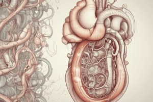
Liver Development
- The liver forms from endoderm within the foregut.
- Surrounding tissues (ectoderm, mesoderm, and notochord) release inhibitors to locally establish the expression of liver-specific genes.
- Fibroblast Growth Factor 2 (FGF2) secreted by cardiac mesoderm and blood vessels initiates liver gene expression.
- Bone Morphogenetic Proteins (BMPs) produced by the septum transversum prepare the endoderm to respond to FGF2.
Pancreas Development
- The pancreas develops from two separate endodermal buds: dorsal and ventral.
- The dorsal bud forms first within the dorsal mesentery.
- The ventral bud is close to the bile duct and shifts during gut rotation to merge with the dorsal bud.
- The main pancreatic duct of Wirsung forms from the fusion of both buds.
- Proximal parts of the dorsal bud may persist as accessory pancreatic duct of Santorini.
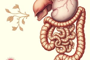
Midgut Development
- The midgut extends from the bile duct to the junction of the transverse and descending colon.
- It forms a significant portion of the small intestine and proximal large intestine.
- During development, the superior mesenteric artery is the axis of a 90-degree counter-clockwise rotation of the midgut.
Hindgut Development
- The hindgut extends and forms the distal transverse colon, descending colon, sigmoid colon, rectum, and upper anal canal.
- It plays a role in the internal lining formation of the bladder and urethra.
- The urorectal septum divides the hindgut and the urogenital sinus.
Cloacal Development
- The cloaca is a terminal portion of the hindgut.
- The cloaca is divided into:
- Anorectal canal (rectum and upper anal canal),
- Urogenital sinus(bladder and urethra).
- The development of the cloaca includes the rupturing of the cloacal membrane between the ectoderm and endoderm and the creation of separate openings for the anal canal and the urogenital sinus with the help of the urorectal septum.
Studying That Suits You
Use AI to generate personalized quizzes and flashcards to suit your learning preferences.