Podcast
Questions and Answers
What are the two main types of Acute Coronary Syndrome based on ECG findings?
What are the two main types of Acute Coronary Syndrome based on ECG findings?
- Septal and Inferior
- ST Elevation and Non-ST Elevation (correct)
- Transmural and Subendocardial
- Hyperacute and Convex
Which leads on an ECG primarily indicate septic wall infarction?
Which leads on an ECG primarily indicate septic wall infarction?
- V3 and V4
- I and aVL
- V1 and V2 (correct)
- II and III
What does ST segment elevation typically indicate in a myocardial infarction?
What does ST segment elevation typically indicate in a myocardial infarction?
- Lateral Wall infarction
- Transmural injury (correct)
- Ventricular hypertrophy
- Subendocardial injury
Which of the following leads would show signs of an inferior wall infarction?
Which of the following leads would show signs of an inferior wall infarction?
Which of the following is an early sign of Acute ST-Elevation Myocardial Infarction?
Which of the following is an early sign of Acute ST-Elevation Myocardial Infarction?
Where on the ECG would you expect to see changes for Anterior Wall infarction?
Where on the ECG would you expect to see changes for Anterior Wall infarction?
Which of the following statements is true about ST segment changes?
Which of the following statements is true about ST segment changes?
Which leads are associated with lateral wall infarction?
Which leads are associated with lateral wall infarction?
Which of the following describes the state of the ST segment during an acute myocardial infarction?
Which of the following describes the state of the ST segment during an acute myocardial infarction?
How can the localization of myocardial infarction be determined?
How can the localization of myocardial infarction be determined?
What does ST depression in leads remote from an acute infarct typically indicate?
What does ST depression in leads remote from an acute infarct typically indicate?
Which of the following describes the characteristic shape of depressed ST segments?
Which of the following describes the characteristic shape of depressed ST segments?
What is indicated by the presence of pathological Q waves?
What is indicated by the presence of pathological Q waves?
Which feature is characteristic of left ventricular hypertrophy on an ECG?
Which feature is characteristic of left ventricular hypertrophy on an ECG?
What are the two primary changes observed in Bundle Branch Blocks on an ECG?
What are the two primary changes observed in Bundle Branch Blocks on an ECG?
In which form of tachycardia are the QRS complexes wide and abnormal?
In which form of tachycardia are the QRS complexes wide and abnormal?
Which description best represents the early beats in premature ventricular contractions (PVCs)?
Which description best represents the early beats in premature ventricular contractions (PVCs)?
What is the primary characteristic that differentiates first-degree heart block from other types?
What is the primary characteristic that differentiates first-degree heart block from other types?
Which of the following is true regarding the trend of ST and T changes as an infarction evolves?
Which of the following is true regarding the trend of ST and T changes as an infarction evolves?
Which feature indicates right ventricular hypertrophy on an ECG?
Which feature indicates right ventricular hypertrophy on an ECG?
Flashcards
Reciprocal ST Depression
Reciprocal ST Depression
ST depression in leads away from the site of an acute infarct, indicating a high probability (90%) of infarction. Often seen with inferior (70%) and anterior (30%) infarctions. The depressed ST segment typically appears horizontal or downsloping.
Pathological Q Waves
Pathological Q Waves
Loss of R wave amplitude and emergence of new Q waves, indicating loss of viable myocardium. Firm evidence of myocardial necrosis. May resolve with scar tissue contraction during healing.
Left Ventricular Hypertrophy
Left Ventricular Hypertrophy
Increased voltage of the QRS complex, specifically a large S wave in lead V1 and a large R wave in lead V6, indicating enlargement of the left ventricle.
Right Ventricular Hypertrophy
Right Ventricular Hypertrophy
Signup and view all the flashcards
Premature Atrial Contractions (PACs)
Premature Atrial Contractions (PACs)
Signup and view all the flashcards
Premature Ventricular Contractions (PVCs)
Premature Ventricular Contractions (PVCs)
Signup and view all the flashcards
Sinus Tachycardia
Sinus Tachycardia
Signup and view all the flashcards
Ventricular Tachycardia
Ventricular Tachycardia
Signup and view all the flashcards
Atrial Fibrillation (AF)
Atrial Fibrillation (AF)
Signup and view all the flashcards
First Degree Heart Block
First Degree Heart Block
Signup and view all the flashcards
Hyperacute T waves
Hyperacute T waves
Signup and view all the flashcards
ST segment elevation
ST segment elevation
Signup and view all the flashcards
Localization of STEMI
Localization of STEMI
Signup and view all the flashcards
Septal Wall Infarction Localization
Septal Wall Infarction Localization
Signup and view all the flashcards
Anterior Wall Myocardial Infarction Localization
Anterior Wall Myocardial Infarction Localization
Signup and view all the flashcards
Lateral Wall Infarction Localization
Lateral Wall Infarction Localization
Signup and view all the flashcards
Inferior Wall Infarction Localization
Inferior Wall Infarction Localization
Signup and view all the flashcards
Progression of STEMI
Progression of STEMI
Signup and view all the flashcards
Importance of ECG in STEMI
Importance of ECG in STEMI
Signup and view all the flashcards
Differentiating STEMI and NSTEMI
Differentiating STEMI and NSTEMI
Signup and view all the flashcards
Study Notes
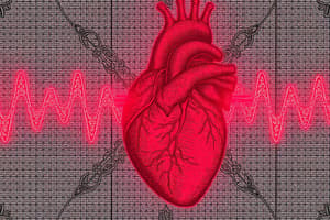
ECG Practical Session (Second Year)
- The presentation covers ECG interpretation for myocardial infarction (MI) and other cardiac conditions.
- Myocardial infarction is a critical cardiac condition where blood flow to the heart muscle is blocked resulting in cell death.
- The coronary system delivers blood to the heart muscle and comprises the aorta, pulmonary artery, and various coronary arteries (including the right coronary artery, left main artery, left anterior descending coronary artery, and circumflex coronary artery).
- Acute Coronary Syndromes (ACS) are conditions that fall under myocardial infarctions. ACS can be categorized into ST-Elevation Myocardial Infarction (STEMI) & Non-ST-Elevation Myocardial Infarction (NSTEMI).
- STEMI includes transmural (Q-wave) and NSTEMI includes subendocardial (non-Q-wave) types.
- ECG changes associated with MI include hyperacute T waves, ST segment elevation, and pathological Q waves.
- Hyperacute T waves are the earliest sign and characterized by prominent, symmetrical T waves that typically precede ST elevation. They are usually transient.
- ST segment elevation occurs when the ST segment is raised above the baseline level, usually in leads related to the particular muscle damage. This is a crucial diagnostic feature for determining the location and severity of the MI.
- Pathological Q waves develop as a result of myocardial necrosis. The R wave loses amplitude, and a new Q wave forms. Q waves can be a lasting/permanent marker after MI healing.
- The location of the ST segment elevation or other ECG changes can indicate the affected heart wall (inferior, anterior, or lateral).
- Specific leads on an ECG relate to specific locations in the heart. (e.g., leads II, III, and aVF are associated with the inferior wall, and leads V1-V4 with the anterior wall).
- Reciprocal ST depression in leads remote from the site of the infarction can frequently be a highly sensitive indicator of infarction. This depression is frequently horizontal or downsloping.
- Infarction resolution is characterized by diminishing ST elevation and T wave inversion.
- Chamber enlargements (such as left ventricular hypertrophy or right ventricular hypertrophy) can be identified through changes in the QRS complex size and shape (voltage) in an ECG.
- Bundle branch blocks (e.g., right bundle branch block (RBBB) and left bundle branch block (LBBB)) result in characteristic QRS complex widening and morphology changes. Key features to recognize on an ECG include the presence or absence of an rSR′ or qsR′ appearance in the respective leads.
- Arrhythmias (e.g., premature atrial contractions (PACs), premature ventricular contractions (PVCs), sinus tachycardia, atrial fibrillation (AF), supraventricular tachycardia (SVT), ventricular tachycardia, heart block (first-degree, second-degree, and third-degree)) can be identified in an ECG rhythm strip.
- Specific arrhythmia types are associated with distinct characteristics in the ECG rhythm strip, such as irregular R-R intervals in atrial fibrillation or various QRS morphologies.
Studying That Suits You
Use AI to generate personalized quizzes and flashcards to suit your learning preferences.