Podcast
Questions and Answers
How does Profilin contribute to actin filament polymerization?
How does Profilin contribute to actin filament polymerization?
- By capping the ends of actin filaments, preventing further elongation.
- By severing existing actin filaments, creating more free ends.
- By aiding the addition of actin monomers to the filament. (correct)
- By cross-linking actin filaments, forming complex structures.
What distinguishes the function of Kinesin from that of Dynein in intracellular transport?
What distinguishes the function of Kinesin from that of Dynein in intracellular transport?
- Kinesin transports cargo towards the negative pole, while Dynein moves towards the positive pole.
- Kinesin moves cargo towards the positive pole, while Dynein moves towards the negative pole. (correct)
- Kinesin is responsible for transporting vesicles, while Dynein transports organelles.
- Kinesin functions in muscle cell contraction, while Dynein functions in cellular migration.
How do cells ensure controlled growth and shrinkage of microtubules during cell division?
How do cells ensure controlled growth and shrinkage of microtubules during cell division?
- By exclusively polymerizing microtubules to ensure robust chromosomal separation.
- By halting both polymerization and depolymerization after chromosomes align.
- By precisely balancing microtubule polymerization and depolymerization. (correct)
- By evenly distributing tubulin dimers, ensuring no net change in microtubule length.
What implications does the phosphorylation of MAPs (Microtubule-Associated Proteins) have on microtubule stability?
What implications does the phosphorylation of MAPs (Microtubule-Associated Proteins) have on microtubule stability?
How do intermediate filaments differ from microfilaments and microtubules in terms of their structural role within cells?
How do intermediate filaments differ from microfilaments and microtubules in terms of their structural role within cells?
How do diseases related to keratin mutations, such as Epidermolysis Bullosa Simplex, manifest structurally?
How do diseases related to keratin mutations, such as Epidermolysis Bullosa Simplex, manifest structurally?
How does Tropomyosin prevent muscle contraction in its resting state?
How does Tropomyosin prevent muscle contraction in its resting state?
What is the functional role of ATP hydrolysis in the cross-bridge cycle during muscle contraction?
What is the functional role of ATP hydrolysis in the cross-bridge cycle during muscle contraction?
How do Vincristine function as a cancer treatment?
How do Vincristine function as a cancer treatment?
How does taxol's mechanism of action differ from that of vincristine in cancer treatment?
How does taxol's mechanism of action differ from that of vincristine in cancer treatment?
Flashcards
Microfilaments
Microfilaments
Microfilaments consisting of actin are the flexible, primary drivers of cell movement. Found beneath the plasma membrane.
Motor Proteins
Motor Proteins
A family of proteins that enable muscle cell contraction, cellular migration and intracelluar transport.
Dynein
Dynein
Moves towards the negative pole (retrograde) during intracellular transport.
Kinesin
Kinesin
Signup and view all the flashcards
MAPs
MAPs
Signup and view all the flashcards
Dynamic Instability
Dynamic Instability
Signup and view all the flashcards
Intermediate Filaments
Intermediate Filaments
Signup and view all the flashcards
Thin Filaments (Actin)
Thin Filaments (Actin)
Signup and view all the flashcards
Troponin Complex
Troponin Complex
Signup and view all the flashcards
Vincristine
Vincristine
Signup and view all the flashcards
Study Notes
- The cytoskeleton provides the basis for cell movement and intracellular transport
- Actin cytoskeleton also has a role in skeletal muscle function
Key Components of the Cytoskeleton
- Microfilaments (Actin filaments) are primary drivers of movement
- Intermediate filaments
- Microtubles
- They make up around 3-6% of the cell, specifically beneath the plasma membrane
Microfilaments
- Possess nucleotide-binding sites which bind to ATP and ADP
- They polymerize into actin
- They have polarity with barbed (+) and pointed (-) ends
- Specific mechanisms include cell crawling, immune cell migration, and wound healing
Cellular Movement
- Actin filaments polymerize in the direction of intended movement
- Lamellipodia (cell protrusions) form and attach to a substrate
- Myosin II then contracts, pulling along newly formed actin filaments
Polymerization Regulators
- Profilin aids polymerization
- Capping proteins prevent further growth
- Severing proteins, like gelsolin and ADF/cofilin will break existing filaments
Functional Dynamics
- The functional dynamics of the cytoskeleton include cross-linking or bundling into complex structures
- They are critical for maintaining cell shape, muscle contraction, and cell crawling mechanisms
Intracellular Transport
- Microtubule-based transport is the primary mechanism
- Motor proteins enable muscle cell contraction and cellular migration
- Dynein moves towards the negative pole (retrograde)
- Kinesin moves towards the positive pole (anterograde)
- The process is ATP-dependent for the transport of vesicles and organelles
Specialized Microtubule Structures
- Cilia are projections that move from the cell membrane
- Respiratory cilia move mucus
- Flagella only exist in sperm cells
- Centrioles do cell division
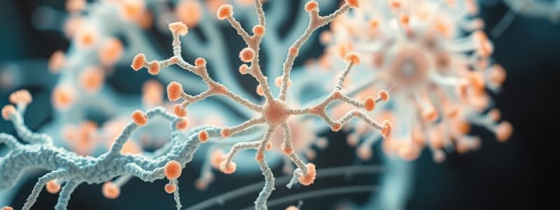
Intermediate Filaments
- There is structural diversity among them
- They can be divided into 6 classes
- They are roughly 10nm wide
- Main component of hair
Intermediate Filament Classes
- Over 70 distinct intermediate filament proteins exist
- Keratins (Acidic and Basic) are in Epithelia
- Some are called "keratins"
- Desmin exist in muscle
- GFAP exists in glial cells
- Neurofilaments exist in neurons
- Lamins exist in the nuclear envelope
- Nestin exists in neural stem cells
Intermediate Filament Characteristics
- Semi-permanent tissue structures
- Phosphorylation controls disassembly
- They are involved in cell-cell adhesion
- Provide mechanical strength throughout the cell
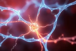
Microtubules
- Intracellular transport
- Microtubule Associated Proteins (MAP) attach to the microtubule and stop them from deep memorizing
Instability
- The MAPS stabilize their dynamic so it will have higher memory
- MAPs stabilize microtubules through protein interaction e.g. (TAU)
Regulating Function
- Function tightly regulated by phosphorylation
- Alzheimer's disease will cause them to not associate adding a phosphate group to proteins
GTP Hydrolysis
- Gtp is hydrolyzed during polymerization immediately following polymerization
- Intracellular transport
Microtubule Dimensions
- There are roughly around 25nm
- Microtubule Associated Protein (MAP)
- Stabilize microtubules with protein interaction e.g., tau
- Alzheimer's disease
Tubulin Dimers
- Microtubules are composed of Tubulin Dimers that are subunits of the protein "tubulin"
- They consist of α-tubulin and β-tubulin
- They have a polar structure
- 13 polymers form the circumference
- Alpha-tubulin is at the (-) end
- Beta-tubulin is always added at the (+) end
- All microtubules are attached to a centrioles in the centrosome
- The centrosome us made up of microtubules
Actin and Muscle Function
- Actin cytoskeleton is also the basis of skeletal muscle function
Structural Muscle Levels
- Muscle fiber
- Myofibril
- Sarcomere (functional unit)
- Contains primary contractile elements
- There are Key Molecular components:
- Thin filaments (Actin)
- Actins Regulatory Proteins controls muscle contraction: -Tropomyosin -Troponin complex -Calcium sensitive protein triggers muscle contraction, and consists of 3 subunits
Contractile Proteins
Key Molecular components:
- Primary contractile protein, runs entire length of sarcomere, contains regulatory proteins
- Thick filaments (Myosin)
- Motor protein, generates mechanical force, interacts with actin .
Mechanism During Resting State
- Tropomyosin blocks myosin-binding sites
- Calcium channels closed
- Actin filaments held in the inhibited configuration
Calcium Release
- Binds to troponin C, causes conformational protein changes - ATP-powered moves -Sarcomere shortens
- Precise/ coordinated Molecular movement.
Muscle Contraction Requires
- Sliding theory
- Myosin head pulls actin Muscle contraction
Mutations
- Mutations or the poisoning of the cytoskeleton can result in specific diseases. Also a significant drug target (i.e. vincristine, taxol)
Microfilament (Actin) Related Diseases
- Disrupted positive and negative cellular movements
- Negative movements include:
- Cancer metastasis, abnormal cell migration, uncontrolled spread Angiogenesis in diabetic Retinopathy which leads to Blood cells proliferation & disperse into the Retina & displace retina -Neuropathic pain related Multiple sclerosis
MICROTUBULES disorders
Neurodegeneration
Six Major Disrupted II Classes
1 Kevatins epithelial 2 Desmin muscle 3 GFAP glial cells 4 Neurofilaments nervous 5 Launius nuclear envelope Type 1 11 disorders Hmin
Neurodegeneration disorders
-parkninsons’s, Alzheimer's
. Frontotemporal dementia and other tauopathies
Disrupted Systems
- Respiration
- Reproductive
Microtubule pathologies examples
Neurodegeneration:
Drug Action
Vinicistine& Taxol effect drug:
-Vinicistine
action: destabilizesmicotables
prevents: Microtubulepolymerisation
Disrupts Mitoticspindle
Molecular action:
-Binds to - tublin protein prevents
MICROTUBULEDESTABILIZERS action:
- Stops cell division
- Triggers apoptosis in rapidly dividing
- Binds to: Microtubule
Stabilizesexistingstructures - Preventsmicrotubuledisassembly Locksmicrotubulesinfixedconfiguation disrupts normal cellular division. Interrupts:
- rapidlydyning
- PreciseMolculartargeting
- Interruptcellular
Studying That Suits You
Use AI to generate personalized quizzes and flashcards to suit your learning preferences.