Podcast
Questions and Answers
Which of the following is a primary characteristic of intermediate filaments that distinguishes them from actin filaments and microtubules?
Which of the following is a primary characteristic of intermediate filaments that distinguishes them from actin filaments and microtubules?
- They undergo constant assembly and disassembly.
- They facilitate intracellular transport via motor proteins.
- They are semi-permanent tissue structures providing mechanical strength. (correct)
- They are the primary drivers of cell movement.
How does profilin regulate actin polymerization?
How does profilin regulate actin polymerization?
- By cross-linking actin filaments into complex structures.
- By preventing further growth at the plus end of actin filaments.
- By severing existing actin filaments, promoting disassembly.
- By aiding the addition of actin monomers to the growing filament. (correct)
How do Dynein and Kinesin motor proteins differ in their function within a cell?
How do Dynein and Kinesin motor proteins differ in their function within a cell?
- Dynein is ATP-independent, while Kinesin requires ATP to function.
- Dynein moves towards the positive end of microtubules, while Kinesin moves towards the negative end.
- Dynein facilitates muscle contraction, while Kinesin is involved in cell crawling.
- Dynein moves towards the negative end of microtubules, while Kinesin moves towards the positive end. (correct)
Which characteristic of dynamic instability in microtubules describes the consequence of GTP hydrolysis?
Which characteristic of dynamic instability in microtubules describes the consequence of GTP hydrolysis?
In muscle contraction, what is the role of tropomyosin?
In muscle contraction, what is the role of tropomyosin?
How do drugs like Vincristine, used in cancer treatment, target microtubules?
How do drugs like Vincristine, used in cancer treatment, target microtubules?
What process is most directly affected by mutations in keratin genes?
What process is most directly affected by mutations in keratin genes?
How does the mechanism of action of Taxol differ from that of Vincristine in cancer treatment?
How does the mechanism of action of Taxol differ from that of Vincristine in cancer treatment?
Alzheimer's disease is associated with dysfunction of a Microtubule Associated Protein (MAP). Which statement correctly describes this relationship?
Alzheimer's disease is associated with dysfunction of a Microtubule Associated Protein (MAP). Which statement correctly describes this relationship?
How does calcium binding to troponin C trigger muscle contraction?
How does calcium binding to troponin C trigger muscle contraction?
Flashcards
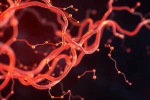
Microfilaments (Actin filament)
Microfilaments (Actin filament)
Primary drivers of cell movement, cell crawling, immune cell migration, and wound healing.
Profilin
Profilin
Aids actin polymerisation for cellular movement and structure.
Dynein
Dynein
Move toward the negative pole (retrograde) during intracellular transport.
Microtubule Associated Protein (MAP)
Microtubule Associated Protein (MAP)
Signup and view all the flashcards
GTP
GTP
Signup and view all the flashcards
Thin Filaments (Actin)
Thin Filaments (Actin)
Signup and view all the flashcards
Tropomyosin
Tropomyosin
Signup and view all the flashcards
Troponin complex
Troponin complex
Signup and view all the flashcards
Vincristine
Vincristine
Signup and view all the flashcards
Taxol (Taxanes)
Taxol (Taxanes)
Signup and view all the flashcards
Study Notes
- The cytoskeleton is essential for cell movement and intracellular transport, and the actin cytoskeleton plays a crucial role in skeletal muscle function.
Key components of the Cytoskeleton
- Microfilaments (actin filaments)
- Intermediate filaments
- Microtubules
Microfilaments (Actin Filaments)
- Made of actin monomers.
- Located beneath the plasma membrane.
- They are 3-6nm .
- Primarily drives cell movement
- Requires nucleotide binding (ATP or ADP)
- Polymerizes into filaments with positive (barbed) and negative (pointed) ends
Specific Mechanisms
- Cell crawling
- Immune cell migration
- Wound healing
Cellular Movement
- Involves actin filaments polymerizing in the direction of movement.
- Lamellipodia (cell protrusions) form and attach to the substrate, followed by myosin II contraction, which pulls the cell along newly formed actin filaments.
Polymerization Regulators
- Profilin enhances polymerization.
- Capping proteins prevent further growth.
- Severing proteins (e.g., gelsolin, ADF/cofilin) break existing filaments.
Functional Dynamics
- The cytoskeleton cross-links or bundles into structures and is crucial for maintaining cell shape, muscle contraction, and cell crawling mechanisms.
Intracellular Transport
- Accomplished primarily through microtubule-based transport.
- Motor proteins such as dynein (retrograde transport towards the negative pole) and kinesin (anterograde transport towards the positive pole) enable muscle cell contraction and cellular migration.
- This is an ATP-dependent process for vesicles and organelles.
- Specialized microtubule structures include cilia (projections that enable movement) and flagella (e.g., in sperm).
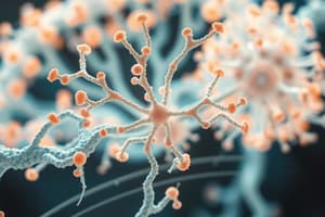
Intermediate Filaments
- Offer structural diversity
- They are 10nm
- Are divided into six classes.
Intermediate Filament Classes
- Keratins (acidic and basic) found in epithelial cells.
- Desmin in muscle tissue, especially skeletal muscle.
- GFAP (glial fibrillary acidic protein) in glial cells.
- Neurofilaments in neurons.
- Lamins in the nuclear envelope.
- Nestin in neural stem cells.
Characteristics of Intermediate FIlaments
- Semi-permanent tissue structures
- Stabilize the structure of the nuclear membrane
- Involved in cell-cell adhesion
- Provide mechanical strength.
- Controlled via phosphorylation
Microtubules Structural Composition
- Consists of tubulin dimers (α-tubulin and β-tubulin).
- Polar structure with 13 polymers forming the circumference.
- Alpha-tubulin is at the negative (-) end.
- Beta-tubulin is at the positive (+) end.
- All microtubules attach to a structure within the centrosome
Centrosome
- Specialized organelle composed of microtubules
Dynamic Instability
- GTP is hydrolyzed
- Constant assembly/disassembly mechanism
- This allows for rapid changes in structural support
Microtubule-Associated Proteins (MAPs)
- Attach to microtubules and prevent them from disassembling.
- Stabilize their dynamic instability through protein interactions (e.g., TAU).
- Function tightly, with regulation by phosphorylation.
Alzheimer's Disease
- Associated dynamic instability from the addition of a phosphate group.
Muscle Tissue Structure
- Structural levels: muscle fiber, myofibril, and sarcomere (the functional unit containing contractile elements).
Sarcomere Components
- Thin Filaments (Actin)
- Thick Filaments (Myosin)
- Regulatory Proteins
Thin Filaments (Actin)
- Primarily contractile protein
- extends along the sarcomere
- Contains regulatory proteins
Thick Filaments (Myosin)
- Motor protein that generates mechanical force through interaction with actin
Regulatory Proteins
- Tropomyosin wraps around actin filaments, blocking myosin-binding sites.
- Troponin complex: Calcium-sensitive protein triggering contraction, consisting of troponin C (calcium binding), troponin I (inhibitory), and troponin T (tropomyosin binding) subunits.
Muscle Contraction Mechanism
- Resting State: Tropomyosin blocks binding sites
- Calcium activation: Channels floods muscle cells, calcium binds to troponin C, exposing myosin binding sites.
- Cross-Bridge Cycling: ATP hydrolysis powers myosin heads to move along actin, shortening the sarcomere through coordinated molecular movement.
- Energy Requirements: Sliding filament theory enables force generation and muscle contraction through a precise, coordinated movement.
Microfilament (Actin) Related Diseases
- Impact muscle contraction mechanisms.
- Disrupt immune cell migration, compromising the immune response.
Wound Healing
- Wound repair is also comprimised
- Repair protein generation is impacted
- Time regeneration is comprimised
Cancer Metastasis
- Abnormal cell migration
- Uncontrolled cellular spread
Angiogenesis in Diabetic Retinopathy
- Epithelial cell migration
- Multiple sclerosis
- Neuronal Sprouting: Resulting in neuropathic pain and spinal cord issues.
Microtubule Disorders
- Neurodegeneration
- Parkinson's disease, Alzheimer's disease, frontotemporal dementia, related tauopathies
TAU
- A stabilizing protein for microtubules.
Ciliopathies
- Affects the respiratory & reproductive systems
- Involves impaired cilia/flagella function
- Compromises sensory system
- Causes developmental abnormalities.
Muscular Dystrophy
- Duchenne muscular dystrophy results from a mutation in MAP.
Intermediate Filament Related Disorder
Type I & II Disorders (Keratins & Desmin)
- Epidermolysis Bullosa Simplex
- Skin fragility
- Cellular structural weakness
Type III Disorders (GFAP)
- Amyotrophic Lateral Sclerosis (ALS)
- Neurological degeneration
- Motor neuron dysfunction
Type IV Disorders (Neurofilaments
- Parkinson's disease
- Alzheimer's disease
Type V Disorders (Lamins)
- Divine systemic condition
- Lipodystrophy
- Muscle laminopathies
- Neurological disorders
- Systemic laminopathies
Type VI Disorders (Nestin)
- Related category
- Affects neural stem cells
Vincristine and Taxol
- Used as drug targets
Vincristine Mechanism
- Destabilizes microtubules, preventing polymerization and disrupting mitotic spindle formation, leading to cell cycle arrest and apoptosis in rapidly dividing cells.
Taxol (Taxanes)
- Stabilizes existing microtubule structures
- Prevents disassembly, locking microtubules in a fixed configuration
- Interrupts normal cellular division.
Clinical Applications of Vincristine and Taxol
- Used in lymphoma, leukemia, hodgkin's lymphoma, ovarian cancer and breast cancer
Shared Characteristics of Vincristine and Taxol
- Target rapidly dividing cells
- Primarily used in cancer treatment
- Interrupt cellular division processes
- Precise molecular targeting occurs in both.
Studying That Suits You
Use AI to generate personalized quizzes and flashcards to suit your learning preferences.