Podcast
Questions and Answers
What structural feature allows myosin II to interact with actin during muscle contraction?
What structural feature allows myosin II to interact with actin during muscle contraction?
- The neck region
- The globular heads (correct)
- The light chains
- The long rod-shaped tail
What role does calcium play in muscle contraction?
What role does calcium play in muscle contraction?
- It releases ADP from the myosin head
- It activates myosin directly
- It caps actin filaments
- It triggers a conformational shift in troponin (correct)
Which protein is responsible for stabilizing F-actin by capping its plus end?
Which protein is responsible for stabilizing F-actin by capping its plus end?
- Cofilin
- Profilin
- CapZ (correct)
- Nebulin
How do myosins facilitate muscle contraction?
How do myosins facilitate muscle contraction?
What occurs when ATP binds to the myosin head?
What occurs when ATP binds to the myosin head?
Which of the following proteins helps to prevent tearing of muscle during contraction?
Which of the following proteins helps to prevent tearing of muscle during contraction?
What is the primary function of the Arp2/3 complex?
What is the primary function of the Arp2/3 complex?
What occurs to the muscle when there is no ATP available?
What occurs to the muscle when there is no ATP available?
Which structure caps the minus end of actin to regulate its length?
Which structure caps the minus end of actin to regulate its length?
What is the main structural component of a sarcomere?
What is the main structural component of a sarcomere?
Which protein binds to ADP-actin and severs filaments to promote depolymerization?
Which protein binds to ADP-actin and severs filaments to promote depolymerization?
How does profilin help regulate actin polymerization?
How does profilin help regulate actin polymerization?
What is a key characteristic of myosin in the context of muscle contraction?
What is a key characteristic of myosin in the context of muscle contraction?
What distinguishes intermediate filaments from other cytoskeletal elements?
What distinguishes intermediate filaments from other cytoskeletal elements?
Which of the following statements about intermediate filaments is correct?
Which of the following statements about intermediate filaments is correct?
What role do plectin molecules play in the cytoskeleton?
What role do plectin molecules play in the cytoskeleton?
What is the primary function of keratins within the intermediate filament family?
What is the primary function of keratins within the intermediate filament family?
How do actin filaments differ from intermediate filaments in terms of their assembly?
How do actin filaments differ from intermediate filaments in terms of their assembly?
What happens during the phosphorylation of lamin during prophase?
What happens during the phosphorylation of lamin during prophase?
Which condition is associated with mutations in keratin polypeptides?
Which condition is associated with mutations in keratin polypeptides?
What is the basic building block of intermediate filaments?
What is the basic building block of intermediate filaments?
Which type of myosin is primarily responsible for muscle contraction?
Which type of myosin is primarily responsible for muscle contraction?
What determines the directionality of muscle contraction in myosin II?
What determines the directionality of muscle contraction in myosin II?
Which accessory proteins influence actin polymerization?
Which accessory proteins influence actin polymerization?
What characterizes the assembly of actin filaments in vivo?
What characterizes the assembly of actin filaments in vivo?
How does desmin contribute to muscle cell function?
How does desmin contribute to muscle cell function?
Which structural characteristic is unique to actin filaments?
Which structural characteristic is unique to actin filaments?
Flashcards
Intermediate Filaments (IFs)
Intermediate Filaments (IFs)
Strong, flexible fibers providing mechanical strength to animal cells under stress. They're chemically diverse, encoded by many genes, and organized by different classes.
IF Assembly (mechanism)
IF Assembly (mechanism)
Intermediate filament assembly occurs through the association of rodlike tetramers; eight tetramers form a filament, then filaments link end-to-end, without using ATP or GTP.
Plectin
Plectin
An elongated dimeric protein that cross-links intermediate filaments to each other and other cytoskeletal elements.
Keratins (IF type)
Keratins (IF type)
Signup and view all the flashcards
IF Subunits
IF Subunits
Signup and view all the flashcards
IF Assembly Regulation
IF Assembly Regulation
Signup and view all the flashcards
Actin Filaments (Microfilaments)
Actin Filaments (Microfilaments)
Signup and view all the flashcards
G-actin
G-actin
Signup and view all the flashcards
F-actin
F-actin
Signup and view all the flashcards
Actin Filament Assembly
Actin Filament Assembly
Signup and view all the flashcards
Myosin
Myosin
Signup and view all the flashcards
Myosin II
Myosin II
Signup and view all the flashcards
Nuclear Lamina
Nuclear Lamina
Signup and view all the flashcards
Epidermolysis Bullosa Simplex (EBS)
Epidermolysis Bullosa Simplex (EBS)
Signup and view all the flashcards
Desmin-related myopathy
Desmin-related myopathy
Signup and view all the flashcards
Myosin II structure
Myosin II structure
Signup and view all the flashcards
Myosin II fiber assembly
Myosin II fiber assembly
Signup and view all the flashcards
Muscle fiber
Muscle fiber
Signup and view all the flashcards
Myofibril
Myofibril
Signup and view all the flashcards
Sarcomere
Sarcomere
Signup and view all the flashcards
Sliding filament model
Sliding filament model
Signup and view all the flashcards
Contractile cycle
Contractile cycle
Signup and view all the flashcards
Rigor mortis
Rigor mortis
Signup and view all the flashcards
Troponin-tropomyosin complex
Troponin-tropomyosin complex
Signup and view all the flashcards
Calcium triggering contraction
Calcium triggering contraction
Signup and view all the flashcards
Actin polymerization regulation
Actin polymerization regulation
Signup and view all the flashcards
Capping proteins function
Capping proteins function
Signup and view all the flashcards
Arp2/3 complex
Arp2/3 complex
Signup and view all the flashcards
Rho family GTPases
Rho family GTPases
Signup and view all the flashcards
GDI (guanine nucleotide dissociation inhibitor)
GDI (guanine nucleotide dissociation inhibitor)
Signup and view all the flashcards
Cell motility
Cell motility
Signup and view all the flashcards
Study Notes
Cytoskeleton and Cell Motility
- The cytoskeleton is a network of protein filaments that provides structural support and allows for cell motility.
Actin and Intermediate Filaments
-
Actin filaments (microfilaments) are flexible structures composed of actin monomers. They have a diameter of 5-9 nanometers.
-
Actin filaments are involved in cell motility, shape, structural support, and muscle contraction. They are highly concentrated just beneath the plasma membrane.
-
Intermediate filaments (IFs) are rope-like fibers, strong, and flexible. They have a diameter of 10-12 nanometers.
-
IFs are found in animal cells, providing mechanical strength where cells are subjected to physical stress.
-
IFs are chemically heterogeneous, existing in multiple classes based on various factors (e.g., cell type, biochemical, genetic). About 70 different genes encode them.
-
IF assembly does not involve ATP or GTP hydrolysis.
Intermediate Filaments (continued)
- IFs extend throughout the cytoplasm in a variety of animal cells, often interconnected by wispy cross-bridges.
- Plectin is a protein involved in cross-bridging IFs to other cytoskeletal filaments.
- A single plectin molecule has a binding site for an intermediate filament at one end and, depending on the isoform, a binding site for another intermediate filament, microfilament, or microtubule at the other end.
- IF assembly involves the association of eight tetramers in a lateral arrangement, forming filaments with a length of approximately 60 nanometers.
- Unit lengths of filaments then associate end-to-end to form longer intermediate filaments.
- IF assembly does not involve ATP or GTP hydrolysis. Polarity doesn't exist in IFs.
Actin and Myosin
- G-actin is the globular form of actin, and F-actin is a filamentous form of actin.
- In the presence of ATP, G-actin monomers polymerize into F-actin filaments via a flexible helical structure.
- The ATP binding cleft is oriented in the same direction in all actin subunits (monomers) within the filament.
- Plus and minus ends of actin filaments have different polymerization rates.
- The minus end of the filament contains an exposed binding cleft (pointed end) and the plus end (also called the barbed end) is where the minus end of G-actin binds.
- Only ADP-actin filaments dissociate.
- In vivo, polymerization only occurs at the plus end of ATP-actin filaments.
Actin Binding Proteins
- Many accessory proteins bind to actin, regulating actin polymerization and organization.
- Cofilin promotes depolymerisation at the minus end of actin filaments.
- Profilin binds to ADP-actin at the plus end, affecting actin monomer binding.
- Thymosin sequesters G-actin to prevent polymerization.
- CapZ caps plus ends, preventing further growth and capping at the minus end prevents loss of subunits during F-actin.
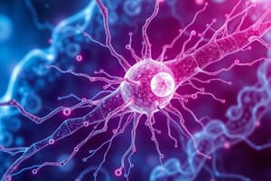
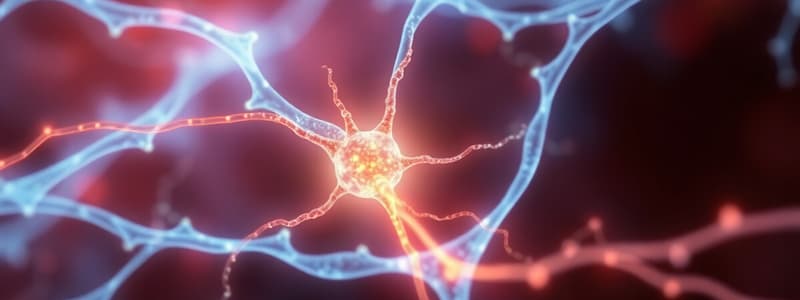
Myosins
- Myosins are actin-based motor proteins.
- All myosins have a characteristic motor domain that have ATPase activity.
- Different types of myosins are involved in different tasks, such as muscle contraction, cytokinesis, and cell migration.
- Type II myosins are best studied in muscle contraction.
- Conventional myosins (Type II) consist of two heavy chains, each with a globular head and a tail; two light chains are part of each heavy chain's head, forming a highly asymmetric structure.
- Myosin II assembly is into fibers with tails pointing toward the center and globular heads pointing outwards.
Muscle Organization and Contraction
- Muscle consists of parallel fibers (cells) joined by tendons.
- Muscle fibers are multinucleate cells formed during embryogenesis, specializing in contraction.
- Myofibrils are thinner cylindrical strands composing each muscle fiber.
- Myofibrils consist of repeating sarcomeres.
- A sarcomere includes the area between adjacent Z lines.
- Thin filaments (actin) extend from one Z line to the next, and thick filaments (myosin) are also found between adjacent Z lines.
- A sarcomere is composed of an A band (where thick filaments are present), and I bands (where thin filaments are located).
- The H zone is a region of the sarcomere where only thick filaments are present.
- The M line consists of proteins that connect adjacent thick filaments.
- Titin is a protein extending through the myosin filaments (thick filaments) and attaches to the Z line, preventing muscle tearing.
- Tropomyosin blocks myosin binding sites on actin.
- Troponin is a complex with three subunits (TnT, TnI, TnC) that regulates the interaction of troponin with tropomyosin and actin.
Calcium ions
- Calcium ions trigger muscle contraction by binding to troponin.
- This binding releases the tropomyosin blockage, allowing myosin to bind to actin, initiating contraction.
Other Myosin-Related Pathologies
Myosin mutations can cause diseases ranging from deafness to familial hypertrophic cardiomyopathy.
Contractile cycle
- Myosin head undergoes conformational changes driven by ATP hydrolysis.
- ATP binding causes the myosin head to detach from actin.
- Hydrolysis activates the myosin head for a new power stroke.
- The power stroke causes actin filaments to slide relative to each other.
- In the absence of ATP, rigor mortis occurs due to the inability for myosin heads to detach from actin filaments.
###Actin in endocytosis
- Actin plays a crucial role in endocytosis, including membrane invagination, pinching, and scission.
- Components are recycled and the process of endocytosis occurs with different phases.
Actin polymerization regulation
- Actin polymerization is regulated by several proteins, allowing for diverse cellular functions and complex organization.
Rho family GTPases
- Rho family of small GTPases regulate the actin cytoskeleton, influencing cellular functions like membrane remodeling and motility.
- GDI (guanine nucleotide dissociation inhibitor) protein associates with these GTPases and may affect their functions at the cellular membrane.
Studying That Suits You
Use AI to generate personalized quizzes and flashcards to suit your learning preferences.