Podcast
Questions and Answers
Which staining method is used to distinguish between different types of extracellular tissue components?
Which staining method is used to distinguish between different types of extracellular tissue components?
- Trichrome staining (correct)
- Periodic acid-Schiff (PAS) reaction
- Sudan black staining
- Hematoxylin and eosin (H&E) staining
What is the primary purpose of the Periodic acid-Schiff (PAS) reaction in histology?
What is the primary purpose of the Periodic acid-Schiff (PAS) reaction in histology?
- To stain lipids in tissue samples
- To stain nuclei and cytoplasm
- To visually enhance certain extracellular matrix fibers
- To stain carbohydrate-rich tissue structures (correct)
How does Sudan black staining differ from other histological staining techniques?
How does Sudan black staining differ from other histological staining techniques?
- It stains the nucleus blue and the cytoplasm pink.
- It uses a three-color system to emphasize support fibers in connective tissue.
- It stains lipids by avoiding the processing steps that remove lipids, such as heat and organic solvents. (correct)
- It uses silver salts to visually enhance specific cellular elements in nervous tissue.
What is the primary purpose of metal impregnation techniques in histology?
What is the primary purpose of metal impregnation techniques in histology?
Which staining method uses a three-color system to emphasize support fibers in connective tissue?
Which staining method uses a three-color system to emphasize support fibers in connective tissue?
What is the primary function of hematoxylin and eosin (H&E) staining in histology?
What is the primary function of hematoxylin and eosin (H&E) staining in histology?
What is the primary purpose of the pinhole aperture in a confocal microscope?
What is the primary purpose of the pinhole aperture in a confocal microscope?
Which of the following is a characteristic of basic dyes used in histology?
Which of the following is a characteristic of basic dyes used in histology?
Which of the following cell components would be stained by an acidic dye in histology?
Which of the following cell components would be stained by an acidic dye in histology?
How does the use of a small point of high-intensity light, often from a laser, in confocal microscopy improve resolution?
How does the use of a small point of high-intensity light, often from a laser, in confocal microscopy improve resolution?
What is the primary difference between the staining properties of basic and acidic dyes in histology?
What is the primary difference between the staining properties of basic and acidic dyes in histology?
How does the use of a confocal microscope improve the localization of specimen components compared to traditional fluorescence microscopy?
How does the use of a confocal microscope improve the localization of specimen components compared to traditional fluorescence microscopy?
Which type of microscopy allows for the visualization of thin optical sections within a thick specimen?
Which type of microscopy allows for the visualization of thin optical sections within a thick specimen?
Which staining technique is commonly used in fluorescence microscopy to visualize specific cellular components?
Which staining technique is commonly used in fluorescence microscopy to visualize specific cellular components?
Which type of dyes are generally used to stain acidic components in cells and tissues?
Which type of dyes are generally used to stain acidic components in cells and tissues?
Which cellular component is typically stained by the basic dye hematoxylin in histological preparations?
Which cellular component is typically stained by the basic dye hematoxylin in histological preparations?
Which staining technique is commonly used to visualize glycogen and mucopolysaccharides in tissues?
Which staining technique is commonly used to visualize glycogen and mucopolysaccharides in tissues?
What is the primary purpose of the fixation step in tissue preparation for light microscopy?
What is the primary purpose of the fixation step in tissue preparation for light microscopy?
Flashcards are hidden until you start studying
Study Notes
Staining and Stains
- Trichrome stains allow for greater distinctions among various extracellular tissue components, such as Masson trichrome.
- The periodic acid-Schiff (PAS) reaction utilizes the hexose rings of polysaccharides and other carbohydrate-rich tissue structures, staining them purple or magenta.
- Sudan black is a lipid-soluble dye that stains lipids, avoiding processing steps that remove lipids.
- Metal impregnation using solutions of silver salts can visualize specific ECM fibers and cellular elements in nervous tissue.
Commonly Used Histological Stains
- Hematoxylin and eosin (H&E): nucleus/blue, cytoplasm/pink.
- Van Gieson method: collagen/pink, muscle/yellow.
- Trichrome method: three-color system to emphasize support fibers: connective tissue/blue, cytoplasm/pink, nuclei/dark brown.
Tissue Types
- Epithelium
- Connective tissue
- Muscular tissue
- Nervous tissue
Electron Microscopy
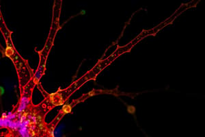
- Confocal microscopy achieves high resolution and sharp focus using a small point of high-intensity light and a plate with a pinhole aperture.
- The point light source, focal point of the lens, and detector's pinpoint aperture are optically conjugated.
Staining
- Most cells and extracellular material are completely colorless.
- Dyes form electrostatic (salt) linkages with ionizable radicals of macromolecules in tissues.
- Basic dyes (e.g., toluidine blue, alcian blue, methylene blue) stain basophilic components (e.g., nucleic acids).
- Acid dyes (e.g., eosin, orange G, acid fuchsin) stain acidophilic components (e.g., proteins).
Histology
- Histology is the study of the tissues of the body and how these tissues are arranged to constitute organs.
- Tissue is composed of cells and ECM (extracellular matrix).
- Levels of organization: tissue → organ → organism.
Tissue Processing for Histology
- Fixation: small pieces of tissue are placed in solutions that cross-link proteins and inactivate degradative enzymes.
- Dehydration: the tissue is transferred through a series of increasingly concentrated alcohol solutions, removing all water.
- Clearing: alcohol is removed in organic solvents in which both alcohol and paraffin are miscible.
- Infiltration: the tissue is placed in melted paraffin until it becomes completely infiltrated.
- Embedding: the paraffin-infiltrated tissue is placed in a small mold with melted paraffin and allowed to harden.
Studying That Suits You
Use AI to generate personalized quizzes and flashcards to suit your learning preferences.


