Podcast
Questions and Answers
Why are CT scans useful for detecting small changes in tissue type?
Why are CT scans useful for detecting small changes in tissue type?
CT scans have extremely good low contrast resolution, which allows for the detection of small changes in tissue type.
What does the acronym 'CT' stand for?
What does the acronym 'CT' stand for?
Computerized Axial Tomography
What is the function of a CT scanner?
What is the function of a CT scanner?
A CT scanner is an X-ray device capable of cross-sectional imaging, creating images of slices through the patient.
What is the primary difference between a CT scan and a conventional X-ray?
What is the primary difference between a CT scan and a conventional X-ray?
The intensity of X-ray beams remains constant as they travel through matter.
The intensity of X-ray beams remains constant as they travel through matter.
What does the term 'attenuation' refer to in the context of CT scans?
What does the term 'attenuation' refer to in the context of CT scans?
How are CT numbers related to X-ray attenuation?
How are CT numbers related to X-ray attenuation?
What is the Hounsfield unit (HU) scale used for, in the context of CT scans?
What is the Hounsfield unit (HU) scale used for, in the context of CT scans?
What is the general range of Hounsfield units (HU) in a CT scan?
What is the general range of Hounsfield units (HU) in a CT scan?
Match the following structures with their respective Hounsfield unit (HU) ranges:
Match the following structures with their respective Hounsfield unit (HU) ranges:
What is windowing in the context of CT scans?
What is windowing in the context of CT scans?
What does window width determine in CT image processing?
What does window width determine in CT image processing?
What are the two main components of a CT scanner?
What are the two main components of a CT scanner?
What is the typical aperture size of a CT scanner's gantry?
What is the typical aperture size of a CT scanner's gantry?
Flashcards
Computed Tomography
Computed Tomography
A medical imaging technique that uses X-rays and computer processing to create cross-sectional images (slices) of the body.
Tomography
Tomography
Imaging technique where an object is analyzed by examining its slices.
CT Scanner
CT Scanner
X-ray device capable of creating images of slices through the patient.
Conventional Radiography
Conventional Radiography
Signup and view all the flashcards
Why CT is Better
Why CT is Better
Signup and view all the flashcards
CT Image Matrix
CT Image Matrix
Signup and view all the flashcards
Voxel
Voxel
Signup and view all the flashcards
Pixel
Pixel
Signup and view all the flashcards
Field of View (FOV)
Field of View (FOV)
Signup and view all the flashcards
CT Pixel Size
CT Pixel Size
Signup and view all the flashcards
Voxel Size Dependencies
Voxel Size Dependencies
Signup and view all the flashcards
Attenuation
Attenuation
Signup and view all the flashcards
CT Numbers
CT Numbers
Signup and view all the flashcards
Hounsfield Units (HU)
Hounsfield Units (HU)
Signup and view all the flashcards
Windowing
Windowing
Signup and view all the flashcards
Narrow Window
Narrow Window
Signup and view all the flashcards
Wide Window
Wide Window
Signup and view all the flashcards
Window Level
Window Level
Signup and view all the flashcards
Window Width
Window Width
Signup and view all the flashcards
CT Scanner Components
CT Scanner Components
Signup and view all the flashcards
Gantry
Gantry
Signup and view all the flashcards
Patient Couch
Patient Couch
Signup and view all the flashcards
Gantry Characteristics
Gantry Characteristics
Signup and view all the flashcards
Tilting Range
Tilting Range
Signup and view all the flashcards
Aperture
Aperture
Signup and view all the flashcards
CT Image Acquisition
CT Image Acquisition
Signup and view all the flashcards
Image Reconstruction
Image Reconstruction
Signup and view all the flashcards
CT Scan Applications
CT Scan Applications
Signup and view all the flashcards
Study Notes
Computed Tomography (CT)
- CT scanners use X-rays and computer processing to create cross-sectional images (slices) of the body.
- CT was introduced in 1971, with a single detector, for brain studies.
- CT technology evolved with an increase in the number of detectors and a decrease in scan time.
- The technology is by AHMED JASEM ABASS (MSC of Medical Imaging).
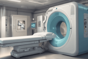
CT Scanners
- An X-ray device for cross-sectional imaging.
- It creates images of slices through the patient.
Why CT?
- Conventional radiography shows collapsing 3D structures as 2D images.
- CT has lower resolution, but excellent low-contrast resolution.
- Enables detecting small changes in tissue type.
- Provides accurate diagnostic information about the distribution of structures within the body.
Tomography
- Non-invasive medical imaging modality.
- Combines X-rays and computer processing to create tomographic slices.
- Tomos = slice; Graphein = to write.
- Definition: imaging of an object by analyzing its slices.
- Different types of CT include: Computerized Axial Tomography (CAT), Spiral CT, Multi-Slice CT.
Matrix
- The CT image is a matrix of numbers.
- Matrix: A two-dimensional array of numbers arranged in rows and columns.
- Each element in the matrix represents a 3D volume element (voxel).
- Voxel is represented as a 2D picture element (pixel).
- Field of view (FOV): The diameter of the body region being imaged.
- Pixel size in CT = FOV / (matrix size).
Pixel vs. Voxel
- Pixel size depends on matrix size and FOV.
- Voxel size depends on FOV, matrix size, and slice thickness.
Attenuation
- Attenuation: Reduction of X-ray beam intensity as it passes through matter.
- Attenuation is caused by absorption or deflection of photons.
- Factors affecting attenuation include beam energy and atomic number of the absorber.
CT Numbers
- CT numbers are values in the image matrix indicating X-ray attenuation.
- Different shades of gray represent varying CT numbers on a gray scale.
- Hounsfield Units (HU): Units for displaying CT numbers; Water = 0 HU.
- Range of CT numbers: approximately -1000 to +3000 HU. (Modern scanners can extend beyond 4000.)
Hounsfield Values (HU)
- Provides a standardized scale for converting CT numbers to gray shades.
- Different tissues have specific HU values. (e.g., air, lung, fat, water, soft tissue, bone).
Windowing
- Process of adjusting CT image display to enhance specific structures.
- Maps Hounsfield units to 256 gray shades.
- Wide window: Overview of structures, spans a broad range of HU values.
- Narrow window: Focusing on a specific structure, centers gray shades around a particular HU value, highlighting variations in tissue composition.
Windowing - Details
- Window level (WL): CT number for the center of the displayed range of numbers.
- Window width (WW): Total range of values selected for display.
- Narrow window enhances inherent contrast (distinguishes subtle differences).
- Window level influences brightness.
Imaging System
- Scanner: Contains the gantry, patient couch, and other components: X-ray tube, generator, filters, collimators, and detectors.
- Patient couch: 450 pounds (204 kg) weight limit, scannable range from head to thigh (162 cm).
Gantry Characteristics
- Tilting range: +/- 30 degrees.
- Aperture: 70 cm, on average.
Studying That Suits You
Use AI to generate personalized quizzes and flashcards to suit your learning preferences.