Podcast
Questions and Answers
What is the primary purpose of the oil surrounding the evacuated X-ray vessel?
What is the primary purpose of the oil surrounding the evacuated X-ray vessel?
- For cooling and electrical isolation (correct)
- To increase the X-ray output
- To enhance image quality
- To minimize radiation exposure
What is the range of potential difference applied between the anode and cathode during X-ray production?
What is the range of potential difference applied between the anode and cathode during X-ray production?
- 25 to 140 kV (correct)
- 50 to 200 kV
- 75 to 125 kV
- 10 to 50 kV
Why is tungsten the preferred metal for the anode in X-ray tubes?
Why is tungsten the preferred metal for the anode in X-ray tubes?
- It is cost-effective compared to other metals
- It has a low atomic number
- It possesses high thermal conductivity and a high atomic number (correct)
- It melts at low temperatures
What additional component is often used in tungsten anodes to enhance mechanical stability?
What additional component is often used in tungsten anodes to enhance mechanical stability?
Which of the following describes the role of the cathode in an X-ray tube?
Which of the following describes the role of the cathode in an X-ray tube?
What effect does a higher atomic number of the target metal have on X-ray production?
What effect does a higher atomic number of the target metal have on X-ray production?
How does the vacuum within the X-ray tube benefit the electron path?
How does the vacuum within the X-ray tube benefit the electron path?
What is the typical melting point of tungsten, making it suitable for high-temperature applications in X-ray tubes?
What is the typical melting point of tungsten, making it suitable for high-temperature applications in X-ray tubes?
What is the primary function of the cathode in an X-ray tube?
What is the primary function of the cathode in an X-ray tube?
Which component of the X-ray tube is responsible for converting electron energy into X-ray energy?
Which component of the X-ray tube is responsible for converting electron energy into X-ray energy?
Why is it important for the X-ray tube to maintain an interior vacuum?
Why is it important for the X-ray tube to maintain an interior vacuum?
What material is commonly used for the filament in the cathode of an X-ray tube?
What material is commonly used for the filament in the cathode of an X-ray tube?
What is the role of the focusing cup in the X-ray tube's cathode?
What is the role of the focusing cup in the X-ray tube's cathode?
Which of the following options does not describe a component of an X-ray tube?
Which of the following options does not describe a component of an X-ray tube?
What is the purpose of the tube envelope in an X-ray tube?
What is the purpose of the tube envelope in an X-ray tube?
Which statement about the X-ray energy spectrum generated by the tube is true?
Which statement about the X-ray energy spectrum generated by the tube is true?
Which material is used for the anode in digital mammography X-ray tubes?
Which material is used for the anode in digital mammography X-ray tubes?
What is the effect of decreasing the bevel angle of the anode?
What is the effect of decreasing the bevel angle of the anode?
What range of effective focal spot sizes is typically found for digital mammography?
What range of effective focal spot sizes is typically found for digital mammography?
What phenomenon describes the intensity variation of the X-ray beam between the cathode-end and anode-end?
What phenomenon describes the intensity variation of the X-ray beam between the cathode-end and anode-end?
Which of the following parameters does NOT affect the performance of an X-ray tube?
Which of the following parameters does NOT affect the performance of an X-ray tube?
What is the typical range for tube current (mA) in planar radiography?
What is the typical range for tube current (mA) in planar radiography?
Which mechanism for X-ray production is primarily responsible for producing a wide range of energies?
Which mechanism for X-ray production is primarily responsible for producing a wide range of energies?
What kilovoltage peak (kVp) is typically used for digital mammography applications?
What kilovoltage peak (kVp) is typically used for digital mammography applications?
Flashcards
X-ray Planar Radiography
X-ray Planar Radiography
A common medical imaging technique used to screen for injuries and diseases. It uses X-rays to create images of the inside body.
Bone Fracture Assessment
Bone Fracture Assessment
Using X-rays to determine the extent or severity of a bone fracture.
Lung Cancer/Emphysema Diagnosis
Lung Cancer/Emphysema Diagnosis
X-ray imaging can detect masses and changes associated with lung diseases like lung cancer or emphysema.
Kidney Stone Detection
Kidney Stone Detection
Signup and view all the flashcards
Gastrointestinal (GI) Diseases
Gastrointestinal (GI) Diseases
Signup and view all the flashcards
X-ray Tube
X-ray Tube
Signup and view all the flashcards
X-ray Tube Components
X-ray Tube Components
Signup and view all the flashcards
X-ray Imaging Contrast
X-ray Imaging Contrast
Signup and view all the flashcards
X-ray production
X-ray production
Signup and view all the flashcards
Cathode function
Cathode function
Signup and view all the flashcards
Accelerating voltage (kVp)
Accelerating voltage (kVp)
Signup and view all the flashcards
Anode material selection
Anode material selection
Signup and view all the flashcards
Tungsten selection
Tungsten selection
Signup and view all the flashcards
Tungsten-rhenium alloy
Tungsten-rhenium alloy
Signup and view all the flashcards
Oil (x-ray tube) purpose
Oil (x-ray tube) purpose
Signup and view all the flashcards
Digital Mammography X-ray Anode Material
Digital Mammography X-ray Anode Material
Signup and view all the flashcards
Anode Bevel Angle
Anode Bevel Angle
Signup and view all the flashcards
Effective Focal Spot Size
Effective Focal Spot Size
Signup and view all the flashcards
Heel Effect
Heel Effect
Signup and view all the flashcards
X-ray Imaging Parameters
X-ray Imaging Parameters
Signup and view all the flashcards
Tube Current Range
Tube Current Range
Signup and view all the flashcards
Kilovoltage Peak (kVp)
Kilovoltage Peak (kVp)
Signup and view all the flashcards
Bremsstrahlung X-rays
Bremsstrahlung X-rays
Signup and view all the flashcards
Study Notes
X-ray Imaging and Computed Tomography
- X-ray planar radiography is a key tool in radiology departments, providing initial screening for acute and chronic injuries/diseases.
- Planar radiography (general X-rays) are used to assess bone fractures in acute injuries, presence of masses in lung cancer/emphysema, airway pathologies, kidney stones, and gastrointestinal (GI) tract diseases.
- X-rays from a source are directed towards the patient, and those passing through are detected by a solid-state flat panel detector placed below the patient.
- Detected X-ray energy is converted into light, voltage, and then digitized.
- High contrast exists between bones (white, absorb X-rays) and lung tissue (dark, absorbs few X-rays).
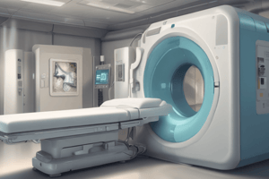
X-ray Tube
- X-ray source for both planar radiography and computed tomography (CT) is a specialized piece of equipment called an X-ray tube.
- X-ray tubes consist of an electron source (heated tungsten filament), a focusing cup (cathode), a target/anode, and an envelope to maintain a vacuum inside.
- Components are housed within an evacuated vessel surrounded by oil for cooling and electrical insulation, and a lead shield with a glass window for X-ray emission.
- The X-ray tube's target (anode) must be efficient at producing X-rays and resistant to high temperatures.
- Tungsten (atomic number 74) is the typical anode material due to high melting point (3370°C) and good thermal conductivity. A tungsten-rhenium alloy enhances mechanical stability.
- For applications like digital mammography, molybdenum is sometimes used in the anode, as it requires lower X-ray energies.
X-ray Beam
- The angle of the anode beveling affects the effective focal spot size (f) and coverage of the X-ray beam. Smaller bevel angles result in smaller focal spot sizes.
- Effective focal spot size ranges from 0.3 mm (digital mammography) to 0.6-1.2 mm (planar radiography/CT).
- Coverage is given by 2 x (source-patient distance) x tan(θ), where θ is the bevel angle.
- X-ray beam intensity is higher at the cathode end than the anode end (Heel effect). This effect is due to differences in distances X-rays travel through the target.
X-ray Imaging Parameters
- Three parameters for X-ray imaging are chosen by the operator: accelerating voltage (kVp), tube current (mA), and exposure time.
- Typical tube current for planar radiography is 50-400 mA, and for CT up to 1000 mA.
- kVp values vary from 25 kV (digital mammography) to ~140 kV (bone/chest).
- X-ray tubes produce X-rays with a range of energies, up to a maximum determined by the kVp.
X-ray Production Mechanisms
- X-ray production occurs through two mechanisms: Bremsstrahlung X-rays and characteristic X-rays.
- Bremsstrahlung X-rays are produced when electrons passing near atomic nuclei lose kinetic energy and emit X-rays.
- Characteristic X-rays are produced when an electron colliding with a tightly bound inner shell (e.g., K-shell) electron ejects it and an outer shell electron fills the vacancy, releasing the difference in energy as an X-ray.
Studying That Suits You
Use AI to generate personalized quizzes and flashcards to suit your learning preferences.