Podcast
Questions and Answers
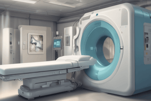
What is a CT scanner?
What is a CT scanner?
An X-ray device capable of cross-sectional imaging.
What does CAT stand for?
What does CAT stand for?
- Computed Axial Tomography (correct)
- Computerized Automated Tomography
- Computerized Artificial Tomography
- Cross-sectional Axial Tomography
CT imaging provides a better representation of 3D structures compared to conventional radiography.
CT imaging provides a better representation of 3D structures compared to conventional radiography.
False (B)
A matrix is a ______ of numbers arranged in rows and columns.
A matrix is a ______ of numbers arranged in rows and columns.
What does Voxel stand for?
What does Voxel stand for?
What is the purpose of windowing in CT imaging?
What is the purpose of windowing in CT imaging?
What are Hounsfield units (HU)?
What are Hounsfield units (HU)?
CT imaging can detect very small changes in tissue type.
CT imaging can detect very small changes in tissue type.
In CT imaging, what does a pixel represent?
In CT imaging, what does a pixel represent?
Flashcards are hidden until you start studying
Study Notes
Computed Tomography
- CT scanners were first introduced in 1971 with a single detector
- The first CT scanner was used for brain studies
- CT scanners underwent multiple improvements with an increase of detectors and a decrease in scan time
What is CT?
- A device that uses X-rays to create cross-sectional images of the body
- Creates images of slices through the patient
Why use CT?
- It provides accurate diagnostic information
- It overcomes the limitations of conventional radiography, which suffers from collapsing of 3D structures into a 2D image
- Although the resolution of CT is lower, it has extremely good low contrast resolution
Tomography
- A medical imaging technique that combines X-rays and computer processing to generate tomographic slices of the scanned area
- Tomos means slice and Graphein means to write
- Defined as the imaging of an object by analyzing its slices
CT Scanner Types
- CAT: Computerized Axial Tomography
- Spiral CT
- Multi-Slice CT or Multi-Detector
CT Matrix
- The image is represented as a matrix of numbers
- A matrix is a two-dimensional array of numbers arranged in rows and columns
- Each element in the image matrix represents a three-dimensional volume element called a voxel
- The voxel is represented on the image as a two-dimensional element called a pixel (picture element)
Field of View (FOV)
- The diameter of the area being imaged
- For example, 25 cm for a head and 40 cm for an abdomen
- CT pixel size is determined by dividing the FOV by the matrix size, which is generally 512 x 512 in CT
Pixel vs Voxel
- Pixel size depends on:
- Matrix size
- FOV
- Voxel size depends on:
- FOV
- Matrix size
- Slice thickness
Attenuation
- The reduction of the intensity of the X-ray beam as it passes through matter
- It can be caused by absorption or deflection of photons
- Different factors affect attenuation, such as the beam energy and atomic number of the absorber
CT Numbers
- The numbers in the image matrix
- Each pixel has a number that represents the X-ray attenuation in the corresponding voxel
- These numbers are assigned different shades of gray on a gray scale to obtain a visual image
- Each shade of gray represents the X-ray attenuation within the corresponding voxel
- CT numbers are compared to the attenuation value of water and displayed on a scale of units named Hounsfield units (HU)
- The range of CT numbers is 2000 HU wide, although some modern scanners have a greater range up to 4000
- Each number represents a shade of gray with +1000 (white) and –1000 (black) at either end of the spectrum
Windowing
- The process of using calculated Hounsfield units to make an image
- The various radio density amplitudes are mapped to 256 shades of gray
- These shades of gray can be distributed over a wide range of HU values or a narrow range
- A narrow window is commonly used for evaluation of structures in the image known as contrast compression
Windowing Example
- To evaluate the abdomen, a liver window can be used
- A liver window would have an average of 70 HU, and would extend from -15 HU to +155 HU
- 70 HU is the average HU value for the liver
- The shades of gray are distributed over a narrow range of HU values, allowing for variations in liver tissue to be identified
Studying That Suits You
Use AI to generate personalized quizzes and flashcards to suit your learning preferences.


