Podcast
Questions and Answers
Which structure is NOT considered a primary part of the central nervous system?
Which structure is NOT considered a primary part of the central nervous system?
- Brain stem
- Cerebellum
- Peripheral nerves (correct)
- Cerebrum
The Corpus callosum facilitates communication between what?
The Corpus callosum facilitates communication between what?
- The cerebrum and cerebellum
- The left and right cerebral hemispheres (correct)
- The brain and spinal cord
- The brain stem and diencephalon
Which part of the brain is most associated with coordination and balance?
Which part of the brain is most associated with coordination and balance?
- Spinal cord
- Brain stem
- Cerebrum
- Cerebellum (correct)
The point where the brain connects to the spinal cord is called what?
The point where the brain connects to the spinal cord is called what?
Which term describes structures that appear as ridges on the surface of the brain?
Which term describes structures that appear as ridges on the surface of the brain?
What is the primary component of white matter in the brain?
What is the primary component of white matter in the brain?
Which direction aligns with 'anterior' in most parts of the brain, due to the neural tube's curvature?
Which direction aligns with 'anterior' in most parts of the brain, due to the neural tube's curvature?
Which term describes the direction toward the back of the head?
Which term describes the direction toward the back of the head?
The ventral direction can also be referred to as?
The ventral direction can also be referred to as?
Which structure develops from the prosencephalon?
Which structure develops from the prosencephalon?
The cerebral aqueduct connects which two structures?
The cerebral aqueduct connects which two structures?
From which primary vesicle does the medulla oblongata develop?
From which primary vesicle does the medulla oblongata develop?
The fourth ventricle is located within what?
The fourth ventricle is located within what?
What is the tentorium cerebelli?
What is the tentorium cerebelli?
Which of the following is a space between the dura mater layers that collects venous blood and CSF?
Which of the following is a space between the dura mater layers that collects venous blood and CSF?
Which structure is responsible for draining CSF into the internal jugular vein?
Which structure is responsible for draining CSF into the internal jugular vein?
Which sinus runs along the inferior border of the falx cerebri?
Which sinus runs along the inferior border of the falx cerebri?
The straight sinus is formed by the convergence of which two sinuses?
The straight sinus is formed by the convergence of which two sinuses?
What is a characteristic feature of the arachnoid mater?
What is a characteristic feature of the arachnoid mater?
Subarachnoid cisterns are formed because the arachnoid mater does not extend into what structures?
Subarachnoid cisterns are formed because the arachnoid mater does not extend into what structures?
Which artery supplies blood to the anterior portion of the arterial circle of Willis?
Which artery supplies blood to the anterior portion of the arterial circle of Willis?
Which arteries unite to form the basilar artery?
Which arteries unite to form the basilar artery?
What area does the posterior inferior cerebellar artery (PICA) supply?
What area does the posterior inferior cerebellar artery (PICA) supply?
The anterior spinal artery supplies which structure?
The anterior spinal artery supplies which structure?
The largest cranial nerve is the what?
The largest cranial nerve is the what?
Which cranial nerve exits from the dorsal part of the brainstem?
Which cranial nerve exits from the dorsal part of the brainstem?
Which cranial nerve is responsible for controlling the muscles of facial expression?
Which cranial nerve is responsible for controlling the muscles of facial expression?
The trochlear nerve innervates which of the following muscles?
The trochlear nerve innervates which of the following muscles?
The central sulcus separates which lobes?
The central sulcus separates which lobes?
Which sulcus is often used as a landmark for identifying the precentral and postcentral gyri?
Which sulcus is often used as a landmark for identifying the precentral and postcentral gyri?
What is located anterior to the central sulcus?
What is located anterior to the central sulcus?
One of the defining characteristics of the central sulcus is that it opens slightly where?
One of the defining characteristics of the central sulcus is that it opens slightly where?
Which gyrus is bounded anteriorly by the central sulcus and contains the primary somatosensory cortex?
Which gyrus is bounded anteriorly by the central sulcus and contains the primary somatosensory cortex?
The cell bodies of most lower motor neurons are located in which of the following areas of the spinal cord?
The cell bodies of most lower motor neurons are located in which of the following areas of the spinal cord?
The parieto-occipital sulcus and calcarine sulcus help to define which structure?
The parieto-occipital sulcus and calcarine sulcus help to define which structure?
What does the Fornix connect the hippocampus to?
What does the Fornix connect the hippocampus to?
Which of the following layers of the cerebral cortex forms the inner surface of structures?
Which of the following layers of the cerebral cortex forms the inner surface of structures?
A patient presents with damage in the medulla and has lost motor control of all of his muscles, but can move his eyes. What is the cause?
A patient presents with damage in the medulla and has lost motor control of all of his muscles, but can move his eyes. What is the cause?
Flashcards
What is the Cerebrum?
What is the Cerebrum?
The largest part of the brain, divided into two hemispheres.
What is the Cerebellum?
What is the Cerebellum?
Located behind and beneath the cerebrum, responsible for coordination and balance.
What is the Brain Stem?
What is the Brain Stem?
Connects the brain to the spinal cord; controls basic life functions.
What is the Spinal Cord?
What is the Spinal Cord?
Signup and view all the flashcards
What are the Meninges?
What are the Meninges?
Signup and view all the flashcards
What is the Corpus Callosum?
What is the Corpus Callosum?
Signup and view all the flashcards
What are Gyri?
What are Gyri?
Signup and view all the flashcards
What are Sulci?
What are Sulci?
Signup and view all the flashcards
What is Rostral?
What is Rostral?
Signup and view all the flashcards
What is Caudal?
What is Caudal?
Signup and view all the flashcards
What is Ventral?
What is Ventral?
Signup and view all the flashcards
What is Dorsal?
What is Dorsal?
Signup and view all the flashcards
What is the Subdural Space?
What is the Subdural Space?
Signup and view all the flashcards
What is the Subarachnoid Space?
What is the Subarachnoid Space?
Signup and view all the flashcards
What is the Dura Mater?
What is the Dura Mater?
Signup and view all the flashcards
What is the Arachnoid Mater?
What is the Arachnoid Mater?
Signup and view all the flashcards
What is the Pia Mater?
What is the Pia Mater?
Signup and view all the flashcards
What is the Dura folds?
What is the Dura folds?
Signup and view all the flashcards
What is the Tentorium cerebelli?
What is the Tentorium cerebelli?
Signup and view all the flashcards
What is the Superior Sagittal Sinus?
What is the Superior Sagittal Sinus?
Signup and view all the flashcards
What is the Inferior Sagittal Sinus?
What is the Inferior Sagittal Sinus?
Signup and view all the flashcards
What is the Straight Sinus?
What is the Straight Sinus?
Signup and view all the flashcards
What are Transverse Sinuses?
What are Transverse Sinuses?
Signup and view all the flashcards
What are the carotid arteries?
What are the carotid arteries?
Signup and view all the flashcards
What are vertebral arteries?
What are vertebral arteries?
Signup and view all the flashcards
What is the Circle of Willis?
What is the Circle of Willis?
Signup and view all the flashcards
What are the Anterior and posterior inferior cerebellar arteries?
What are the Anterior and posterior inferior cerebellar arteries?
Signup and view all the flashcards
What is the Superior cerebellar artery?
What is the Superior cerebellar artery?
Signup and view all the flashcards
What is the Anterior cerebral artery?
What is the Anterior cerebral artery?
Signup and view all the flashcards
What is the Middle cerebral artery?
What is the Middle cerebral artery?
Signup and view all the flashcards
What are cranial nerves?
What are cranial nerves?
Signup and view all the flashcards
What is the Olfactory nerve?
What is the Olfactory nerve?
Signup and view all the flashcards
What is the Optic Nerve?
What is the Optic Nerve?
Signup and view all the flashcards
What is the Oculomotor Nerve?
What is the Oculomotor Nerve?
Signup and view all the flashcards
What is the Trochlear Nerve?
What is the Trochlear Nerve?
Signup and view all the flashcards
What is the Trigeminal Nerve?
What is the Trigeminal Nerve?
Signup and view all the flashcards
What is the Abducens Nerve?
What is the Abducens Nerve?
Signup and view all the flashcards
What is the Facial Nerve?
What is the Facial Nerve?
Signup and view all the flashcards
What is the Vestibulocochlear Nerve?
What is the Vestibulocochlear Nerve?
Signup and view all the flashcards
Study Notes
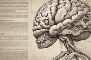
General Structure
- The central nervous system has several main structures
- These include the cerebrum, cerebellum, brain stem, and spinal cord
Cerebrum
- The cerebrum, or large brain, divides into right and left hemispheres
- The left hemisphere is usually dominant, controlling language and speech (90% of right-handed and 60-70% of left-handed people)
- The right hemisphere relates to spatial vision, musical talent and drawing
- The corpus callosum, a network of fibers, connects the hemispheres
- The brain has two tones, reflecting white versus gray matter
- Gray matter contains neuron bodies
- White matter contains long, myelinated extensions
Cerebellum
- The cerebellum, or small brain, sits behind and below the cerebrum and is related to coordination and balance
Brain Stem
- The brain stem connects the brain to the spinal cord
- It consists of three parts to be discussed
Spinal Cord
- The spinal cord extends through the segments of the spinal column to vertebra L2
Gyri and Sulci
- The brain has ridges (gyri) and grooves (sulci)
Directions in the Brain
- All standard directional terms apply, but there are also specific terms
- Rostral: toward the “beak," or nose or forehead; superiorly and anteriorly
- Caudal: toward the tail; inferiorly and posteriorly
- Ventral: toward the belly; usually anteriorly; inferiorly in the brain
- Dorsal: toward the back; usually posteriorly; superiorly in the brain
Sections
- Coronal or frontal, horizontal or transverse and midsagittal
Embryology
- Brain and spinal cord develop from a hollow tube sealed at both ends and fluid-filled
- The caudal part becomes the spinal cord
- Its walls thicken, shrinking the tube, which becomes the central canal of spinal cord
- The rostral part of the tube develops differently
- Walls thicken, interior expands and then divides into three parts
Prosencephalon
- Further divides into two parts
- The upper part is called the telencephalon
- The lower part is called the diencephalon
- Two sides of the telencephalon sprout two bulges that grow and extend down almost covering the diencephalon from two sides
- These create the cerebral hemispheres, or right and left hemispheres
- Ventricles of the brain then form
Diencephalon
- As the diencephalon differentiates, its walls thicken and form two crucial nuclei or right and left thalami
- A third ventricle is formed between these nuclei
Midbrain
- The midbrain stays in its original form with a cerebral aqueduct
Rhombencephalon
- The rhombencephalon develops into two parts
- The upper part or metencephalon becomes the pons
- Also becomes cerebellum
- It closely resembles pons in structure but smaller without any cavity
- The lower part or myelencephalon forms the medulla oblongata, which is contiguous with the spinal cord
- This makes up the fourth ventricle
Meninges
- The brain is wrapped in three membranes, from outer to inner: dura mater, arachnoid mater, and pia mater
- Dura mater usually remains attached to the skull and creates folds
- Falx cerebri: between the cerebral hemispheres within the longitudinal fissure
- Tentotium cerebelli: Separates the cerebellum and the cerebrum
- The two layers separate, and sinuses are formed, draining veins of the brain, and CSF
- Super sagittal sinus: Along the superior boarder
- Interior sagittal sinus: Boarding the bottom
- Straight sinus: At bottom and Falx cerebri
- Transverse sinus : Side
Layers of Membranes
- Arachnoid mater is a delicate, thin and transparent layer detached
- Separates dura mater with Subdural space and Pia Mater
- Trabeculae connect the two named after spider webs and are the Arachnoid
- In addition Subarachnoid space flows CSF then drains to structures,
- Meningeal structure is age Arachnoid villi at a more advanced
- Blood vessels can be seen in the pia mater Subarachnoid
Key cistern
- cisterna magna is main point in the inferior view and is a Cerebellomedullary cistern
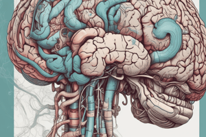
Vessels In The Brain
- Review the map and follow the time through the various tools
- Pay special attention in particular the lobe where the lateral structure
- All temporal and general brain topography will be covered
Veins in brain
- Superficial Middle cerebral Vein flows along the lateral structure, and links to Superior structure, with Superior anastomosis vein-
- Innumerable veins that are connected in all veins with Cerebelar and Superficial veins make for Inferior structure
- Deep Cerebral cord flows through deep veins for Inferior views to get the deep Middle Cerebelar Vein and the Anterior structure connected to Basal Vein to get to Galen
Brain arteries
- In the chest, one to three from the aorta
- From the Internal Catorid arteries -Subclavian
Internal -Arteries
- Right,Left Arterial - Circle allows blood despite blockage Internal Middle Posterior
- 4 splitings
Basilar
- Artery smaller and connects through Pons laterally to the structure cerebalus where it is part of the PICA- smaller areries that connect on Cerebal cord
- Pontine Arteries then Superior connecting all Cerebal Artery
Internal carotid arteries
- Posterior connecting cord the internal -Arteries Small then all parts- connecting with the Pericallosal/Callosonmarging vessel which then creates the cycle
Overview
- Review the previous map and get a better understanding
Cranial Nerves
- Exit cranium and Lead motor nerves from the brain, to muscles and carry sense information to all parts are associated to cranium
- Nerves lead to different areas -Sensory -Motor
CN1- Olfactory Nerve
- Related to smell from bulbs with fibers leading into tract
- Then is when Lateral
CN2- Optic Nerve
- Associated with sight From nerves that reach with Optic Chiasma and tract leading to area of Central nervous system
CN3- CN5
- Oculomotor is cranial Motor, Trochlear related to muscles
- Tigeminal is Large connected to pons to interior area
CN6 and CN7
- Abducens to muscle from side of border to pons
CN8
- Vestibulocochlear tied to all other facial areas. that are connected to the hearing system
CN9-CN11
- Glossopharynygeal to oral area
- CN9 connected from head
- Nerve accesory
CN12: Hypoglossal
- Connected to language all are all linked in area
Studying That Suits You
Use AI to generate personalized quizzes and flashcards to suit your learning preferences.