Podcast
Questions and Answers
How does the amphipathic nature of phospholipids contribute to the selective permeability of the cell membrane?
How does the amphipathic nature of phospholipids contribute to the selective permeability of the cell membrane?
The hydrophobic tails form a barrier to charged or polar molecules, restricting their passage, while the hydrophilic heads interact with water, allowing small polar molecules to pass.
Explain how the Na+/K+ ATPase contributes to maintaining cellular integrity, and what type of transport does it utilize?
Explain how the Na+/K+ ATPase contributes to maintaining cellular integrity, and what type of transport does it utilize?
It maintains the electrochemical gradient necessary for cell volume, secondary transport, and nerve and muscle function. This pump uses active transport.
Describe how microfilaments and microtubules contribute differently to cell movement and structural support?
Describe how microfilaments and microtubules contribute differently to cell movement and structural support?
Microfilaments, made of actin, support cell shape and movement via interactions with motor proteins like myosin. Microtubules, composed of tubulin, provide tracks for intracellular transport and contribute to the structure of cilia and flagella, enabling movement.
How does cholesterol affect membrane fluidity at different temperatures?
How does cholesterol affect membrane fluidity at different temperatures?
Describe the role of the cell membrane in cell signaling. Please give an example.
Describe the role of the cell membrane in cell signaling. Please give an example.
Briefly explain how co-transport mechanisms utilize existing ion gradients to facilitate the movement of other molecules across the cell membrane. Provide an example.
Briefly explain how co-transport mechanisms utilize existing ion gradients to facilitate the movement of other molecules across the cell membrane. Provide an example.
Describe the key structural features common to most cell membrane receptors that bind extracellular signals, and briefly explain how these features contribute to signal transduction.
Describe the key structural features common to most cell membrane receptors that bind extracellular signals, and briefly explain how these features contribute to signal transduction.
In the context of cellular function, explain why it is advantageous for organelles to be enclosed by membranes rather than being 'open' to the cytosol.
In the context of cellular function, explain why it is advantageous for organelles to be enclosed by membranes rather than being 'open' to the cytosol.
Differentiate between co-transport and counter-transport, highlighting the directional movement of molecules and the energy source driving each process. Provide an example of counter-transport.
Differentiate between co-transport and counter-transport, highlighting the directional movement of molecules and the energy source driving each process. Provide an example of counter-transport.
Explain how a cell's response to a specific extracellular signal is determined, even when multiple cell types are exposed to the same signal, and give an example.
Explain how a cell's response to a specific extracellular signal is determined, even when multiple cell types are exposed to the same signal, and give an example.
Describe how the high concentration of sodium ions ([Na+]) in the extracellular fluid (ECF) contributes to both passive and active transport mechanisms in cells.
Describe how the high concentration of sodium ions ([Na+]) in the extracellular fluid (ECF) contributes to both passive and active transport mechanisms in cells.
Explain the difference between facilitated transport and co-transport, highlighting the role of membrane proteins in each process.
Explain the difference between facilitated transport and co-transport, highlighting the role of membrane proteins in each process.
Consider a cell that needs to import glucose against its concentration gradient. Describe a mechanism involving both active and passive transport that could accomplish this.
Consider a cell that needs to import glucose against its concentration gradient. Describe a mechanism involving both active and passive transport that could accomplish this.
If a drug inhibits the function of the Na+/K+ ATPase, how might this indirectly affect the transport of other molecules into the cell? Give one specific example.
If a drug inhibits the function of the Na+/K+ ATPase, how might this indirectly affect the transport of other molecules into the cell? Give one specific example.
Explain why small hydrophobic molecules can diffuse directly across the cell membrane, while larger or charged molecules require the assistance of membrane proteins.
Explain why small hydrophobic molecules can diffuse directly across the cell membrane, while larger or charged molecules require the assistance of membrane proteins.
Describe how the semi-permeable nature of cell membranes contributes to the necessity of energy expenditure by the cell.
Describe how the semi-permeable nature of cell membranes contributes to the necessity of energy expenditure by the cell.
How does the Na+/K+ ATPase contribute to preventing cell swelling due to osmosis?
How does the Na+/K+ ATPase contribute to preventing cell swelling due to osmosis?
Explain how the sodium gradient, established by the Na+/K+ ATPase, can be used to transport other substances across the cell membrane.
Explain how the sodium gradient, established by the Na+/K+ ATPase, can be used to transport other substances across the cell membrane.
What would happen to the concentration gradient of sodium and potassium ions if a cell's ATP production was significantly reduced? Why?
What would happen to the concentration gradient of sodium and potassium ions if a cell's ATP production was significantly reduced? Why?
If a cell suddenly loses its ability to produce ATP, explain why the cell starts to swell.
If a cell suddenly loses its ability to produce ATP, explain why the cell starts to swell.
Describe the significance of aquaporins in maintaining cellular homeostasis, and explain how they function in conjunction with osmosis.
Describe the significance of aquaporins in maintaining cellular homeostasis, and explain how they function in conjunction with osmosis.
How would a drug that inhibits aquaporin function affect cells in a hypotonic solution?
How would a drug that inhibits aquaporin function affect cells in a hypotonic solution?
Explain how the Na+/K+ ATPase contributes to establishing a gradient of charge across the cell membrane, and why this is important for the cell.
Explain how the Na+/K+ ATPase contributes to establishing a gradient of charge across the cell membrane, and why this is important for the cell.
How do microtubules contribute to cellular organization, and what is the role of the MTOC in this process?
How do microtubules contribute to cellular organization, and what is the role of the MTOC in this process?
Describe how dyneins and kinesins facilitate cellular movement using microtubules, and what is the key difference in their movement?
Describe how dyneins and kinesins facilitate cellular movement using microtubules, and what is the key difference in their movement?
What are the structural differences between centrioles and the microtubules that radiate from the MTOC?
What are the structural differences between centrioles and the microtubules that radiate from the MTOC?
Explain how the dynamic instability of F-actin and microtubules is related to the function of molecular motors like myosin, dynein and kinesin.
Explain how the dynamic instability of F-actin and microtubules is related to the function of molecular motors like myosin, dynein and kinesin.
How does the structure of intermediate filaments contribute to their stability, and why is this stability important for cells?
How does the structure of intermediate filaments contribute to their stability, and why is this stability important for cells?
Describe the role of the primary cilium in cellular signaling and its importance for cellular function or localization.
Describe the role of the primary cilium in cellular signaling and its importance for cellular function or localization.
How do keratins contribute to the properties of epithelial cells, hair, and nails?
How do keratins contribute to the properties of epithelial cells, hair, and nails?
Explain the distinct roles of microtubules during cell division.
Explain the distinct roles of microtubules during cell division.
What characteristics differentiate intermediate filaments from microfilaments and microtubules, in terms of protein structure and energy requirements?
What characteristics differentiate intermediate filaments from microfilaments and microtubules, in terms of protein structure and energy requirements?
Describe the function of lamins and their location within the cell.
Describe the function of lamins and their location within the cell.
How do tight junctions contribute to the compartmentalization of tissues, and what is the significance of their varying selectivity?
How do tight junctions contribute to the compartmentalization of tissues, and what is the significance of their varying selectivity?
Describe the roles of cadherins and intermediate filaments in the structural integrity provided by desmosomes.
Describe the roles of cadherins and intermediate filaments in the structural integrity provided by desmosomes.
How do hemidesmosomes differ from desmosomes in terms of their extracellular components and the structures they connect?
How do hemidesmosomes differ from desmosomes in terms of their extracellular components and the structures they connect?
Adherens junctions can connect to another cell via _______, or to a basement membrane via _______?
Adherens junctions can connect to another cell via _______, or to a basement membrane via _______?
Explain how the cytoskeleton contributes to both cellular movement and the organization of intracellular components.
Explain how the cytoskeleton contributes to both cellular movement and the organization of intracellular components.
Describe the roles of microtubules, microfilaments, and intermediate filaments in maintaining the overall structural integrity of a cell.
Describe the roles of microtubules, microfilaments, and intermediate filaments in maintaining the overall structural integrity of a cell.
How does the dynamic nature of the cytoskeleton allow cells to respond to changing environmental conditions or signals?
How does the dynamic nature of the cytoskeleton allow cells to respond to changing environmental conditions or signals?
Explain how the regulation of actin filament assembly and disassembly can influence cell shape and movement.
Explain how the regulation of actin filament assembly and disassembly can influence cell shape and movement.
Actin monomers, known as _______, polymerize to form _______.
Actin monomers, known as _______, polymerize to form _______.
Describe how ATP hydrolysis affects the stability of F-actin filaments and the likelihood of G-actin monomers dissociating from the filament.
Describe how ATP hydrolysis affects the stability of F-actin filaments and the likelihood of G-actin monomers dissociating from the filament.
How do capping proteins influence the stability of actin filaments, and what effect does this have on cell behavior?
How do capping proteins influence the stability of actin filaments, and what effect does this have on cell behavior?
How do the organization and stability of actin filaments differ in muscle cells compared to fibroblasts, and why are these differences important for their respective functions?
How do the organization and stability of actin filaments differ in muscle cells compared to fibroblasts, and why are these differences important for their respective functions?
What are the two types of tubulin that form dimers?
What are the two types of tubulin that form dimers?
How does GTP hydrolysis affect the stability of microtubules, and what is this phenomenon known as?
How does GTP hydrolysis affect the stability of microtubules, and what is this phenomenon known as?
How does dynamic instability contribute to the diverse functions of microtubules in cellular processes?
How does dynamic instability contribute to the diverse functions of microtubules in cellular processes?
Flashcards
Phospholipid Bilayer
Phospholipid Bilayer
A double layer of phospholipids forming the cell membrane's structural foundation, providing barrier and fluidity.
Membrane Proteins
Membrane Proteins
Proteins embedded in the cell membrane serving various functions like transport and signaling.
Na+/K+ ATPase
Na+/K+ ATPase
An enzyme that uses ATP to pump sodium out and potassium into the cell, maintaining cellular integrity.
Diffusion
Diffusion
Signup and view all the flashcards
Cytoskeleton
Cytoskeleton
Signup and view all the flashcards
Active Transport
Active Transport
Signup and view all the flashcards
Passive Transport
Passive Transport
Signup and view all the flashcards
Facilitated Transport
Facilitated Transport
Signup and view all the flashcards
Co-Transport
Co-Transport
Signup and view all the flashcards
Ion Channels
Ion Channels
Signup and view all the flashcards
Counter-transport
Counter-transport
Signup and view all the flashcards
Receptor signaling
Receptor signaling
Signup and view all the flashcards
Hydrophobic domains
Hydrophobic domains
Signup and view all the flashcards
Membrane protein functions
Membrane protein functions
Signup and view all the flashcards
Tight Junctions
Tight Junctions
Signup and view all the flashcards
Anchoring Junction - Desmosome
Anchoring Junction - Desmosome
Signup and view all the flashcards
Hemi-desmosome
Hemi-desmosome
Signup and view all the flashcards
Adherens Junction
Adherens Junction
Signup and view all the flashcards
Cytoskeleton Functions
Cytoskeleton Functions
Signup and view all the flashcards
Microtubules
Microtubules
Signup and view all the flashcards
Microfilaments
Microfilaments
Signup and view all the flashcards
Intermediate Filaments
Intermediate Filaments
Signup and view all the flashcards
Dynamic Filament Assembly
Dynamic Filament Assembly
Signup and view all the flashcards
G-actin vs F-actin
G-actin vs F-actin
Signup and view all the flashcards
Dynamic Instability of Microtubules
Dynamic Instability of Microtubules
Signup and view all the flashcards
Integrins
Integrins
Signup and view all the flashcards
Cadherins
Cadherins
Signup and view all the flashcards
Basement Membrane
Basement Membrane
Signup and view all the flashcards
Microtubule Organizing Centre (MTOC)
Microtubule Organizing Centre (MTOC)
Signup and view all the flashcards
Centrioles
Centrioles
Signup and view all the flashcards
Tubulin Triplet Structure
Tubulin Triplet Structure
Signup and view all the flashcards
Cilia and Flagella
Cilia and Flagella
Signup and view all the flashcards
Cell Division
Cell Division
Signup and view all the flashcards
Primary Cilium
Primary Cilium
Signup and view all the flashcards
F-actin
F-actin
Signup and view all the flashcards
Molecular Motors
Molecular Motors
Signup and view all the flashcards
Types of Intermediate Filaments
Types of Intermediate Filaments
Signup and view all the flashcards
Osmosis
Osmosis
Signup and view all the flashcards
Semi-permeable membrane
Semi-permeable membrane
Signup and view all the flashcards
Cellular swelling prevention
Cellular swelling prevention
Signup and view all the flashcards
Aquaporins
Aquaporins
Signup and view all the flashcards
Concentration gradients
Concentration gradients
Signup and view all the flashcards
Study Notes
Physiology 1.04 - Foundational Physiology Cell Membranes in Physiology
- This course covers foundational cell membrane concepts within physiology.
- The course is part of BMS 100, a broader study on specific modules within physiology.
- The Canadian College of Naturopathic Medicine (CCNM) is the provider of this course material.
Learning Outcomes
- Students will understand the structural components of cell membranes including phospholipids, cholesterol, sphingolipids, and membrane proteins.
- They will examine the basic structure of the phospholipid bilayer and its function as a molecular barrier for homeostasis.
- Students will identify different types of transport across the plasma membrane, focusing on diffusion, osmosis, and the Na+/K+ ATPase's role in maintaining cellular integrity.
- The function of the plasma membrane in cell signaling will also be analyzed.
- Students will understand the cytoskeleton, including detailed descriptions of microfilaments, microtubules, and intermediate filaments.
- Cellular motors associated with F-actin and microtubules are to be described.
Overview - Cell Membranes
- Key lipid components of cell membranes include phospholipids, cholesterol, and sphingolipids.
- Types of membrane proteins and their function are examined.
- Comparisons of the plasma membrane and the endomembrane system are made for a deeper understanding.
- Cytoskeleton components (microfilaments, microtubules, and intermediate filaments) are a key concept.
- Functions of cell membranes, including molecular partitioning, homeostasis, transport, movement, cellular integrity, and cell signaling, are covered.
The Cell Membrane - General Structure
- The cell membrane's components, such as glycoproteins, glycolipids, peripheral membrane proteins, integral membrane proteins, cholesterol, and channel proteins, each contribute to its unique function.
- Diagram of a typical cell membrane showing these components is presented.
Lipid Components
- Glycerophospholipids: Phosphatidylcholine, phosphatidylethanolamine, phosphatidylserine, and phosphatidylinositol are the most common glycerophospholipids in the cell membrane. Phosphatidylinositol plays a role in signaling.
- Sphingolipids: Slightly differing shape compared to glycerophospholipids, and sphingolipids can decrease membrane fluidity. They are involved in cellular signaling and the formation of lipid rafts and myelin. Contribution to glycocalyx.
- Glycocalyx: Carbohydrates covalently attached to protein and lipid components, which are more prominent on the outer leaflet of the cell membrane. Key roles include protective structures, structural interactions for signaling.
Cholesterol
- Cholesterol, belonging to the isoprenoid class of lipids, intercalates between phospholipids.
- It helps stabilize membrane fluidity.
Structure and Function of Membrane Lipids
- The amphipathic nature of lipids in cell membranes leads to bilayer formation.
- Hydrophobic tails interact with one another, while hydrophilic heads interact with the aqueous environment.
- The phospholipid bilayer functions as a barrier for certain molecules.
- The integrity of the plasma membrane is essential for cell survival and function, including maintaining homeostasis, keeping vital molecules within the cell, and cellular movement.
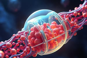
Importance of the Na+/K+ ATPase
- Na+/K+ ATPase is a critical plasma membrane transporter.
- Its operation involves 3 sodium ions pumped out and 2 potassium ions pumped in, powered by ATP hydrolysis, regulating cellular volume and charge gradients.
- It plays a significant role in osmotic balance and transmembrane transport.
Transport and the Cell Membrane
- Transport across membranes can be passive (e.g., diffusion) or active (requiring energy).
- Passive transport moves material along concentration gradients; active requires energy.
- Specific types of transport are discussed including active transport (e.g., Na+/K+ ATPase), passive transport (e.g., aquaporins), facilitated transport (e.g., glucose transporter), cotransport, and countertransport.
Other Cellular Membranes and Gradients
- Different organelles have specialized membranes that are important for their function.
- The membrane keeps the organelle's internal components separate from the cytosol.
Plasma Membrane and Signaling
- Cells respond to extracellular signals, often through receptors on the cell membrane.
- Extracellular signals, such as hormones and growth factors, bind to receptors, triggering intracellular signaling pathways.
- Receptors typically have domains that span the lipid bilayer and bind extracellular signals while interacting intracellularly to amplify signals
Membrane Proteins
- Membrane proteins have a wide range of functions relating to signaling, transport and general homeostasis, protection, and structure/movement.
- Different proteins have different functions.
- Membrane proteins can link the cell membrane to both extracellular and intracellular structures and are important for stability.
Tight Junctions
- Tight junctions separate cells into apical and basal compartments.
- They regulate the movement of molecules and substances across cell membranes and in other epithelial structures.
Anchoring Junctions
- Intracellular components of anchoring junctions, such as desmosomes, involve molecules associated with cadherins and intermediate filaments.
- Extracellular components include cadherins on neighboring cells.
- Purpose: Structural integrity in cells and tissue.
Anchoring Junction - Hemi-Desmosome
- Similar to desmosomes, but the extracellular component involves integrins.
- Typically link epithelial cells to the basement membrane.
Adherens Junction
- These junctions use a plaque that can connect to another cell or a basement via microfilaments.
The Cytoskeleton
- The cytoskeleton is crucial for cellular functions like movement, organization, maintenance of cellular structure, and interaction with organelles and vesicles.
Microfilaments
- Microfilaments are primarily composed of actin.
- Their structure and function are discussed.
- Actin filaments are involved in cell movement and force production, such as cell crawling and muscle contraction.
Microtubules
- Microtubules are more complex structures than actin and are composed of dimers of alpha and beta-tubulin.
- They are involved in cellular organization, movement (e.g., cilia, flagella), and cell division.
Microtubules - Important aspects
- Microtubules are composed of alpha and beta tubulin, and are involved in the formation of cilia, flagella, and the cell division process.
Microfilaments, Microtubules, and Cellular Motors
- F-actin and microtubules, coupled with molecular motors like myosin, dynein, and kinesin, are essential for cellular movement, transport, and overall organization.
Intermediate Filaments
- Intermediate filaments provide structural integrity to cells and tissues, resisting mechanical stress, including those found in neurons, skin, and other areas.
- Many different types of intermediate filaments exist with varied, complex structures.
Intermediate Filaments – Some Examples
- Lamins are found beneath the nuclear membrane, contributing to nuclear structure and stability.
- Keratins function in epithelial cells, nails, and hair, providing strength and resistance to stress.
- Vimentin family provides stability in mesenchymal cells.
- Neurofilaments are found in neurons and support the cell structure there.
Studying That Suits You
Use AI to generate personalized quizzes and flashcards to suit your learning preferences.
Related Documents
Description
Explore the structure and functions of cell membranes, including selective permeability, transport mechanisms like Na+/K+ ATPase, and the roles of microfilaments and microtubules. Understand how cholesterol affects membrane fluidity and the cell membrane's role in cell signaling. Discover the importance of membrane-bound organelles.