Podcast
Questions and Answers
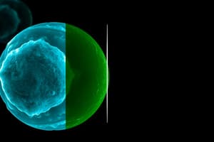
You are investigating maternal factors that regulate the cell cycle during early development. A mouse embryo is flushed from the uterine tube, treated with acid Tyrode solution to remove its zona pellucida, and examined by phase microscopy (shown in the image). The embryo exhibits a cleavage furrow and appears to be undergoing cytokinesis. These events take place during what phase of mitosis?
You are investigating maternal factors that regulate the cell cycle during early development. A mouse embryo is flushed from the uterine tube, treated with acid Tyrode solution to remove its zona pellucida, and examined by phase microscopy (shown in the image). The embryo exhibits a cleavage furrow and appears to be undergoing cytokinesis. These events take place during what phase of mitosis?
- Metaphase
- Anaphase
- Interphase
- Prophase
- Telophase (correct)
What intracellular protein complex links microtubules of the spindle apparatus to sister chromatids during mitosis and meiosis?
What intracellular protein complex links microtubules of the spindle apparatus to sister chromatids during mitosis and meiosis?
- Astral fibers
- Centrosome
- Centrioles
- Kinetochore (correct)
- Centromere
As part of your research, you examine integral membrane proteins in cleavage-stage mouse embryos using fluorescence microscopy (shown in the image). A pulse of high-intensity UV light is directed at a small patch on the surface of one blastomere, thereby causing an immediate loss of fluorescence emission (photobleaching). Over the next 10 minutes, fluorescence emission from this patch of membrane recovers. Which of the following cellular properties/processes best explains these experimental findings?
As part of your research, you examine integral membrane proteins in cleavage-stage mouse embryos using fluorescence microscopy (shown in the image). A pulse of high-intensity UV light is directed at a small patch on the surface of one blastomere, thereby causing an immediate loss of fluorescence emission (photobleaching). Over the next 10 minutes, fluorescence emission from this patch of membrane recovers. Which of the following cellular properties/processes best explains these experimental findings?
- Lipid raft assembly
- Receptor-mediated endocytosis
- Membrane fluidity (correct)
- Patching and capping
- Protein trafficking
You are studying cell migration during embryonic development. Neural tubes are harvested from post-implantation mouse embryos and placed in culture on plastic dishes coated with fibronectin. Time-lapse imaging reveals neural crest cells migrating away from the explanted tissue. The cells are observed to undergo continuous changes in cell shape, including the formation and retraction of lamellipodia. What protein is the principal mediator of membrane ruffling and locomotion in these cultured cells?
You are studying cell migration during embryonic development. Neural tubes are harvested from post-implantation mouse embryos and placed in culture on plastic dishes coated with fibronectin. Time-lapse imaging reveals neural crest cells migrating away from the explanted tissue. The cells are observed to undergo continuous changes in cell shape, including the formation and retraction of lamellipodia. What protein is the principal mediator of membrane ruffling and locomotion in these cultured cells?
A skin biopsy is examined at a double-headed microscope. The surgical pathologist directs your attention to waxy/lipid material filling the cytoplasm of secretory cells forming a sebaceous gland (shown in the image). Secretion of this waxy material to the pilosebaceous canal involves programmed cell death (apoptosis). Which of the following cytologic features provides evidence of apoptosis in this gland?
A skin biopsy is examined at a double-headed microscope. The surgical pathologist directs your attention to waxy/lipid material filling the cytoplasm of secretory cells forming a sebaceous gland (shown in the image). Secretion of this waxy material to the pilosebaceous canal involves programmed cell death (apoptosis). Which of the following cytologic features provides evidence of apoptosis in this gland?
A sample of adrenal cortex obtained at autopsy is fixed with formalin, embedded in paraffin, sectioned at 6 µm, stained with H&E, and examined by light microscopy (shown in the image). Cells of the zona fasciculata appear washed out and “spongy” due to an accumulation of cholesterol and other precursors for steroid hormone biosynthesis. Electron microscopic examination of these "steroid factory" cells would be expected to show an abundance of which of the following organelles?
A sample of adrenal cortex obtained at autopsy is fixed with formalin, embedded in paraffin, sectioned at 6 µm, stained with H&E, and examined by light microscopy (shown in the image). Cells of the zona fasciculata appear washed out and “spongy” due to an accumulation of cholesterol and other precursors for steroid hormone biosynthesis. Electron microscopic examination of these "steroid factory" cells would be expected to show an abundance of which of the following organelles?
A portion of the small intestine is collected at autopsy, and sections are stained with periodic acid-Schiff (PAS) and counterstained with hematoxylin. The mucosa of the intestine is examined by light microscopy (shown in the image). PAS is particularly useful for identifying which of the following biological materials?
A portion of the small intestine is collected at autopsy, and sections are stained with periodic acid-Schiff (PAS) and counterstained with hematoxylin. The mucosa of the intestine is examined by light microscopy (shown in the image). PAS is particularly useful for identifying which of the following biological materials?
You are asked to lead a seminar on intracellular protein trafficking. What organelle provides a microenvironment for the posttranslational modification and sorting of membrane and secretory proteins?
You are asked to lead a seminar on intracellular protein trafficking. What organelle provides a microenvironment for the posttranslational modification and sorting of membrane and secretory proteins?
Hematopoietic stem cells are cultured in vitro at 37°C in the presence of recombinant erythropoietin. A photomicrograph of a typical “burst-forming unit” committed to the erythrocyte pathway of differentiation is shown in the image. Which of the following histochemical stains can be used as a "vital dye" to distinguish viable from nonviable cells in your cell culture?
Hematopoietic stem cells are cultured in vitro at 37°C in the presence of recombinant erythropoietin. A photomicrograph of a typical “burst-forming unit” committed to the erythrocyte pathway of differentiation is shown in the image. Which of the following histochemical stains can be used as a "vital dye" to distinguish viable from nonviable cells in your cell culture?
Hepatocytes in a liver biopsy are examined by electron microscopy. The parallel lines with knob-like features (arrows, shown in the image) represent which of the following intracellular organelles?
Hepatocytes in a liver biopsy are examined by electron microscopy. The parallel lines with knob-like features (arrows, shown in the image) represent which of the following intracellular organelles?
A small muscular artery is examined in the pathology department. Smooth muscle fibers in the tunica media appear red, whereas collagen bundles in the tunica adventitia appear blue (shown in the image). This slide was most likely colored using which of the following histochemical stains?
A small muscular artery is examined in the pathology department. Smooth muscle fibers in the tunica media appear red, whereas collagen bundles in the tunica adventitia appear blue (shown in the image). This slide was most likely colored using which of the following histochemical stains?
A digital slide of a sympathetic chain ganglion is examined in the histology laboratory. Large multipolar neurons are surrounded by nerve fibers and connective tissue (shown in the image). Identify the dark basophilic region within the nucleus of these ganglion cells.
A digital slide of a sympathetic chain ganglion is examined in the histology laboratory. Large multipolar neurons are surrounded by nerve fibers and connective tissue (shown in the image). Identify the dark basophilic region within the nucleus of these ganglion cells.
A spinal cord smear preparation is obtained at autopsy and stained with Luxol fast blue/cresyl violet. The large octopus-like cells on this slide are multipolar motor neurons (shown in the image). What protein forms intracellular tracts that deliver organelles and vesicles to distant nerve terminals via anterograde axonal transport?
A spinal cord smear preparation is obtained at autopsy and stained with Luxol fast blue/cresyl violet. The large octopus-like cells on this slide are multipolar motor neurons (shown in the image). What protein forms intracellular tracts that deliver organelles and vesicles to distant nerve terminals via anterograde axonal transport?
The motor neurons described in Question 13 are labeled by immunocytochemistry using antibodies directed against a neuron-specific protein that helps maintain the shape of dendrites and axons. This structural protein forms which of the following intracellular organelles?
The motor neurons described in Question 13 are labeled by immunocytochemistry using antibodies directed against a neuron-specific protein that helps maintain the shape of dendrites and axons. This structural protein forms which of the following intracellular organelles?
A soft tissue biopsy is examined in the pathology department. Normal adipocytes are examined at high magnification (shown in the image). The clear space that has pushed the cytoplasm and nucleus to the periphery of these cells is best described by which of the following terms?
A soft tissue biopsy is examined in the pathology department. Normal adipocytes are examined at high magnification (shown in the image). The clear space that has pushed the cytoplasm and nucleus to the periphery of these cells is best described by which of the following terms?
You are studying the role of mitochondrial dysfunction in alcoholic liver disease. Genes for an inner mitochondrial membrane protein and a red fluorescent protein are spliced, and the fusion protein is expressed in mouse embryo fibroblasts. The distribution of mitochondria in the transfected cells is visualized by confocal fluorescence microscopy (shown in the image). Inhibition of the electron transport chain in this organelle leads to which of the following reversible changes in cell behavior?
You are studying the role of mitochondrial dysfunction in alcoholic liver disease. Genes for an inner mitochondrial membrane protein and a red fluorescent protein are spliced, and the fusion protein is expressed in mouse embryo fibroblasts. The distribution of mitochondria in the transfected cells is visualized by confocal fluorescence microscopy (shown in the image). Inhibition of the electron transport chain in this organelle leads to which of the following reversible changes in cell behavior?
Release of cytochrome c from the organelle described in Question 16 activates which of the following cellular processes?
Release of cytochrome c from the organelle described in Question 16 activates which of the following cellular processes?
Fluorescent fusion proteins are used to monitor the distribution of organelles in a myoblast cell line. The distribution of mitochondria and microfilaments is examined by confocal fluorescence microscopy (shown in the image). In this composite image, DNA is colored blue, microfilaments are colored green, and mitochondria are colored red. Which of the following cell adhesion proteins forms anchoring junctions that link actin microfilaments to adhesive glycoproteins on the surface of the culture dish?
Fluorescent fusion proteins are used to monitor the distribution of organelles in a myoblast cell line. The distribution of mitochondria and microfilaments is examined by confocal fluorescence microscopy (shown in the image). In this composite image, DNA is colored blue, microfilaments are colored green, and mitochondria are colored red. Which of the following cell adhesion proteins forms anchoring junctions that link actin microfilaments to adhesive glycoproteins on the surface of the culture dish?
Which of the following cellular processes describes the uptake of extracellular fluids and small particles by the cell described in Question 18?
Which of the following cellular processes describes the uptake of extracellular fluids and small particles by the cell described in Question 18?
The genes for green fluorescent protein and tubulin are spliced, and the fusion protein is expressed in a myoblast cell line. The distribution of microtubules is monitored by confocal fluorescence microscopy (shown in the image). During mitosis, these cytoskeletal proteins are reorganized to coordinate chromosome separation. Which of the following organelles is the principal microtubule-organizing center in these myoblasts?
The genes for green fluorescent protein and tubulin are spliced, and the fusion protein is expressed in a myoblast cell line. The distribution of microtubules is monitored by confocal fluorescence microscopy (shown in the image). During mitosis, these cytoskeletal proteins are reorganized to coordinate chromosome separation. Which of the following organelles is the principal microtubule-organizing center in these myoblasts?
You attend a national meeting on regenerative medicine. One of the talks focuses on cellular senescence and cancer. Reactivation of the gene for which of the following nuclear proteins may enable some cancer cells to escape cellular senescence, continue to proliferate, and maintain genomic stability?
You attend a national meeting on regenerative medicine. One of the talks focuses on cellular senescence and cancer. Reactivation of the gene for which of the following nuclear proteins may enable some cancer cells to escape cellular senescence, continue to proliferate, and maintain genomic stability?
The gene for green fluorescent protein is modified by the addition of a signal sequence that targets the translation product to the lumen of the endoplasmic reticulum (ER). The distribution of the rough ER in a transfected myoblast cell line is monitored by confocal fluorescence microscopy (shown in the image). Which of the following families of proteins facilitates proper protein folding in the ER, cytoplasm, and nucleus of this muscle stem cell?
The gene for green fluorescent protein is modified by the addition of a signal sequence that targets the translation product to the lumen of the endoplasmic reticulum (ER). The distribution of the rough ER in a transfected myoblast cell line is monitored by confocal fluorescence microscopy (shown in the image). Which of the following families of proteins facilitates proper protein folding in the ER, cytoplasm, and nucleus of this muscle stem cell?
Hepatocytes from a liver biopsy are examined by electron microscopy. Identify the elongated organelles shown in the image.
Hepatocytes from a liver biopsy are examined by electron microscopy. Identify the elongated organelles shown in the image.
A 23-year-old man presents with a 6-month history of yellow skin and sclerae. Physical examination shows mild jaundice and peritoneal ascites. The patient is subsequently diagnosed with a-1-antitrypsin deficiency. A liver biopsy stained with PAS reveals globular inclusions of misfolded a-1-antitrypsin (shown in the image). The abundance of these abnormal glycoproteins has apparently overwhelmed normal degradation pathways. Which of the following cellular processes describes the normal mechanism for specifically targeting and degrading misfolded proteins within cells?
A 23-year-old man presents with a 6-month history of yellow skin and sclerae. Physical examination shows mild jaundice and peritoneal ascites. The patient is subsequently diagnosed with a-1-antitrypsin deficiency. A liver biopsy stained with PAS reveals globular inclusions of misfolded a-1-antitrypsin (shown in the image). The abundance of these abnormal glycoproteins has apparently overwhelmed normal degradation pathways. Which of the following cellular processes describes the normal mechanism for specifically targeting and degrading misfolded proteins within cells?
A 42-year-old woman presents with increasing abdominal girth and yellow discoloration of her skin and sclera. Physical examination reveals hepatomegaly and evidence of liver failure (jaundice). A Prussian blue stain of a liver biopsy is shown in the image. This stain identifies which of the following elements?
A 42-year-old woman presents with increasing abdominal girth and yellow discoloration of her skin and sclera. Physical examination reveals hepatomegaly and evidence of liver failure (jaundice). A Prussian blue stain of a liver biopsy is shown in the image. This stain identifies which of the following elements?
A kidney biopsy from a 44-year-old man is examined by electron microscopy. The nucleus of an endothelial cell exhibits a peripheral ring of dark-stained chromatin (arrow, shown in the image). Which of the following best describes the functional significance of the dark-stained ring of marginal chromatin observed in this electron micrograph?
A kidney biopsy from a 44-year-old man is examined by electron microscopy. The nucleus of an endothelial cell exhibits a peripheral ring of dark-stained chromatin (arrow, shown in the image). Which of the following best describes the functional significance of the dark-stained ring of marginal chromatin observed in this electron micrograph?
Which of the following proteins contributes to the structural matrix that anchors chromatin to the nuclear membrane during interphase of the cell cycle?
Which of the following proteins contributes to the structural matrix that anchors chromatin to the nuclear membrane during interphase of the cell cycle?
An 85-year-old woman with Alzheimer disease dies in her sleep. At autopsy, hepatocytes are noted to contain golden cytoplasmic granules that do not stain with Prussian blue (shown in the image). This "wear-and-tear" pigment of aging (lipofuscin) accumulates primarily within which of the following cellular organelles?
An 85-year-old woman with Alzheimer disease dies in her sleep. At autopsy, hepatocytes are noted to contain golden cytoplasmic granules that do not stain with Prussian blue (shown in the image). This "wear-and-tear" pigment of aging (lipofuscin) accumulates primarily within which of the following cellular organelles?
You are involved in a translational research project to develop small-molecule inhibitors of pepsin secretion by chief cells in the stomach mucosa. Chief cells store precursor enzymes within zymogen granules. By electron microscopy, these "protein factory" cells would most likely show an abundance of which of the following intracellular organelles?
You are involved in a translational research project to develop small-molecule inhibitors of pepsin secretion by chief cells in the stomach mucosa. Chief cells store precursor enzymes within zymogen granules. By electron microscopy, these "protein factory" cells would most likely show an abundance of which of the following intracellular organelles?
A 55-year-old woman learns that she has high levels of serum cholesterol (greater than 280 mg/dL; normal less than 200 mg/dL) and is at increased risk for development of ischemic heart disease. The patient asks you to explain the normal pathway for serum cholesterol uptake and clearance. You explain to her that low-density lipoprotein (LDL) receptors present in her liver bind LDL cholesterol and internalize it by forming coated vesicles (endosomes). Which of the following structural proteins mediates LDL receptor internalization by organizing small buds of plasma membrane into endosomes?
A 55-year-old woman learns that she has high levels of serum cholesterol (greater than 280 mg/dL; normal less than 200 mg/dL) and is at increased risk for development of ischemic heart disease. The patient asks you to explain the normal pathway for serum cholesterol uptake and clearance. You explain to her that low-density lipoprotein (LDL) receptors present in her liver bind LDL cholesterol and internalize it by forming coated vesicles (endosomes). Which of the following structural proteins mediates LDL receptor internalization by organizing small buds of plasma membrane into endosomes?
A 23-year-old woman complains of recurrent bone pain and increasing abdominal girth. Physical examination reveals enlargement of the patient's liver and spleen (hepatosplenomegaly). A spleen biopsy reveals large macrophages, with a fibrillar appearance reminiscent of "wrinkled tissue paper" (shown in the image). The patient is subsequently diagnosed with Gaucher disease. She carries mutations in the genes for glucocerebrosidase. Without this hydrolytic enzyme, glucocerebroside accumulates within which of the following cellular organelles?
A 23-year-old woman complains of recurrent bone pain and increasing abdominal girth. Physical examination reveals enlargement of the patient's liver and spleen (hepatosplenomegaly). A spleen biopsy reveals large macrophages, with a fibrillar appearance reminiscent of "wrinkled tissue paper" (shown in the image). The patient is subsequently diagnosed with Gaucher disease. She carries mutations in the genes for glucocerebrosidase. Without this hydrolytic enzyme, glucocerebroside accumulates within which of the following cellular organelles?
You are studying the differentiation of epithelial cells lining the intestinal mucosa and identify a common stem cell for the secretory lineage that gives rise to Paneth cells, enterocytes, and goblet cells. Which of the following terms describes the developmental potential of these gastrointestinal stem cells?
You are studying the differentiation of epithelial cells lining the intestinal mucosa and identify a common stem cell for the secretory lineage that gives rise to Paneth cells, enterocytes, and goblet cells. Which of the following terms describes the developmental potential of these gastrointestinal stem cells?
You join a research laboratory to investigate the growth and differentiation of human embryonic stem (ES) cells. These remarkable cells have been shown to differentiate into a wide variety of somatic cell types including (1) dopamine-producing neurons, (2) cardiac myocytes, and (3) insulin-producing pancreatic islet cells. ES cells are similar or equivalent to which of the following populations of cells/tissues in the early embryo?
You join a research laboratory to investigate the growth and differentiation of human embryonic stem (ES) cells. These remarkable cells have been shown to differentiate into a wide variety of somatic cell types including (1) dopamine-producing neurons, (2) cardiac myocytes, and (3) insulin-producing pancreatic islet cells. ES cells are similar or equivalent to which of the following populations of cells/tissues in the early embryo?
As part of your research, you investigate the role of cyclins and cyclin-dependent kinases in regulating ES cell growth in vitro. These rapidly dividing cells spend most of their time in which phase of the mitotic cell cycle?
As part of your research, you investigate the role of cyclins and cyclin-dependent kinases in regulating ES cell growth in vitro. These rapidly dividing cells spend most of their time in which phase of the mitotic cell cycle?
You are invited to give a seminar on the molecular mechanisms of lineage formation and cell differentiation. During the seminar, you are asked to list the primary germ layers of the embryo and discuss their derivatives. Blood vessels and hematopoietic stem cells originate from which of the following tissues/structures during embryogenesis?
You are invited to give a seminar on the molecular mechanisms of lineage formation and cell differentiation. During the seminar, you are asked to list the primary germ layers of the embryo and discuss their derivatives. Blood vessels and hematopoietic stem cells originate from which of the following tissues/structures during embryogenesis?
The principal investigator of your laboratory asks you whether pluripotent ES cells can differentiate into neural crest cells or primordial germ cells. You suggest that cellular and molecular markers would help you answer that question. Markers for which of the following cells could be used to monitor neural crest cell differentiation in vitro?
The principal investigator of your laboratory asks you whether pluripotent ES cells can differentiate into neural crest cells or primordial germ cells. You suggest that cellular and molecular markers would help you answer that question. Markers for which of the following cells could be used to monitor neural crest cell differentiation in vitro?
A cervical biopsy is obtained from a 42-year-old woman with a history of abnormal Pap smears. The tissue is tested for human papillomavirus (HPV) by in situ hybridization using cDNA probes. Evidence of HPV viral genome is detected in cells in the cervical biopsy (dark blue spots, shown in the image). The patient is told that she is at increased risk for the development of cervical cancer. She asks you to elaborate. You explain that HPV encodes an early gene (E6) that activates a cellular protein that, in turn, accelerates the degradation of the p53 tumor suppressor protein. Name the protein that is activated by HPV E6.
A cervical biopsy is obtained from a 42-year-old woman with a history of abnormal Pap smears. The tissue is tested for human papillomavirus (HPV) by in situ hybridization using cDNA probes. Evidence of HPV viral genome is detected in cells in the cervical biopsy (dark blue spots, shown in the image). The patient is told that she is at increased risk for the development of cervical cancer. She asks you to elaborate. You explain that HPV encodes an early gene (E6) that activates a cellular protein that, in turn, accelerates the degradation of the p53 tumor suppressor protein. Name the protein that is activated by HPV E6.
Flashcards
Cytokinesis
Cytokinesis
Division of the cytoplasm that occurs in late mitosis to create two separate cells.
Membrane fluidity
Membrane fluidity
The capacity of membrane lipids to move laterally within the lipid bilayer.
Actin
Actin
A protein that mediates membrane ruffling and locomotion in migrating cells.
Golgi apparatus
Golgi apparatus
Signup and view all the flashcards
Golgi apparatus
Golgi apparatus
Signup and view all the flashcards
PAS (Periodic acid-Schiff)
PAS (Periodic acid-Schiff)
Signup and view all the flashcards
Masson trichrome
Masson trichrome
Signup and view all the flashcards
Tubulin
Tubulin
Signup and view all the flashcards
Chaperones
Chaperones
Signup and view all the flashcards
Lysosomes
Lysosomes
Signup and view all the flashcards
Telophase
Telophase
Signup and view all the flashcards
Kinetochore
Kinetochore
Signup and view all the flashcards
Apoptosis
Apoptosis
Signup and view all the flashcards
Smooth Endoplasmic Reticulum
Smooth Endoplasmic Reticulum
Signup and view all the flashcards
Totipotency
Totipotency
Signup and view all the flashcards
Trypan Blue
Trypan Blue
Signup and view all the flashcards
Cytoskeleton
Cytoskeleton
Signup and view all the flashcards
Inclusion
Inclusion
Signup and view all the flashcards
Hydropic Swelling
Hydropic Swelling
Signup and view all the flashcards
Mitochondrial Permeability Transition Pore
Mitochondrial Permeability Transition Pore
Signup and view all the flashcards
Integrins
Integrins
Signup and view all the flashcards
Pinocytosis
Pinocytosis
Signup and view all the flashcards
Centrosomes
Centrosomes
Signup and view all the flashcards
Telomerase
Telomerase
Signup and view all the flashcards
Mitochondria
Mitochondria
Signup and view all the flashcards
Ubiquitin-Proteasome Pathway
Ubiquitin-Proteasome Pathway
Signup and view all the flashcards
Heterochromatin
Heterochromatin
Signup and view all the flashcards
Lamins
Lamins
Signup and view all the flashcards
Rough Endoplasmic Reticulum
Rough Endoplasmic Reticulum
Signup and view all the flashcards
Receptor-Mediated Endocytosis
Receptor-Mediated Endocytosis
Signup and view all the flashcards
Multipotent
Multipotent
Signup and view all the flashcards
Epiblast
Epiblast
Signup and view all the flashcards
G1 Phase
G1 Phase
Signup and view all the flashcards
Mesoderm
Mesoderm
Signup and view all the flashcards
Melanocytes
Melanocytes
Signup and view all the flashcards
Ubiquitin Ligase
Ubiquitin Ligase
Signup and view all the flashcards
p53
p53
Signup and view all the flashcards
HPV E6
HPV E6
Signup and view all the flashcards
In situ hybridization
In situ hybridization
Signup and view all the flashcards
Study Notes
- HPV E6 activates Ubiquitin ligase, which leads to the degradation of the p53 tumor suppressor protein.
Studying That Suits You
Use AI to generate personalized quizzes and flashcards to suit your learning preferences.