Podcast
Questions and Answers
Which vessels bring de-oxygenated blood to the right atrium?
Which vessels bring de-oxygenated blood to the right atrium?
- Superior vena cava (correct)
- Inferior vena cava (correct)
- Pulmonary arteries
- Aorta
What is the primary function of the heart valves mentioned in the content?
What is the primary function of the heart valves mentioned in the content?
- To allow flow in only one direction (correct)
- To increase blood pressure
- To separate the chambers of the heart
- To pump oxygenated blood
What is the relationship between blood flow (Q), pressure gradient ( extdelta-P), and resistance (R)?
What is the relationship between blood flow (Q), pressure gradient ( extdelta-P), and resistance (R)?
- Q = extdelta-P / R (correct)
- Q = R / extdelta-P
- Q = extdelta-P + R
- Q = extdelta-P x R
Which valve separates the right atrium from the right ventricle?
Which valve separates the right atrium from the right ventricle?
Which chamber of the heart receives oxygenated blood from the pulmonary circulation?
Which chamber of the heart receives oxygenated blood from the pulmonary circulation?
What type of valve is located between the left atrium and left ventricle?
What type of valve is located between the left atrium and left ventricle?
What structure maintains the one-way flow of blood from the right ventricle to the pulmonary arteries?
What structure maintains the one-way flow of blood from the right ventricle to the pulmonary arteries?
Which factor primarily influences blood flow through the circulatory system?
Which factor primarily influences blood flow through the circulatory system?
What function does the sinoatrial node serve in the heart?
What function does the sinoatrial node serve in the heart?
Which structure is the only point allowing electrical current to pass from the atria to the ventricles?
Which structure is the only point allowing electrical current to pass from the atria to the ventricles?
What is the primary role of the atrioventricular node in the heart's conduction system?
What is the primary role of the atrioventricular node in the heart's conduction system?
How does the conduction speed through the Purkinje fibers compare to that through the atrioventricular node?
How does the conduction speed through the Purkinje fibers compare to that through the atrioventricular node?
What is the significance of the electrical insulation between the atria and ventricles?
What is the significance of the electrical insulation between the atria and ventricles?
Which of the following structures conducts current very rapidly through the right and left atria?
Which of the following structures conducts current very rapidly through the right and left atria?
What is the role of the mitral and tricuspid valves during cardiac contraction?
What is the role of the mitral and tricuspid valves during cardiac contraction?
Where does the electrical conduction from the atrioventricular node primarily lead?
Where does the electrical conduction from the atrioventricular node primarily lead?
What initiates the electrical currents measured by an ECG?
What initiates the electrical currents measured by an ECG?
Where are electrodes typically placed for the classical three-lead ECG?
Where are electrodes typically placed for the classical three-lead ECG?
What is the purpose of the earth lead in a three-lead ECG setup?
What is the purpose of the earth lead in a three-lead ECG setup?
Which statement correctly describes a 'lead' in the context of an ECG?
Which statement correctly describes a 'lead' in the context of an ECG?
What is the alternative placement for electrodes when the subject is exercising?
What is the alternative placement for electrodes when the subject is exercising?
What does the term 'bipolar' signify in a three-lead ECG?
What does the term 'bipolar' signify in a three-lead ECG?
Which lead configuration is associated with the right arm being positive?
Which lead configuration is associated with the right arm being positive?
What physiological basis underlies the recording of an ECG?
What physiological basis underlies the recording of an ECG?
During diastole, which valves are open?
During diastole, which valves are open?
What happens during systole with respect to the valves in the heart?
What happens during systole with respect to the valves in the heart?
What is the state of blood flow during mid-diastole?
What is the state of blood flow during mid-diastole?
What is the pressure in the left ventricle during mid-diastole?
What is the pressure in the left ventricle during mid-diastole?
What indicates that the heart is in a relaxing state during mid-diastole?
What indicates that the heart is in a relaxing state during mid-diastole?
What is the pressure of the blood in the aorta during mid-diastole?
What is the pressure of the blood in the aorta during mid-diastole?
Which phase of the cardiac cycle allows for the passive filling of the ventricles?
Which phase of the cardiac cycle allows for the passive filling of the ventricles?
What occurs after the T wave during the cardiac cycle?
What occurs after the T wave during the cardiac cycle?
What does the QT interval represent in an ECG?
What does the QT interval represent in an ECG?
Which of the following factors can affect the length of the QT interval?
Which of the following factors can affect the length of the QT interval?
What condition can result from a prolonged QT interval?
What condition can result from a prolonged QT interval?
What does a depressed ST segment on an ECG suggest?
What does a depressed ST segment on an ECG suggest?
What effect does increased preload have on stroke volume?
What effect does increased preload have on stroke volume?
If the ST segment is elevated, what condition may this indicate?
If the ST segment is elevated, what condition may this indicate?
Which electrolyte imbalances can affect the QT interval?
Which electrolyte imbalances can affect the QT interval?
What is the primary relationship described by Starling's Law of the Heart?
What is the primary relationship described by Starling's Law of the Heart?
Which factor primarily determines the afterload on the heart?
Which factor primarily determines the afterload on the heart?
How might one remember the relationship between depressed ST segments and ischemia?
How might one remember the relationship between depressed ST segments and ischemia?
Which of the following is a potential impact of ventricular fibrillation?
Which of the following is a potential impact of ventricular fibrillation?
What happens to stroke volume when end diastolic volume decreases?
What happens to stroke volume when end diastolic volume decreases?
How does increased end diastolic volume affect the heart's contractile force?
How does increased end diastolic volume affect the heart's contractile force?
What does the Starling curve illustrate?
What does the Starling curve illustrate?
In heart physiology, what does the term 'preload' refer to?
In heart physiology, what does the term 'preload' refer to?
What role does peripheral resistance play in heart function?
What role does peripheral resistance play in heart function?
Flashcards
Blood flow
Blood flow
The movement of blood through the circulatory system, driven by the pressure difference between the heart and the rest of the circulation.
Pressure Gradient (Blood)
Pressure Gradient (Blood)
The difference in pressure between the heart and other parts of the circulatory system.
Resistance (Bloodflow)
Resistance (Bloodflow)
Opposition to blood flow in the blood vessels.
Right Heart
Right Heart
Signup and view all the flashcards
Left Heart
Left Heart
Signup and view all the flashcards
Atrioventricular Valves
Atrioventricular Valves
Signup and view all the flashcards
Tricuspid Valve
Tricuspid Valve
Signup and view all the flashcards
Bicuspid (Mitral) Valve
Bicuspid (Mitral) Valve
Signup and view all the flashcards
Sinoatrial node (SA node)
Sinoatrial node (SA node)
Signup and view all the flashcards
Atrioventricular (AV) node
Atrioventricular (AV) node
Signup and view all the flashcards
Atrial contraction
Atrial contraction
Signup and view all the flashcards
Conduction Delay
Conduction Delay
Signup and view all the flashcards
Purkinje fibers
Purkinje fibers
Signup and view all the flashcards
Heart rate
Heart rate
Signup and view all the flashcards
Inter-nodal pathways
Inter-nodal pathways
Signup and view all the flashcards
Atrioventricular bundle (Bundle of His)
Atrioventricular bundle (Bundle of His)
Signup and view all the flashcards
What is an ECG?
What is an ECG?
Signup and view all the flashcards
How many leads are in a 3-lead ECG?
How many leads are in a 3-lead ECG?
Signup and view all the flashcards
What is a lead in an ECG?
What is a lead in an ECG?
Signup and view all the flashcards
Where are the electrodes placed in a 3-lead ECG?
Where are the electrodes placed in a 3-lead ECG?
Signup and view all the flashcards
What is the purpose of the earth electrode in an ECG?
What is the purpose of the earth electrode in an ECG?
Signup and view all the flashcards
Can electrodes be placed on the abdomen for a 3-lead ECG?
Can electrodes be placed on the abdomen for a 3-lead ECG?
Signup and view all the flashcards
What does the ECG recording show?
What does the ECG recording show?
Signup and view all the flashcards
What is Lead 1 in a 3-lead ECG?
What is Lead 1 in a 3-lead ECG?
Signup and view all the flashcards
Ventricular Diastole
Ventricular Diastole
Signup and view all the flashcards
Semilunar Valves During Diastole
Semilunar Valves During Diastole
Signup and view all the flashcards
Atrioventricular Valves During Diastole
Atrioventricular Valves During Diastole
Signup and view all the flashcards
Passive Blood Flow (Diastole)
Passive Blood Flow (Diastole)
Signup and view all the flashcards
Aortic Pressure During Diastole
Aortic Pressure During Diastole
Signup and view all the flashcards
Left Ventricle Pressure During Diastole
Left Ventricle Pressure During Diastole
Signup and view all the flashcards
Left Atrial Pressure During Diastole
Left Atrial Pressure During Diastole
Signup and view all the flashcards
Absence of Heart Sounds During Diastole
Absence of Heart Sounds During Diastole
Signup and view all the flashcards
What is the QT interval?
What is the QT interval?
Signup and view all the flashcards
What factors affect the QT interval?
What factors affect the QT interval?
Signup and view all the flashcards
What is prolonged QT syndrome?
What is prolonged QT syndrome?
Signup and view all the flashcards
What are the risks of prolonged QT syndrome?
What are the risks of prolonged QT syndrome?
Signup and view all the flashcards
How do electrolytes affect the QT interval?
How do electrolytes affect the QT interval?
Signup and view all the flashcards
What are the ST segment changes?
What are the ST segment changes?
Signup and view all the flashcards
What is ischemia?
What is ischemia?
Signup and view all the flashcards
What is a myocardial infarct?
What is a myocardial infarct?
Signup and view all the flashcards
What is preload?
What is preload?
Signup and view all the flashcards
How does preload affect stroke volume?
How does preload affect stroke volume?
Signup and view all the flashcards
What is Starling's Law of the Heart?
What is Starling's Law of the Heart?
Signup and view all the flashcards
What is afterload?
What is afterload?
Signup and view all the flashcards
What determines afterload?
What determines afterload?
Signup and view all the flashcards
How does afterload affect stroke volume?
How does afterload affect stroke volume?
Signup and view all the flashcards
What is the relationship between venous return and stroke volume?
What is the relationship between venous return and stroke volume?
Signup and view all the flashcards
What is a Starling curve?
What is a Starling curve?
Signup and view all the flashcards
Study Notes
Cardiovascular System Function
- Homeostasis: The cardiovascular system maintains a stable internal environment for life by transporting materials and removing waste.
- Material Transport: Delivers nutrients (oxygen, glucose, amino acids, fats) and removes waste (carbon dioxide, lactic acid, urea) to/from interstitial fluid surrounding tissues.
- Signaling: Carries hormones between tissues and organs, assisting in thermoregulation, immunity, and responses to infection.
- Plasma Flow: Maintains proper nutrient levels, delivering materials in plasma and removing waste substances from interstitial fluid.
- Diffusion: Oxygen diffuses across capillaries into interstitial fluid. Carbon dioxide moves in opposite direction.
- Convection: Substances are carried by the plasma (e.g., oxygen, nutrients).
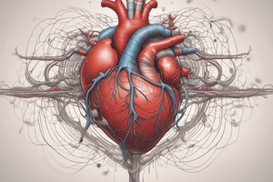
Heart Function
- Two Sides & Four Chambers: The heart has a right and left side (two atria and two ventricles), where the right side receives deoxygenated blood and the left receives oxygenated blood.
- Pulmonary Circulation: Right side pumps blood to the lungs for oxygen uptake.
- Systemic Circulation: Left side pumps oxygenated blood to the rest of the body.
- Valves: Prevent backflow: Atrioventricular valves (tricuspid & bicuspid) between atria & ventricles; semilunar valves (pulmonary and aortic) between ventricles and arteries.
- Pressure Gradient: The pressure gradient between the heart and circulation drives blood flow.
- Resistance: Blood flow depends on blood vessels' resistance to the blood flow.
Cardiac Cycle: Electrical Events
- Sinoatrial Node (SA): The heart's natural pacemaker, initiating electrical signals.
- Action Potentials: Depolarization leads to contraction. Repolarization leads to relaxation.
- Conduction System: Special conducting pathways transmit signals rapidly through the heart allowing all cells to contract simultaneously.
- Atrioventricular Node (AV): Delays signal and coordinate atrial and ventricular contraction.
- Internodal pathways: rapidly transmit signals through the atria.
- Electrocardiogram (ECG): A recording of the heart's electrical activity.
Cardiac Cycle: Mechanical Events
- Diastole: Heart relaxation and filling.
- Systole: Heart contraction and emptying.
- Four Phases: Diastolic filling, isovolumetric contraction, ventricular ejection, isovolumetric relaxation.
- Electrolyte balance: Potassium and calcium levels affect cardiac function.
- Ventricular Filling: AV valves open; blood passively flows from atria to ventricles.
- Ventricular Ejection: Pressure in ventricles exceeds pressure in arteries, causing semilunar valves to open and blood ejection.
Cardiac Output
- Cardiac output: The volume of blood pumped by the heart per minute (product of heart rate x stroke volume).
- Stroke volume (SV): The volume of blood ejected by a ventricle in each contraction.
- Preload: Degree of stretch on ventricular walls before contraction.
- Afterload: Resistance to blood flow from the ventricles.
- Control: Autonomic nervous system (sympathetic and parasympathetic) and hormones (adrenaline) regulate heart rate and contractility.
- Exercise or other physical activity increases the output needed.
Studying That Suits You
Use AI to generate personalized quizzes and flashcards to suit your learning preferences.