Podcast
Questions and Answers
What is a characteristic feature of myocardium?
What is a characteristic feature of myocardium?
- It contains intercalated discs for communication. (correct)
- It consists solely of smooth muscle fibers.
- It is under voluntary control.
- It serves as the thickest tissue in all heart chambers.
Which chamber of the heart is the thickest part of the myocardium located?
Which chamber of the heart is the thickest part of the myocardium located?
- Left atrium
- Left ventricle (correct)
- Right ventricle
- Right atrium
What role do chordae tendinae play in the heart's function?
What role do chordae tendinae play in the heart's function?
- They facilitate the flow of blood from ventricles to arteries.
- They prevent valves from opening upward into the atria. (correct)
- They provide electrical impulses for heart contractions.
- They connect the atria with the ventricles.
Which statement accurately describes the flow of blood through the heart?
Which statement accurately describes the flow of blood through the heart?
What structures are responsible for supporting the heart valves?
What structures are responsible for supporting the heart valves?
What significant role does the pericardium serve in relation to the heart?
What significant role does the pericardium serve in relation to the heart?
Which structure serves as the primary pacemaker of the heart?
Which structure serves as the primary pacemaker of the heart?
Which of the following statements accurately describes the position of the heart?
Which of the following statements accurately describes the position of the heart?
What complication arises from bleeding into the pericardial cavity?
What complication arises from bleeding into the pericardial cavity?
What is the significance of the AV nodal delay?
What is the significance of the AV nodal delay?
Which layer of the heart is responsible for the contraction and is the thickest?
Which layer of the heart is responsible for the contraction and is the thickest?
Which division of the nervous system predominantly influences the SA and AV nodes?
Which division of the nervous system predominantly influences the SA and AV nodes?
The serous membrane that secretes fluid into the pericardial cavity consists of which two layers?
The serous membrane that secretes fluid into the pericardial cavity consists of which two layers?
What is the primary role of the Purkinje fibers in the heart?
What is the primary role of the Purkinje fibers in the heart?
How do the ventricles differ from the atria in their function?
How do the ventricles differ from the atria in their function?
Which anatomical structure is located superiorly to the heart?
Which anatomical structure is located superiorly to the heart?
What is the main purpose of the fibrous sac of the pericardium?
What is the main purpose of the fibrous sac of the pericardium?
Which of the following statements about the coronary arteries is true?
Which of the following statements about the coronary arteries is true?
Which structure is responsible for holding a secondary pacemaker function in case of failure of the primary pacemaker?
Which structure is responsible for holding a secondary pacemaker function in case of failure of the primary pacemaker?
Which structures are located posteriorly to the heart?
Which structures are located posteriorly to the heart?
What role do circulating chemicals, nerve impulses, and hormones have in the heart's conducting system?
What role do circulating chemicals, nerve impulses, and hormones have in the heart's conducting system?
Flashcards
Fibrous pericardium
Fibrous pericardium
A tough, fibrous sac that encloses the heart, preventing overstretching and providing protection.
Serous pericardium
Serous pericardium
The inner layer of the pericardium, composed of thin epithelial cells that secrete lubricating fluid.
Pericardial cavity
Pericardial cavity
The potential space between the parietal and visceral layers of the serous pericardium, filled with lubricating fluid.
Pericarditis
Pericarditis
Signup and view all the flashcards
Cardiac tamponade
Cardiac tamponade
Signup and view all the flashcards
Myocardium
Myocardium
Signup and view all the flashcards
Endocardium
Endocardium
Signup and view all the flashcards
Fibrous skeleton of the heart
Fibrous skeleton of the heart
Signup and view all the flashcards
Intercalated Discs
Intercalated Discs
Signup and view all the flashcards
Atrioventricular (AV) Valves
Atrioventricular (AV) Valves
Signup and view all the flashcards
Chordae Tendinae
Chordae Tendinae
Signup and view all the flashcards
Autorhythmicity
Autorhythmicity
Signup and view all the flashcards
Sinoatrial (SA) Node
Sinoatrial (SA) Node
Signup and view all the flashcards
Atrioventricular (AV) Node
Atrioventricular (AV) Node
Signup and view all the flashcards
AV Bundle of His
AV Bundle of His
Signup and view all the flashcards
Purkinje fibers
Purkinje fibers
Signup and view all the flashcards
Vagus Nerve
Vagus Nerve
Signup and view all the flashcards
Sympathetic Nerves
Sympathetic Nerves
Signup and view all the flashcards
Coronary Arteries
Coronary Arteries
Signup and view all the flashcards
Study Notes
Cardiovascular System Lecture Notes (L16)
- The cardiovascular system is a complex system of organs responsible for transporting blood throughout the body.
- The heart is a hollow muscular organ, roughly the size of the owner's fist.
- The heart is located in the thoracic cavity, within the mediastinum.
- It lies obliquely, positioned more towards the left side of the body.
- The base of the heart is situated above, and the apex is positioned below.
- The heart is positioned between the 1/3 and 2/3 of the body's long axis, from ribs 2 to 5.
- Anteriorly, the heart is protected by the sternum and ribs.
- Posteriorly, structures such as the esophagus, trachea, and descending aorta are situated behind the heart.
- Inferiorly, the central tendon of the diaphragm is located below the heart.
- Superiorly, large blood vessels like the great vessels are above the heart
- Laterally, the heart is flanked by the lungs.
Heart Structure
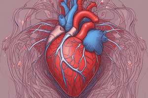
- The heart has three tissue layers: pericardium, myocardium, and endocardium.
- The pericardium is a fibroserous sac, consisting of a fibrous sac (open) and a serous sac (closed).
- The fibrous sac is continuous with the tunica adventitia of the great vessels and adheres to the diaphragm, preventing overdistention.
- The pericardium is made of two layers - parietal (outer) and visceral (inner, or epicardium).
- The fluid-filled space between the layers is called the pericardial cavity and contains serous fluid.
- Serous fluid allows for smooth movement between the heart and surrounding structures.
Heart Chambers and Valves
- The heart is divided into four chambers: two atria and two ventricles.
- The atria are the upper chambers.
- The ventricles are the lower chambers.
- The heart has atrioventricular (AV) valves and semilunar valves.
- AV valves are formed by double folds of endocardium strengthened by fibrous tissue which are the mitral and tricuspid valves.
- The AV valves allow blood to flow from the atria to the ventricles but prevent backflow.
- Semilunar valves (pulmonary and aortic valves) regulate blood flow out of the heart into arteries.
Cardiac Muscle (Myocardium)
- The myocardium is specialized cardiac muscle found only in the heart.
- Cardiac muscle cells are branched and contain a single nucleus.
- Cardiac muscle cells are connected by intercalated discs, enabling coordinated contraction.
- The myocardium is thickest at the heart's apex and thins towards the base.
- Its thickness is greatest in the left ventricle.
The Conduction System of the Heart
- Specialized cells within the heart form the intrinsic conduction system that automatically stimulates cardiac muscle contraction rhythmically without external impulses.
- The system consists of the sinoatrial (SA) node, atrioventricular (AV) node, AV bundle (bundle of His), right and left bundle branches, and Purkinje fibers.
Nerve Supply
- The vagus nerves (parasympathetic) supply the SA and AV nodes and atrial muscle.
- Parasympathetic stimulation slows the heart rate and decreases the force of contraction.
- Sympathetic nerves supply the SA and AV nodes and the myocardium of the atria and ventricles.
- Sympathetic stimulation increases the heart rate and enhances the force of contraction.
Circulatory Pathways
- Blood vessels are functionally divided into two circuits: the pulmonary circuit and the systemic circuit.
- The pulmonary circuit carries blood between the heart and lungs for gas exchange (oxygenation).
- The systemic circuit pumps oxygenated blood to the rest of the body from the heart.
Venous Drainage
- Blood returns to the heart via veins.
- The coronary sinus is a major vessel responsible for collecting deoxygenated blood from the heart wall.
Studying That Suits You
Use AI to generate personalized quizzes and flashcards to suit your learning preferences.


