Podcast
Questions and Answers
Which of the following is NOT a primary component of the cardiovascular system?
Which of the following is NOT a primary component of the cardiovascular system?
- Pump (heart)
- Lungs (correct)
- Liquid (blood)
- Tubes (vessels)
The endocardium is the outermost layer of tissue that lines the chambers of the heart.
The endocardium is the outermost layer of tissue that lines the chambers of the heart.
False (B)
Which layer of the heart is a striated muscle and differs in many aspects from skeletal muscle?
Which layer of the heart is a striated muscle and differs in many aspects from skeletal muscle?
- Myocardium (correct)
- Epicardium
- Endocardium
- Pericardium
The double-walled sac containing the heart and the roots of the great vessels is known as the ______.
The double-walled sac containing the heart and the roots of the great vessels is known as the ______.
Match the heart valves with their descriptions:
Match the heart valves with their descriptions:
Which of the following is NOT a primary function of blood?
Which of the following is NOT a primary function of blood?
Arterioles are blood vessels that carry blood away from the heart, connecting to capillaries.
Arterioles are blood vessels that carry blood away from the heart, connecting to capillaries.
Which of the following best describes the role of capillaries in the cardiovascular system?
Which of the following best describes the role of capillaries in the cardiovascular system?
The cardiac muscle contraction provides a driving force, also known as ______ gradient, for blood flow.
The cardiac muscle contraction provides a driving force, also known as ______ gradient, for blood flow.
Which of the following is NOT considered one of the five cardiac subsystems?
Which of the following is NOT considered one of the five cardiac subsystems?
The cardiac cycle consists of diastole, which is the contraction phase, and systole, which is the relaxation phase.
The cardiac cycle consists of diastole, which is the contraction phase, and systole, which is the relaxation phase.
What does the term 'automaticity' refer to in the context of the heart?
What does the term 'automaticity' refer to in the context of the heart?
The heart's independence from external regulation for the initiation of contraction is referred to as ______.
The heart's independence from external regulation for the initiation of contraction is referred to as ______.
What is the typical range for a resting heart rate?
What is the typical range for a resting heart rate?
In working ventricular myocardial cells, action potentials are generated spontaneously without external stimulation.
In working ventricular myocardial cells, action potentials are generated spontaneously without external stimulation.
What is the approximate resting membrane potential (RMP) of working ventricular myocardial cells?
What is the approximate resting membrane potential (RMP) of working ventricular myocardial cells?
Pacemaker cells within the SA node are self-excitable and have a maximum diastolic potential (MDP) of ______ mV.
Pacemaker cells within the SA node are self-excitable and have a maximum diastolic potential (MDP) of ______ mV.
During which phase of the working cardiomyocyte action potential does the upstroke occur, driven by rapid $Na^+$ influx?
During which phase of the working cardiomyocyte action potential does the upstroke occur, driven by rapid $Na^+$ influx?
The plateau phase (Phase 2) of the working cardiomyocyte action potential is primarily due to the rapid influx of potassium ions.
The plateau phase (Phase 2) of the working cardiomyocyte action potential is primarily due to the rapid influx of potassium ions.
During which phase of the working cardiomyocyte action potential does repolarization primarily occur?
During which phase of the working cardiomyocyte action potential does repolarization primarily occur?
Transient outward current (Ito) is predominantly active during action potential phase ______.
Transient outward current (Ito) is predominantly active during action potential phase ______.
What is a key characteristic of Phase 0 depolarization in working cardiomyocytes?
What is a key characteristic of Phase 0 depolarization in working cardiomyocytes?
Inactivation of $Na^+$ channels is faster than their activation during Phase 0 of the action potential.
Inactivation of $Na^+$ channels is faster than their activation during Phase 0 of the action potential.
What is the primary function of the $I_{CaL}$ current during Phase 2 (plateau) of the action potential?
What is the primary function of the $I_{CaL}$ current during Phase 2 (plateau) of the action potential?
During phase 3, $I_K$ increases after inactivation of ______ channels, acting as a delayed rectifier.
During phase 3, $I_K$ increases after inactivation of ______ channels, acting as a delayed rectifier.
The duration of the action potential is uniform throughout the heart; there is no regional variation.
The duration of the action potential is uniform throughout the heart; there is no regional variation.
Which specific channel is crucial for spontaneous slow diastolic depolarization (SDD) in the sinoatrial (SA) node?
Which specific channel is crucial for spontaneous slow diastolic depolarization (SDD) in the sinoatrial (SA) node?
Unlike working cardiomyocytes, the sinoatrial (SA) node lacks a stable resting potential and instead exhibits a maximum diastolic potential (MDP) of approximately ______ mV.
Unlike working cardiomyocytes, the sinoatrial (SA) node lacks a stable resting potential and instead exhibits a maximum diastolic potential (MDP) of approximately ______ mV.
In sinoatrial node cells, Phase 0 (rapid depolarization) is primarily due to the influx of sodium ions ($Na^+$).
In sinoatrial node cells, Phase 0 (rapid depolarization) is primarily due to the influx of sodium ions ($Na^+$).
What role does the transient calcium current ($I_{CaT}$) play in sinoatrial node cells?
What role does the transient calcium current ($I_{CaT}$) play in sinoatrial node cells?
The fastest normal pacemaker activity of the heart occurs in the ______ node.
The fastest normal pacemaker activity of the heart occurs in the ______ node.
Select the substance that decreases conduction velocity.
Select the substance that decreases conduction velocity.
The AV node conducts the electrical impulse faster than the Purkinje Fibers.
The AV node conducts the electrical impulse faster than the Purkinje Fibers.
What event is triggered and initiated by the conducting system?
What event is triggered and initiated by the conducting system?
The ______ node, located at the base of the right atrium, slows propagation of action potentials.
The ______ node, located at the base of the right atrium, slows propagation of action potentials.
Which factor enhances the speed of depolarization?
Which factor enhances the speed of depolarization?
The conduction velocity is the same throughout the heart.
The conduction velocity is the same throughout the heart.
Match the component of the heart and the rate / velocity value:
Match the component of the heart and the rate / velocity value:
What is one factor that effects conduction velocity?
What is one factor that effects conduction velocity?
What best describes Automaticity?
What best describes Automaticity?
Flashcards
Cardiovascular System Organization
Cardiovascular System Organization
The heart, blood, and vessels ensure blood circulation.
Heart Definition
Heart Definition
Hollow muscular organ that ensures blood circulation through regular contractions.
Endocardium
Endocardium
Innermost layer of tissue lining the heart chambers, similar to endothelial cells.
Myocardium
Myocardium
Signup and view all the flashcards
Pericardium
Pericardium
Signup and view all the flashcards
Heart Valves
Heart Valves
Signup and view all the flashcards
Blood Definition
Blood Definition
Signup and view all the flashcards
Arteries
Arteries
Signup and view all the flashcards
Arterioles
Arterioles
Signup and view all the flashcards
Capillaries
Capillaries
Signup and view all the flashcards
Venules
Venules
Signup and view all the flashcards
Veins
Veins
Signup and view all the flashcards
4-Chambered Heart
4-Chambered Heart
Signup and view all the flashcards
Intrinsic Pacemaker Activity
Intrinsic Pacemaker Activity
Signup and view all the flashcards
Secondary Heart Pumps
Secondary Heart Pumps
Signup and view all the flashcards
Driving Force
Driving Force
Signup and view all the flashcards
Cardiac Subsystems
Cardiac Subsystems
Signup and view all the flashcards
Cardiac Cycle
Cardiac Cycle
Signup and view all the flashcards
Systole
Systole
Signup and view all the flashcards
Diastole
Diastole
Signup and view all the flashcards
Regular Electrical Activity
Regular Electrical Activity
Signup and view all the flashcards
Automaticity
Automaticity
Signup and view all the flashcards
Autonomy
Autonomy
Signup and view all the flashcards
Rhythmicity
Rhythmicity
Signup and view all the flashcards
RMP of Myocardial Cell
RMP of Myocardial Cell
Signup and view all the flashcards
SA Node MDP
SA Node MDP
Signup and view all the flashcards
Phase 0
Phase 0
Signup and view all the flashcards
Phase 1
Phase 1
Signup and view all the flashcards
Phase 2
Phase 2
Signup and view all the flashcards
Phase 3
Phase 3
Signup and view all the flashcards
Phase 4
Phase 4
Signup and view all the flashcards
AP Threshold mV
AP Threshold mV
Signup and view all the flashcards
Transient Current (Ito)
Transient Current (Ito)
Signup and view all the flashcards
Voltage gated gates
Voltage gated gates
Signup and view all the flashcards
Sinoatrial Node MDP
Sinoatrial Node MDP
Signup and view all the flashcards
Conduction sequence
Conduction sequence
Signup and view all the flashcards
Peak Conduction Velocity
Peak Conduction Velocity
Signup and view all the flashcards
Sarcolemma
Sarcolemma
Signup and view all the flashcards
Study Notes
- The Cardiovascular system is an organ system consisting of;
- Pump (heart)
- Liquid (blood)
- Tubes (vessels)
Pump - Heart
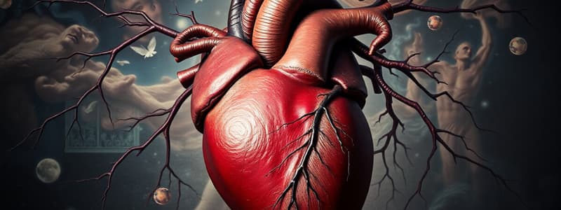
- The heart is a hollow muscular organ that, through regular contractions, ensures the circulation of blood in the body.
- The wall of the heart chambers consists of 3 layers:
- Endocardium is the innermost layer of tissue that lines the chambers of the heart. Its cells are embryologically and biologically similar to the endothelial cells that line blood vessels.
- Myocardium is striated muscle in appearance but with some key differences to skeletal muscle
- Pericardium is a double-walled sac containing the heart and the roots of the great vessels; consists of two layers, a serous (visceral) layer and a fibrous (parietal) layer, which in turn encloses the pericardial cavity, which contains pericardial fluid.
- Valves consist of atrioventricular (mitral, tricuspid) + papillary muscles + chordae tendineae + semilunar (aortic, pulmonary)
Liquid - Blood
- Blood is a viscous fluid, consisting of the plasma and blood cells.
- It delivers necessary substances such as nutrients and oxygen to the cells.
- It transports metabolic waste products away from those same cells.
- The main functions of blood are:
- Transportation
- Protection
- Homeostasis, like osmotic pressure, pH, ions
- Temperature regulation
Tubes - Vessels
- The cardiovascular system includes blood vessels to transport blood;
- Arteries
- Arterioles
- Capillaries
- Venules
- Veins
General Features of The Heart
- It is a 4-chambered organ
- It is self-activated by intrinsic pacemaker activity
- Two separate pumps (RV, LV) are supplied with two secondary pumps (RA, LA)
- Atrial pumps contract prior to the contraction of ventricles
- Cardiac muscle contraction provides the driving force (pressure gradient) for the blood flow
The Five Cardiac Subsystems are:
- The electrical pacemaker and conducting system
- The heart muscle
- The valves of the heart
- The coronary circulation
- The autonomic innervation of the heart
Basic Features of The Heart Function
- Regular electrical activity of the heart originating in the pacemaker cells precedes mechanical activity (contraction)
- The sequence of electrical and mechanical events that repeats rhythmically is called the CARDIAC CYCLE.
- The cardiac cycle consists of contraction (SYSTOLE) and ventricular relaxation (DIASTOLE).
- During systole, blood is ejected from the heart chambers into the aorta and pulmonary artery.
- During diastole, the chambers are filled with blood returning through the venous system.
- Automaticity is the heart's pacemaker activity generating a periodical electrical oscillation
- Autonomy is the heart's independence of external regulation for initiation of contraction
- Rhythmicity refers to the action potential’s regular intervals; average heart rate of between 60 – 90 beats per minute.
Two Major Types of Action Potential in the Heart
- Working ventricular myocardial cell action potential features: RMP -80 mV AP elicited after external stimulation (from conduction system or neighbouring cardiomyocytes)
- Pacemaker cell of SA node action potential features:
- MDP -65 mV
- Self-excitable
Working Cardiomyocytes
-
Resting Membrane Potential is -80 mV
-
Working Cardiomyocytes present 5 phases;
-
Phase 0 – upstroke – INa
-
Phase 1 – brief initial repolarization K (ITO)
-
Phase 2 – plateau phase (ICAL)
-
Phase 3 – repolarization (IK)
-
Phase 4 – resting membrane potential (IK1)
-
Phase 0 consists of depolarization, or upstroke with Na (INa)
-
Phase 1 consists of initial (transient) repolarization through K (Ito)
-
Phase 2 is the plateau phase that flows through ICaL
-
Phase 3 is the terminal repolarization which flows through IK
-
Phase 4 is the resting membrane potential which flows through IK1
Resting Membrane Potential
- Resting membrane potential is around -80 mV, and is stable
- Resting membrane potential is close to Ek
- IK1, INabgr are both crucial background currents, at the value of RMP at which INa and Ik are equal (Ix = gx × driving potential)
AP - Phase 0 – Depolarization – Na – INa
- The threshold for depolarization is around -60 mV, with a high driving potential for Na
- The membrane potential goes up to +30 mV
- INa lasts 1-2 ms, and is a voltage-gated channel that has activation and inactivation gates
- Determines velocity of AP conduction
- Depolarization is a gating signal for neighbouring cardiomyocytes
- IK1 is blocked
AP - Phase 0 – Depolarization – Na – INa (continued)
- Activation occurs once at -60 mV, increasing Na permeability 1000x, and the MP approaches ENa
- Inactivation happens slower than activation, closing a majority of channels around 0 mV
- Recovery happens within the range of -60 to -80 mV
AP - Phase 1 – Initial Repolarization – K (Ito)
- Transient K+ current moves from the cell - Ito
- Voltage gated channel with 2 gates (A, I, R)
- Opens in the course of depolarization
AP - Phase 2 – Plateau – Ca (ICAL)
- This phase consists of slow repolarization, achieved through Ical – voltage gated channel with 2 gates (A, I, R)
- Higher density in sarcolemma of T-tubules
- Opens in the course of depolarization, cca -40 mV
- Inactivation governed by voltage IC and IC Ca2+ concentration
- Channel phosphorylation by cAMP-dependent protein kinase
- Long depolarization essential for refractoriness
- Replenishes IC Ca2+
- CICR
- Recovery starts at -40 mV
AP - Phase 3 – Terminal Repolarization (IK)
- Phase 3 contains also terminal rapid repolarization
- Ik – in reality 2 voltage gated channels with 1 gate (A, D) – Ikr and Iks rapid, slow
- Activation governed by voltage and IC Ca2+ concentration
- It increases after inactivation of Ca channels (delayed rectifier)
- 𝛽-adrenergic stimulation increases Iks
- Deactivation -60 mV
- In the range -60 to -80 mV IK₁ becomes major repolarizing current
Heterogeneity of AP of Working Cardiomyocytes
- Different duration of AP
- Shorter in RV, in outer layers of cardiomyocytes
- The cells activated first should still be refractory when depolarization reaches subepicardial cardiomyocytes
- During a longer plateau phase more Ca2+ enters the cell, so subsequent contraction is stronger
Major Channels in Working Cardiomyocytes
Channel | Gating | Functional Role
- ---------|----------|---------- Fast sodium current | V (-65 to -80 mV) | Phase 0; activation (-65 mV); inactivation; recovery (-60 to -80 mV) Calcium current | V (-40 mV) | Phase 2, activation cca -40 mV, 𝛽-adrenergic stimulation ↑ Transient outward current | V (0 mV) | Phase 1, activation cca 0 mV Delayed rectifier | V | Phase 3 Ligand-activated potassium currents | L | Membrane hyperpolarization (phase 4) Potassium background current | V | Phase 4, closes during phase 0, finishes repolarization
Sinoatrial Node
Pacemaker cells – pale, cca 5 µm
-
Inflow tract of the RA, close crista terminalis and SCV
-
Maximum diastolic potential (MDP) -60 mV
-
Unstable RMP - spontaneous Slow Diastolic Depolarization (SDD) -IbgNa, -I (-60 mV), -Ісат (-60 mV), -ICAL (-35 mV), -Ik deactivated
-
Missing IK1
-
INa inactivated
-
The fastest normal pacemaker maintains between 60-80/min
Sinoatrial Node Pacemaker Cell
- Phase 0 has a rapid depolarization through Ca (ICAL)
- Phase 3 has terminal repolarization through (Ikr,s)
- Phase 4 has slow diastolic depolarization (INabg, Cat, CaL, If)
SDD
• IbgNa • If - hyperpolarization-activated current (-60 mV), can be affected by adrenergic stimulation • Ісат (-60 mV) - transient • ICaL (-35 mV) - long-lasting • Ikr,s deactivated
Major Channels in Sinoatrial Pacemaker Cells
Channel | Gating | Functional Role
- ---------|----------|---------- Sodium background current | V | Phase 4 (SDD) Transient calcium current | V | Phase 4 (SDD), activation cca -60 mV Calcium current | V | End phase 4 + phase 0, activation cca -40 mV, 𝛽-adrenergic stimulation ↑ Hyperpolarization-activated current | V | Beginning phase 4 (SDD), „funny“ current Delayed rectifier | V | Phase 3, deactivates at -60 mV Ligand-gated potassium currents | L | HR regulation Fast sodium current | V | INACTIVATED! Potassium background current | V | ABSENT!
Conducting System
- Automaticity (spontaneous production of AP, self-excitation)
- Conduction of AP
- Synchronization of mechanical activity of the heart
Sequence of Excitation
- Generation:
- Sinoatrial node – right atrium (determines the heart rate)
- Conduction:
- Bachman's bundle and internodal pathways
- Atrioventricular node (base of RA, propagation of AP relatively slow)
- Bundle of His (interventricular septum)
- Right and left bundle branches
- Purkinje fibers
Sinoatrial Node (SA Node)
- Rate 60 – 80/min, sinus rhythm
- Slow conduction (0.05 m/s) – velocity of conduction is further decreased by hypoxia, low temperature
at the center of Koch's triangle
- at the septal leaflet of the tricuspid valve, the coronary sinus, and the membraneous part of the interatrial septum
- Slow conduction (0.02 m/s) – atrioventricular delay
- Frequency filter- max. 230/min
- Zones atrionodal (A-N), Nodal (N), Nodal-His (N-H)
Purkinje Fibres
• Conduction velocity up to 4 m/s • Longer AP • RMP higher (-90 mV), unstable, role of If • Idioventricular rhythm 15 – 30/min
Conduction Velocity (m/s)
- SA node has a velocity of 0.05 m/s
- Atrial pathways have a velocity of 1 m/s
- AV node has a velocity of 0.05 m/s
- Bundle of His has a velocity of 1 m/s
- Purkinje system has a velocity of 4 m/s
- Ventricular muscle has a velocity of 1 m/s
Determinants of Conduction Velocity
- High density of gap junctions (nexus, tight junction) in the sarcolemma within cardiomyocyte connections
- Phosphorylation
- IC Ca2+ concentration
- Value of RMP; affects the speed of depolarization
- Cardiomyocyte diameter - length (space) constant
Studying That Suits You
Use AI to generate personalized quizzes and flashcards to suit your learning preferences.


