Podcast
Questions and Answers
The first heart sound, often described as "lub," is caused by the closing of the ______ valves.
The first heart sound, often described as "lub," is caused by the closing of the ______ valves.
atrioventricular
The second heart sound, "dub," originates from the closing of the ______ valves.
The second heart sound, "dub," originates from the closing of the ______ valves.
semilunar
The ______ node is known as the pacemaker of the heart.
The ______ node is known as the pacemaker of the heart.
sinoatrial
The P wave on an ECG represents ______ depolarization.
The P wave on an ECG represents ______ depolarization.
The QRS complex on an ECG signifies ______ depolarization.
The QRS complex on an ECG signifies ______ depolarization.
The T wave on an ECG corresponds to ______ repolarization.
The T wave on an ECG corresponds to ______ repolarization.
In the heart's conduction system, after the AV node, the electrical impulse travels through the ______.
In the heart's conduction system, after the AV node, the electrical impulse travels through the ______.
[Blank] detect changes in blood pressure in the aortic arch and carotid arteries.
[Blank] detect changes in blood pressure in the aortic arch and carotid arteries.
Blood flows from the right atrium to the right ventricle through the ______ valve.
Blood flows from the right atrium to the right ventricle through the ______ valve.
Blood flows from the left atrium to the left ventricle through the ______ valve.
Blood flows from the left atrium to the left ventricle through the ______ valve.
The ______ carry oxygenated blood from the lungs to the left atrium of the heart.
The ______ carry oxygenated blood from the lungs to the left atrium of the heart.
The ______ carries deoxygenated blood from the right ventricle to the lungs.
The ______ carries deoxygenated blood from the right ventricle to the lungs.
A normal systolic blood pressure reading is typically less than ______ mmHg.
A normal systolic blood pressure reading is typically less than ______ mmHg.
Increased stretch detected by baroreceptors typically results in a ______ in heart rate.
Increased stretch detected by baroreceptors typically results in a ______ in heart rate.
The ______ is the main artery that carries blood away from the left ventricle to the systemic circulation.
The ______ is the main artery that carries blood away from the left ventricle to the systemic circulation.
The ______ is responsible for the electrical events that precede the mechanical events of contraction or relaxation in the heart.
The ______ is responsible for the electrical events that precede the mechanical events of contraction or relaxation in the heart.
The ______ is affected by cardiac output, total peripheral resistance, and blood volume.
The ______ is affected by cardiac output, total peripheral resistance, and blood volume.
The conduction pathway following the AV bundle is the ______.
The conduction pathway following the AV bundle is the ______.
Following the bundle branches, the ______ conduct the electrical impulse to the contractile cells of the ventricles.
Following the bundle branches, the ______ conduct the electrical impulse to the contractile cells of the ventricles.
Identify the oxygenated blood in the pulmonary circuit: ______.
Identify the oxygenated blood in the pulmonary circuit: ______.
In the systemic circuit, blood returns through the ______.
In the systemic circuit, blood returns through the ______.
The heart sound called 'lub' represents the onset of ______.
The heart sound called 'lub' represents the onset of ______.
Describe the amplitude of the various waves in different cardiac cycles within the same individual?
Describe the amplitude of the various waves in different cardiac cycles within the same individual?
Are the amplitudes and durations of the various waves in different individuals
similar or very different? WHY?
Are the amplitudes and durations of the various waves in different individuals similar or very different? WHY?
The P wave and the QRS complex represent depolarization of the atrial and ventricular muscle respectively. Why does the QRS complex have the largest amplitude?
The P wave and the QRS complex represent depolarization of the atrial and ventricular muscle respectively. Why does the QRS complex have the largest amplitude?
The range for a normal resting heart rate is 60 to 90 bpm. A trained athlete could have a resting heart rate of 45 to 60 bpm. Why might a very fit person have a slower heart rate than someone of average fitness?
The range for a normal resting heart rate is 60 to 90 bpm. A trained athlete could have a resting heart rate of 45 to 60 bpm. Why might a very fit person have a slower heart rate than someone of average fitness?
Flashcards
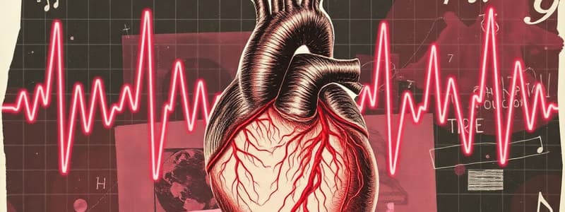
Electrocardiography (ECG)
Electrocardiography (ECG)
The study of the heart's electrical activity using electrodes on the body surface.
ECG Recording
ECG Recording
Represents the composite of all action potentials generated by nodal and contractile cells at a given time.
AV valves
AV valves
Atrioventricular valves prevent backflow into the atria when the ventricles contract.
SL valves
SL valves
Signup and view all the flashcards
"Lub" heart sound
"Lub" heart sound
Signup and view all the flashcards
"Dub" heart sound
"Dub" heart sound
Signup and view all the flashcards
Blood Pressure
Blood Pressure
Signup and view all the flashcards
Baroreceptors
Baroreceptors
Signup and view all the flashcards
Baroreceptor Response (Increased Stretch)
Baroreceptor Response (Increased Stretch)
Signup and view all the flashcards
Baroreceptor Response (Decreased Stretch)
Baroreceptor Response (Decreased Stretch)
Signup and view all the flashcards
Cardiac output
Cardiac output
Signup and view all the flashcards
Total peripheral resistance
Total peripheral resistance
Signup and view all the flashcards
Blood volume
Blood volume
Signup and view all the flashcards
Study Notes
- Study notes for Cardiovascular I focus on electrocardiography and heart sounds.
Today's Lab Focuses
- Anatomical models
- Electrocardiograms
- Heart sounds
- Blood pressure
- Group investigation planning
- Study question/results wrap-up
Heart Anatomy
- Know the structures labeled in figures 4 and 5 of the lab manual (pg. 49) for the lab exam.
Blood Flow
- Blood flows from the venae cavae to the lungs.
- It moves through the right atrium, then the right ventricle before heading to the lungs.
- Blood is oxygenated in the lungs and returns to the heart via the pulmonary veins, entering the left atrium.
- The blood then travels to the left ventricle and out to the systemic circuit through the aorta and its branches.
Conduction Pathway
- Electrical signals pass through the heart starting at the SA node.
- Signals then go to the AV node, AV bundle, bundle branches, and finally to the Purkinje fibers.
Electrocardiogram (ECG)
- The ECG represents the composite of all action potentials generated by nodal and contractile cells, not a tracing of a single action potential.
- Electrical events, such as depolarization and repolarization, precede the mechanical events of contraction or relaxation.
ECG Importance
- Abnormal conduction pathway, damaged regions, or enlarge heart can be determined by comparing ECG records with normal values.
Heart Valves
- Atrioventricular (AV) valves prevent backflow of blood into the atria when the ventricles contract; these valves are the tricuspid and bicuspid/mitral valves.
- Ssemilunar (SL) valves prevent backflow of blood into the ventricles and include the aortic and pulmonary valves.
Heart Sounds
- Dub-like heart sounds are generated by turbulent blood flow during the closure of the AV valves and occur during the onset of systole.
- Lub-like heart sounds occur with turbulent blood flow due to the closure of the aortic and pulmonary valves, marking the onset of diastole.
Blood Pressure
- Blood pressure, a vital sign, measures pressure within large arteries in the systemic circulation.
- Normal resting blood pressure for adults is systolic <120 mmHg and diastolic <80 mmHg.
- Transient alterations in blood pressure can stem from changes in posture, physical exertion, emotional upset, or temperature.
Blood Pressure Regulation
- Nerve endings of the baroreceptors detect stretch in the aortic arch and carotid arteries.
- Information is relayed to the brain for pressure regulation.
- Increased stretch decreases heart rate and promotes vasodilation.
- Conversely, decreased stretch increases heart rate and promotes vasoconstriction.
Factors Affecting blood pressure:
- Cardiac Output
- Total Peripheral Resistance
- Blood Volume
Presentation Planning
- Systolic and Diastolic BP can be presented in 2 to 3 measurements (or plot MAP).
- Include heart rate (HR) in your presentation.
- You must have 2 subjects minimum
- Fill out Idea Form and get it signed by me
Studying That Suits You
Use AI to generate personalized quizzes and flashcards to suit your learning preferences.


