Podcast
Questions and Answers
What is the primary function of atrial cells?
What is the primary function of atrial cells?
- To regulate the heartbeat
- To produce ATP for the heart
- To pump blood into the ventricles
- To monitor changes in intra-atrial pressure (correct)
What percentage of cardiac muscle fibers are autorhythmic?
What percentage of cardiac muscle fibers are autorhythmic?
- 50%
- 5%
- 1% (correct)
- 25%
What is the primary function of the conducting system of the heart?
What is the primary function of the conducting system of the heart?
- To pump blood into the aorta
- To regulate blood pressure
- To produce Atrial Natriuretic Peptide
- To initiate and conduct impulses (correct)
What is the average heart rate in adults?
What is the average heart rate in adults?
What is the sequence of events in the mechanism of heart beating?
What is the sequence of events in the mechanism of heart beating?
What type of pump is the ventricle during systole?
What type of pump is the ventricle during systole?
What is the functional unit of the heart?
What is the functional unit of the heart?
What is the relationship between the pulmonary and systemic circulations?
What is the relationship between the pulmonary and systemic circulations?
What is the main function of the heart valves?
What is the main function of the heart valves?
What is the thickness ratio of the left ventricular wall to the right ventricular wall?
What is the thickness ratio of the left ventricular wall to the right ventricular wall?
What is the name of the inner surface lining of the myocardium?
What is the name of the inner surface lining of the myocardium?
What is the name of the arteries that supply blood to the myocardium?
What is the name of the arteries that supply blood to the myocardium?
What is the dividing point in the capillaries where the pressure changes from high to low?
What is the dividing point in the capillaries where the pressure changes from high to low?
What percentage of ventricular filling is attributed to atrial systole?
What percentage of ventricular filling is attributed to atrial systole?
During which phase of the cardiac cycle do the atria receive venous return?
During which phase of the cardiac cycle do the atria receive venous return?
What would be the consequence of losing atrial function?
What would be the consequence of losing atrial function?
What is the primary characteristic of the cardiac muscle?
What is the primary characteristic of the cardiac muscle?
What is the function of the SA node in the cardiac muscle?
What is the function of the SA node in the cardiac muscle?
What is the characteristic of autorhythmic cells?
What is the characteristic of autorhythmic cells?
What happens during rapid depolarization in the cardiac muscle?
What happens during rapid depolarization in the cardiac muscle?
What is the difference between fast response fibers and slow response fibers?
What is the difference between fast response fibers and slow response fibers?
What is the effect of sympathetic stimulation on autorhythmicity?
What is the effect of sympathetic stimulation on autorhythmicity?
What is the function of the AV node?
What is the function of the AV node?
What is the characteristic of Purkinje fibers?
What is the characteristic of Purkinje fibers?
What is the effect of parasympathetic stimulation on autorhythmicity?
What is the effect of parasympathetic stimulation on autorhythmicity?
What is the primary difference between atrial fibers and ventricular fibers?
What is the primary difference between atrial fibers and ventricular fibers?
Flashcards are hidden until you start studying
Study Notes
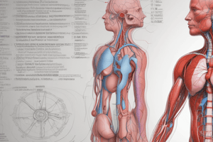
Heart Function and Circulation
- The atrial wall contains stretch receptors that monitor changes in intra-atrial pressure and initiate CV reflexes.
- Atrial cells monitor changes in blood volume, leading to the release of Atrial Natriuretic Peptide, which causes the excretion of Na+ and H2O by the kidneys.
Cardiac Muscle
- Cardiac muscle fibers are divided into two types:
- Contractile fibers (99%): form the atrial and ventricular walls and are specialized for pumping blood.
- Autorrhythmic fibers (1%): form the conducting system of the heart and are specialized for initiation and conduction of impulses.
Circulation
- There are three types of circulation:
- Systemic circulation (greater): starts from the left ventricle, passes through the aorta, and leads to large and small arteries, arterioles, capillaries, venules, and veins.
- Pulmonary circulation (lesser): starts from the right ventricle, passes through the pulmonary artery, and leads to the lungs, pulmonary capillaries, and pulmonary veins.
- Special circulations: include capillary, venous, lymph, coronary, and cerebral circulations.
Mechanism of Heart Beating
- The impulse originates at the sino-atrial node, causing contraction of the atria, then reaches the atrioventricular node, and finally passes to the ventricles through the His-Purkinje system, causing ventricular contraction.
- Heart rate: 60-90 beats/min, with an average of 72 beats/min in adults.
Functional Components of the Vascular System
- Heart: a dual pump, consisting of a compression pump (ventricles pumping blood into arteries during systole) and a suction pump (atria withdrawing blood from the venous system during diastole).
Functional Histology of Cardiac Muscle
- The heart is a functional syncytium, meaning that it is not anatomically a syncytium, but functions as a single unit.
- The pulmonary artery arises from the right ventricle and is guarded by the pulmonary valve.
- The heart valves allow blood to flow in one direction only.
Heart Valves and Endocardium
- The inner surface of the myocardium is lined with endothelial cells called endocardium, which also lines the cardiac valves and the entire vascular system.
- The atrial myocardium is much thinner than that of the ventricles, with the left ventricular wall being 3 times thicker than the right ventricular wall.
Coronary Arteries and Blood Pressure
- Coronary arteries supply the myocardium, arising from the 1st part of the aorta, and branching into small arteries, arterioles, capillaries, venules, and veins.
- The capillaries have a functional capillary pressure of 17 mmHg, dividing the system into high-pressure (arterial) and low-pressure (venous) components.
Autorhythmicity and Excitability
- The heart is characterized by being automatic and rhythmic (autorhythmic), excitable, conductive, and contractile.
- Autorhythmic cells have a low resting membrane potential, are less permeable to K+, and more permeable to Na+ and Ca++.
- The SA node is the normal pacemaker of the heart, firing impulses at a rate of 60-90 impulses/min.
Fast and Slow Response Fibers
- Fast response fibers (atria, ventricles, and Purkinje fibers) have a more negative resting membrane potential, a steeper slope of the upstroke, a greater amplitude of the action potential, and a faster conduction velocity compared to slow response fibers (SAN and AVN).
Factors Affecting Autorhythmicity
- Positive chronotropic factors: sympathetic stimulation, fever, mild alkalosis, and mild hypoxia.
- Negative chronotropic factors: parasympathetic stimulation, hypothermia, mild acidosis, and severe hypoxia.
Studying That Suits You
Use AI to generate personalized quizzes and flashcards to suit your learning preferences.