Podcast
Questions and Answers
How do gap junctions contribute to the coordinated function of cardiomyocytes?
How do gap junctions contribute to the coordinated function of cardiomyocytes?
- They provide structural support, holding cells together against physical stress.
- They store and release large quantities of calcium ions to initiate contraction.
- They enable the direct passage of ions between cells, allowing for rapid electrical communication. (correct)
- They facilitate the exchange of oxygen and carbon dioxide between adjacent cells.
If a drug blocked the function of T-tubules in cardiomyocytes, what effect would this have on muscle contraction?
If a drug blocked the function of T-tubules in cardiomyocytes, what effect would this have on muscle contraction?
- The contraction would be unaffected as T-tubules play no role in muscle contraction.
- The contraction would be weaker and less synchronized throughout the cell. (correct)
- The contraction would be stronger and more prolonged due to increased calcium storage.
- The contraction would occur more rapidly due to increased ion flow between cells.
What would be the immediate effect of administering a drug that inhibits the ryanodine receptors in cardiomyocytes?
What would be the immediate effect of administering a drug that inhibits the ryanodine receptors in cardiomyocytes?
- Increased intracellular calcium levels and enhanced force of contraction.
- Enhanced binding of calcium to troponin C.
- Decreased intracellular calcium levels and reduced force of contraction. (correct)
- Increased reuptake of calcium into the sarcoplasmic reticulum.
During muscle relaxation, what is the primary role of ion transporters in cardiomyocytes?
During muscle relaxation, what is the primary role of ion transporters in cardiomyocytes?
How does the binding of calcium to troponin C lead to muscle contraction?
How does the binding of calcium to troponin C lead to muscle contraction?
What is the role of ATP in the actin-myosin cycle during muscle contraction?
What is the role of ATP in the actin-myosin cycle during muscle contraction?
If blood flow is blocked in a coronary artery, which part of the heart's function would be directly compromised?
If blood flow is blocked in a coronary artery, which part of the heart's function would be directly compromised?
Where do the left and right coronary arteries originate?
Where do the left and right coronary arteries originate?
During the cardiac cycle, what percentage of ventricular filling is typically achieved through passive ventricular filling (diastasis)?
During the cardiac cycle, what percentage of ventricular filling is typically achieved through passive ventricular filling (diastasis)?
Following passive ventricular filling, what contributes to the remaining 10% of ventricular volume?
Following passive ventricular filling, what contributes to the remaining 10% of ventricular volume?
What characteristic is unique to pacemaker cells that allows them to spontaneously generate action potentials?
What characteristic is unique to pacemaker cells that allows them to spontaneously generate action potentials?
What is the primary role of the SA node in the cardiac conduction system?
What is the primary role of the SA node in the cardiac conduction system?
How does the Bachmann's Bundle contribute to atrial contraction?
How does the Bachmann's Bundle contribute to atrial contraction?
Why does the AV node slow down the conduction velocity of the electrical impulse?
Why does the AV node slow down the conduction velocity of the electrical impulse?
What is the main function of the His-Purkinje system?
What is the main function of the His-Purkinje system?
Which of the following statements best describes an ECG?
Which of the following statements best describes an ECG?
What does a positive deflection on an ECG tracing typically indicate?
What does a positive deflection on an ECG tracing typically indicate?
If abnormalities are detected in leads II, III, and aVF on an ECG, which region of the heart might be affected?
If abnormalities are detected in leads II, III, and aVF on an ECG, which region of the heart might be affected?
Which layer of the heart wall is characterized by its direct connection to the cardiac appendages?
Which layer of the heart wall is characterized by its direct connection to the cardiac appendages?
During which phase of the cardiac cycle does the T wave on an ECG begin, indicating ventricular repolarization?
During which phase of the cardiac cycle does the T wave on an ECG begin, indicating ventricular repolarization?
Which event causes the dicrotic notch (small dip) observed in the aortic pressure graph?
Which event causes the dicrotic notch (small dip) observed in the aortic pressure graph?
Which layer of the heart wall contains bundles of cardiac muscle fibers (fascicles) bound by connective tissue?
Which layer of the heart wall contains bundles of cardiac muscle fibers (fascicles) bound by connective tissue?
What is the primary event occurring during the isovolumetric contraction phase of the cardiac cycle?
What is the primary event occurring during the isovolumetric contraction phase of the cardiac cycle?
During which phase of the cardiac cycle is the 'P wave' observed on an ECG?
During which phase of the cardiac cycle is the 'P wave' observed on an ECG?
Which of the following is true regarding the thickness of the myocardium?
Which of the following is true regarding the thickness of the myocardium?
The endocardium is lined with endothelium, which continues into what?
The endocardium is lined with endothelium, which continues into what?
What occurs during the rapid ventricular filling phase?
What occurs during the rapid ventricular filling phase?
The first heart sound (S1) is most directly related to:
The first heart sound (S1) is most directly related to:
What is the main function of mesothelial cells found on the outer surface of the epicardium?
What is the main function of mesothelial cells found on the outer surface of the epicardium?
During which phase of the cardiac cycle does blood volume remain the same because all heart valves are closed?
During which phase of the cardiac cycle does blood volume remain the same because all heart valves are closed?
What is the correct order of blood flow through the heart?
What is the correct order of blood flow through the heart?
What is the relationship between the epicardium and the pericardium?
What is the relationship between the epicardium and the pericardium?
Which of the following best describes the events during ventricular systole?
Which of the following best describes the events during ventricular systole?
Occlusion of which artery commonly leads to severe infarction due to its extensive supply to the ventricles and interventricular septum?
Occlusion of which artery commonly leads to severe infarction due to its extensive supply to the ventricles and interventricular septum?
Which of the following accurately describes the drainage pathway of the great cardiac vein?
Which of the following accurately describes the drainage pathway of the great cardiac vein?
A patient is diagnosed with a blockage in the artery supplying the inferolateral parts of the left ventricle and left atrium. Which artery is most likely affected?
A patient is diagnosed with a blockage in the artery supplying the inferolateral parts of the left ventricle and left atrium. Which artery is most likely affected?
Which of the following cardiac veins does NOT drain directly into the coronary sinus?
Which of the following cardiac veins does NOT drain directly into the coronary sinus?
If the sinuatrial (SA) nodal branch arises from the circumflex artery instead of the right coronary artery, which area's blood supply might be affected if the circumflex artery is compromised?
If the sinuatrial (SA) nodal branch arises from the circumflex artery instead of the right coronary artery, which area's blood supply might be affected if the circumflex artery is compromised?
Which of the following statements correctly describes the function of the inferior interventricular branch (posterior descending artery, or PDA)?
Which of the following statements correctly describes the function of the inferior interventricular branch (posterior descending artery, or PDA)?
The Thebesian veins are part of the smaller cardiac venous system. What is their primary function?
The Thebesian veins are part of the smaller cardiac venous system. What is their primary function?
Which of the following statements accurately describes the coronary sinus?
Which of the following statements accurately describes the coronary sinus?
A cardiologist is examining a patient and notes that the left marginal vein is particularly prominent. Which area of the heart is MOST likely being primarily drained by this vein?
A cardiologist is examining a patient and notes that the left marginal vein is particularly prominent. Which area of the heart is MOST likely being primarily drained by this vein?
Which structure does the conal branch of the right coronary artery supply with blood?
Which structure does the conal branch of the right coronary artery supply with blood?
After a myocardial infarction, a patient exhibits impaired function of the inferior wall of the right ventricle. Which artery is MOST likely to have been affected?
After a myocardial infarction, a patient exhibits impaired function of the inferior wall of the right ventricle. Which artery is MOST likely to have been affected?
The oblique vein of the left atrium plays a role in the formation of which major venous structure?
The oblique vein of the left atrium plays a role in the formation of which major venous structure?
A surgeon needs to identify the vessel that runs close to the inferior border of the heart along the right ventricle. Which vessel are they looking for?
A surgeon needs to identify the vessel that runs close to the inferior border of the heart along the right ventricle. Which vessel are they looking for?
Which of the following represents the correct order in which deoxygenated blood flows from the myocardium back to the heart's right atrium?
Which of the following represents the correct order in which deoxygenated blood flows from the myocardium back to the heart's right atrium?
Which of the following are the two Atrioventricular Valves?
Which of the following are the two Atrioventricular Valves?
What does the PR interval on an ECG represent?
What does the PR interval on an ECG represent?
Which of the following best describes the T wave on an ECG?
Which of the following best describes the T wave on an ECG?
What is the normal duration of the QRS complex?
What is the normal duration of the QRS complex?
Occlusion of which artery would MOST directly affect the lateral surface of the cerebral hemisphere?
Occlusion of which artery would MOST directly affect the lateral surface of the cerebral hemisphere?
Which artery does NOT branch directly from the external carotid artery?
Which artery does NOT branch directly from the external carotid artery?
Which artery is the MOST likely source of an embolus that results in an embolic stroke affecting the brain?
Which artery is the MOST likely source of an embolus that results in an embolic stroke affecting the brain?
A patient exhibits facial drooping, arm weakness, and speech difficulties. Which condition is MOST likely indicated by these symptoms?
A patient exhibits facial drooping, arm weakness, and speech difficulties. Which condition is MOST likely indicated by these symptoms?
A prolonged QT interval can lead to an increased risk of which complication?
A prolonged QT interval can lead to an increased risk of which complication?
The basilar artery is formed by the merging of which two arteries?
The basilar artery is formed by the merging of which two arteries?
Which artery supplies blood to the intrinsic muscles of the tongue?
Which artery supplies blood to the intrinsic muscles of the tongue?
What is the primary target of the superior thyroid artery?
What is the primary target of the superior thyroid artery?
Which of the following is supplied by the thyrocervical trunk?
Which of the following is supplied by the thyrocervical trunk?
Which component of the ECG tracing represents ventricular depolarization?
Which component of the ECG tracing represents ventricular depolarization?
Which artery directly branches off the brachiocephalic trunk?
Which artery directly branches off the brachiocephalic trunk?
The maxillary artery terminates in the:
The maxillary artery terminates in the:
What is the MOST immediate consequence of a blocked artery in the brain during an ischemic stroke?
What is the MOST immediate consequence of a blocked artery in the brain during an ischemic stroke?
The posterior circulation of the brain is supplied by which artery?
The posterior circulation of the brain is supplied by which artery?
Which of the following arteries contributes to the formation of the Circle of Willis?
Which of the following arteries contributes to the formation of the Circle of Willis?
If a patient's ECG shows a significantly prolonged PR interval, which of the following conditions is most likely?
If a patient's ECG shows a significantly prolonged PR interval, which of the following conditions is most likely?
The vertebral artery courses superiorly through the neck via which structures?
The vertebral artery courses superiorly through the neck via which structures?
What is the effect of hypercalcemia on the QT interval?
What is the effect of hypercalcemia on the QT interval?
What is the primary difference between a thrombotic and an embolic ischemic stroke?
What is the primary difference between a thrombotic and an embolic ischemic stroke?
Which of the following best describes the function of the anterior cerebral artery?
Which of the following best describes the function of the anterior cerebral artery?
What does the J point on an ECG represent?
What does the J point on an ECG represent?
Which artery supplies the posterior region of the scalp?
Which artery supplies the posterior region of the scalp?
What is the primary function of the fibrous rings surrounding the atrioventricular orifices?
What is the primary function of the fibrous rings surrounding the atrioventricular orifices?
Which of the following accurately describes the structural difference between the mitral valve and the tricuspid valve?
Which of the following accurately describes the structural difference between the mitral valve and the tricuspid valve?
What is the functional significance of the chordae tendineae and papillary muscles?
What is the functional significance of the chordae tendineae and papillary muscles?
How does the structure of the aortic valve contribute to its function?
How does the structure of the aortic valve contribute to its function?
What would be the most likely consequence of damage to the chordae tendineae associated with the tricuspid valve?
What would be the most likely consequence of damage to the chordae tendineae associated with the tricuspid valve?
Where is the pulmonary valve located?
Where is the pulmonary valve located?
What type of muscle is found in the walls of the heart, and what nervous system regulates it?
What type of muscle is found in the walls of the heart, and what nervous system regulates it?
What are sarcomeres and what is their significance in cardiac muscle cells?
What are sarcomeres and what is their significance in cardiac muscle cells?
What is the key structural difference between cardiomyocyte nuclei and skeletal muscle cell nuclei?
What is the key structural difference between cardiomyocyte nuclei and skeletal muscle cell nuclei?
How do gap junctions within intercalated discs facilitate coordinated heart muscle contraction?
How do gap junctions within intercalated discs facilitate coordinated heart muscle contraction?
What role do desmosomes play within the intercalated discs of cardiac muscle?
What role do desmosomes play within the intercalated discs of cardiac muscle?
Why are intercalated discs important for the mechanical strength and stability of cardiac muscle?
Why are intercalated discs important for the mechanical strength and stability of cardiac muscle?
What is the function of transverse tubules (T-tubules) in cardiomyocytes?
What is the function of transverse tubules (T-tubules) in cardiomyocytes?
What is the primary role of the sarcoplasmic reticulum in cardiomyocytes?
What is the primary role of the sarcoplasmic reticulum in cardiomyocytes?
What are the three layers of the heart wall, from outermost to innermost?
What are the three layers of the heart wall, from outermost to innermost?
A blockage in the anterior cerebral artery would primarily affect the blood supply to which region of the brain?
A blockage in the anterior cerebral artery would primarily affect the blood supply to which region of the brain?
Which artery provides the main blood supply to the retina?
Which artery provides the main blood supply to the retina?
The vertebrobasilar system is formed by the:
The vertebrobasilar system is formed by the:
Which of the following arteries directly supplies the inner ear?
Which of the following arteries directly supplies the inner ear?
What is the role of the Circle of Willis in cerebral circulation?
What is the role of the Circle of Willis in cerebral circulation?
The middle cerebral artery is a direct branch of which other artery?
The middle cerebral artery is a direct branch of which other artery?
Which of the following structures does the anterior choroidal artery supply?
Which of the following structures does the anterior choroidal artery supply?
From which artery does the anterior spinal artery arise?
From which artery does the anterior spinal artery arise?
If a patient exhibits symptoms related to the cerebellum, which artery or arteries would be of primary concern?
If a patient exhibits symptoms related to the cerebellum, which artery or arteries would be of primary concern?
The tunica intima, the innermost layer of a blood vessel, is responsible for which key function?
The tunica intima, the innermost layer of a blood vessel, is responsible for which key function?
After blood flows through the transverse sinuses, which sinus does it enter next?
After blood flows through the transverse sinuses, which sinus does it enter next?
Which of the following anatomical structures does the internal carotid artery pass through on its way to the brain?
Which of the following anatomical structures does the internal carotid artery pass through on its way to the brain?
Which part of the internal carotid artery extends from the carotid canal to the foramen lacerum?
Which part of the internal carotid artery extends from the carotid canal to the foramen lacerum?
What is the primary function of the subendothelium layer in a blood vessel?
What is the primary function of the subendothelium layer in a blood vessel?
Occlusion of the lenticulostriate arteries, branches of the middle cerebral artery, would primarily affect:
Occlusion of the lenticulostriate arteries, branches of the middle cerebral artery, would primarily affect:
According to the Frank-Starling Law, what physiological parameter directly influences the force of muscle contraction during systole?
According to the Frank-Starling Law, what physiological parameter directly influences the force of muscle contraction during systole?
How would administering a beta-blocker affect the stroke volume, assuming all other factors remain constant?
How would administering a beta-blocker affect the stroke volume, assuming all other factors remain constant?
Which artery is the primary contributor to the formation of the superficial palmar arch?
Which artery is the primary contributor to the formation of the superficial palmar arch?
The deep palmar arch primarily originates from which artery?
The deep palmar arch primarily originates from which artery?
If a patient's end-diastolic volume is 130 mL and their end-systolic volume is 60 mL, what is their stroke volume?
If a patient's end-diastolic volume is 130 mL and their end-systolic volume is 60 mL, what is their stroke volume?
A patient has a stroke volume of 60 mL and an end-diastolic volume of 120 mL. What is their ejection fraction?
A patient has a stroke volume of 60 mL and an end-diastolic volume of 120 mL. What is their ejection fraction?
Where do the common palmar digital arteries bifurcate into proper palmar digital arteries?
Where do the common palmar digital arteries bifurcate into proper palmar digital arteries?
Which arteries contribute to the formation of the dorsal carpal arch?
Which arteries contribute to the formation of the dorsal carpal arch?
What is the cardiac output of a patient with a heart rate of 70 beats per minute and a stroke volume of 70 mL?
What is the cardiac output of a patient with a heart rate of 70 beats per minute and a stroke volume of 70 mL?
Which of the following statements correctly compares blood flow to the liver and the kidney?
Which of the following statements correctly compares blood flow to the liver and the kidney?
The abdominal aorta bifurcates into which two arteries at approximately the level of the pelvic brim?
The abdominal aorta bifurcates into which two arteries at approximately the level of the pelvic brim?
If the axillary artery is blocked, what anatomical feature provides an alternative route for blood to reach the upper limb?
If the axillary artery is blocked, what anatomical feature provides an alternative route for blood to reach the upper limb?
Which artery becomes the femoral artery after passing under the inguinal ligament?
Which artery becomes the femoral artery after passing under the inguinal ligament?
Which region of the anterior thigh is supplied by the obturator artery?
Which region of the anterior thigh is supplied by the obturator artery?
During a blood pressure measurement, the cuff is inflated around the arm to compress which artery against the humerus?
During a blood pressure measurement, the cuff is inflated around the arm to compress which artery against the humerus?
What is the anatomical origin of the deep femoral artery (profunda femoris)?
What is the anatomical origin of the deep femoral artery (profunda femoris)?
Which artery is most commonly used to assess the pulse in a clinical setting?
Which artery is most commonly used to assess the pulse in a clinical setting?
The popliteal artery is a continuation of which artery after it passes through the adductor hiatus (opening in adductor magnus)?
The popliteal artery is a continuation of which artery after it passes through the adductor hiatus (opening in adductor magnus)?
The anterior and posterior interosseous arteries, which supply the radius, ulna, and adjacent muscles, originate from which major artery?
The anterior and posterior interosseous arteries, which supply the radius, ulna, and adjacent muscles, originate from which major artery?
What are the terminal branches of the popliteal artery?
What are the terminal branches of the popliteal artery?
What is the primary function of the elastic fibers present in the tunica media of elastic arteries?
What is the primary function of the elastic fibers present in the tunica media of elastic arteries?
How does the structural composition of muscular arteries contribute to their overall function?
How does the structural composition of muscular arteries contribute to their overall function?
What is the functional significance of the tunica externa in blood vessels?
What is the functional significance of the tunica externa in blood vessels?
Which structural feature is unique to veins, aiding in unidirectional blood flow?
Which structural feature is unique to veins, aiding in unidirectional blood flow?
What is the primary role of capillaries in the circulatory system?
What is the primary role of capillaries in the circulatory system?
How do fenestrated capillaries facilitate material exchange, and where are they commonly found?
How do fenestrated capillaries facilitate material exchange, and where are they commonly found?
The common facial vein is formed by the union of which two veins?
The common facial vein is formed by the union of which two veins?
Which vein directly drains into the subclavian vein just before the subclavian vein joins the internal jugular vein?
Which vein directly drains into the subclavian vein just before the subclavian vein joins the internal jugular vein?
What is the role of the vasa vasorum in the walls of large blood vessels?
What is the role of the vasa vasorum in the walls of large blood vessels?
The brachiocephalic vein is formed by the confluence of which two veins?
The brachiocephalic vein is formed by the confluence of which two veins?
During the development of atherosclerosis, what initial event triggers the formation of plaque within the arterial wall?
During the development of atherosclerosis, what initial event triggers the formation of plaque within the arterial wall?
Which of the following veins drains directly into the superior vena cava?
Which of the following veins drains directly into the superior vena cava?
How does the narrowing of the blood vessel lumen due to plaque formation in atherosclerosis affect the tissues supplied by that vessel?
How does the narrowing of the blood vessel lumen due to plaque formation in atherosclerosis affect the tissues supplied by that vessel?
Which vein drains the suboccipital muscles, prevertebral muscles of the neck, and the cervical spine?
Which vein drains the suboccipital muscles, prevertebral muscles of the neck, and the cervical spine?
What is the primary purpose of the internal jugular vein?
What is the primary purpose of the internal jugular vein?
Which venous sinus directly receives blood from the transverse sinuses of the brain?
Which venous sinus directly receives blood from the transverse sinuses of the brain?
How would vasoconstricting factors affect cardiac afterload?
How would vasoconstricting factors affect cardiac afterload?
Which area does the inferior petrosal sinus primarily drain?
Which area does the inferior petrosal sinus primarily drain?
How does increased aortic pressure affect cardiac afterload?
How does increased aortic pressure affect cardiac afterload?
Meningeal veins travel with which blood vessels, and what area do they drain?
Meningeal veins travel with which blood vessels, and what area do they drain?
What direct effect does stimulating muscarinic M2 receptors on cardiomyocytes have on contractility?
What direct effect does stimulating muscarinic M2 receptors on cardiomyocytes have on contractility?
What is the primary role of catecholamines in influencing cardiac contractility?
What is the primary role of catecholamines in influencing cardiac contractility?
What is the destination of the blood collected by the pharyngeal venous plexus?
What is the destination of the blood collected by the pharyngeal venous plexus?
How does increased heart rate, due to sympathetic stimulation, affect cardiac contractility (inotropy)?
How does increased heart rate, due to sympathetic stimulation, affect cardiac contractility (inotropy)?
Considering the structural differences between arteries and veins, which statement accurately describes their composition?
Considering the structural differences between arteries and veins, which statement accurately describes their composition?
Which of the following accurately describes how the sympathetic nervous system influences calcium handling in cardiomyocytes to increase contractility?
Which of the following accurately describes how the sympathetic nervous system influences calcium handling in cardiomyocytes to increase contractility?
If the diastolic blood pressure (DBP) is 80 mmHg and the pulse pressure (PP) is 45 mmHg, what is the Mean Arterial Pressure (MAP)?
If the diastolic blood pressure (DBP) is 80 mmHg and the pulse pressure (PP) is 45 mmHg, what is the Mean Arterial Pressure (MAP)?
If a patient has increased systemic vascular resistance, which of the following compensatory mechanisms would the heart employ to maintain cardiac output?
If a patient has increased systemic vascular resistance, which of the following compensatory mechanisms would the heart employ to maintain cardiac output?
What would be the most likely effect of a drug that selectively blocks beta-1 receptors in the heart?
What would be the most likely effect of a drug that selectively blocks beta-1 receptors in the heart?
Why does pressure drop significantly across the arterioles in the cardiovascular system?
Why does pressure drop significantly across the arterioles in the cardiovascular system?
What is the effect of doubling the radius of a blood vessel on its resistance.?
What is the effect of doubling the radius of a blood vessel on its resistance.?
Which of the following would result in decreased cardiac afterload?
Which of the following would result in decreased cardiac afterload?
How does increased intracellular calcium concentration directly affect cardiac contractility?
How does increased intracellular calcium concentration directly affect cardiac contractility?
How does an increased number of capillaries affect total resistance in the capillary beds?
How does an increased number of capillaries affect total resistance in the capillary beds?
Which of the following conditions would MOST directly lead to an increase in blood viscosity and, consequently, vascular resistance?
Which of the following conditions would MOST directly lead to an increase in blood viscosity and, consequently, vascular resistance?
If a patient's blood vessel length increases due to the growth of new blood vessels, how would this affect vascular resistance, assuming all other factors remain constant?
If a patient's blood vessel length increases due to the growth of new blood vessels, how would this affect vascular resistance, assuming all other factors remain constant?
In a series of blood vessels with varying resistances, if one vessel constricts, causing its resistance to increase significantly, what happens to the total resistance of the series?
In a series of blood vessels with varying resistances, if one vessel constricts, causing its resistance to increase significantly, what happens to the total resistance of the series?
Considering the factors affecting vascular resistance, which of the following physiological responses would MOST effectively reduce resistance in a patient experiencing high blood pressure?
Considering the factors affecting vascular resistance, which of the following physiological responses would MOST effectively reduce resistance in a patient experiencing high blood pressure?
Which artery primarily supplies the muscles in the anterior compartment of the leg?
Which artery primarily supplies the muscles in the anterior compartment of the leg?
The dorsalis pedis artery terminates as which two arteries?
The dorsalis pedis artery terminates as which two arteries?
The lateral plantar artery ultimately contributes to the formation of which structure in the plantar foot?
The lateral plantar artery ultimately contributes to the formation of which structure in the plantar foot?
How does digoxin increase inotropy in cardiomyocytes?
How does digoxin increase inotropy in cardiomyocytes?
What is the primary mechanism by which an increased heart rate can enhance cardiomyocyte contractility?
What is the primary mechanism by which an increased heart rate can enhance cardiomyocyte contractility?
Which vein directly receives venous blood flow from the common digital veins of the hand?
Which vein directly receives venous blood flow from the common digital veins of the hand?
Which vein in the forearm is the most common site for venipuncture?
Which vein in the forearm is the most common site for venipuncture?
During postextrasystolic potentiation, why does the premature beat result in a weaker contraction than normal?
During postextrasystolic potentiation, why does the premature beat result in a weaker contraction than normal?
The basilic vein transitions into which vein as it continues its course up the arm?
The basilic vein transitions into which vein as it continues its course up the arm?
How would a drug that increases venous tone affect cardiac preload?
How would a drug that increases venous tone affect cardiac preload?
What is the clinical relevance of preload in the context of heart function?
What is the clinical relevance of preload in the context of heart function?
Which two veins merge to form the axillary vein?
Which two veins merge to form the axillary vein?
What is the name of the vein formed by the union of the subclavian vein and the internal jugular vein?
What is the name of the vein formed by the union of the subclavian vein and the internal jugular vein?
How does decreased ventricular compliance affect preload?
How does decreased ventricular compliance affect preload?
How does increased resistance in the aortic valve influence preload?
How does increased resistance in the aortic valve influence preload?
What percentage of venous return from the lower limbs is typically handled by the deep veins?
What percentage of venous return from the lower limbs is typically handled by the deep veins?
What is the primary role of the perforating veins in the lower limb?
What is the primary role of the perforating veins in the lower limb?
What is the effect of hypovolemia on preload?
What is the effect of hypovolemia on preload?
How does increased sympathetic activation influence cardiomyocyte contractility?
How does increased sympathetic activation influence cardiomyocyte contractility?
Which artery gives rise to the dorsal metatarsal arteries that supply metatarsals 2-5?
Which artery gives rise to the dorsal metatarsal arteries that supply metatarsals 2-5?
Into which vein does the deep palmar venous arch primarily drain?
Into which vein does the deep palmar venous arch primarily drain?
Which of the following best describes how atrial contraction affects preload?
Which of the following best describes how atrial contraction affects preload?
Which of the following best describes the path of the cephalic vein?
Which of the following best describes the path of the cephalic vein?
If a patient has a narrowed AV valve, how would this affect preload?
If a patient has a narrowed AV valve, how would this affect preload?
How does a significant increase in heart rate typically affect cardiac preload, and why?
How does a significant increase in heart rate typically affect cardiac preload, and why?
Which of the following leg arteries also supplies the fibula bone itself?
Which of the following leg arteries also supplies the fibula bone itself?
How would a blockage of the arcuate artery most directly affect the foot?
How would a blockage of the arcuate artery most directly affect the foot?
Which hemodynamic change would be expected in a patient receiving a drug that significantly increases ventricular compliance?
Which hemodynamic change would be expected in a patient receiving a drug that significantly increases ventricular compliance?
A patient with uncontrolled hypertension develops left ventricular hypertrophy, resulting in a stiffer, less compliant ventricle. How would this condition be expected to affect the patient's preload?
A patient with uncontrolled hypertension develops left ventricular hypertrophy, resulting in a stiffer, less compliant ventricle. How would this condition be expected to affect the patient's preload?
A patient is given a medication that causes significant vasodilation. What is the expected effect on preload and why?
A patient is given a medication that causes significant vasodilation. What is the expected effect on preload and why?
Which of the following describes the correct path of blood flow from the plantar digital veins?
Which of the following describes the correct path of blood flow from the plantar digital veins?
A patient has impaired blood flow in the posterolateral region of their lower leg. Which vein is MOST likely affected?
A patient has impaired blood flow in the posterolateral region of their lower leg. Which vein is MOST likely affected?
What veins merge to form the posterior tibial veins?
What veins merge to form the posterior tibial veins?
Which vein directly drains blood from the top of the foot?
Which vein directly drains blood from the top of the foot?
A thrombus in the popliteal vein would directly impair drainage from which of the following veins?
A thrombus in the popliteal vein would directly impair drainage from which of the following veins?
Which vein transitions into the external iliac vein?
Which vein transitions into the external iliac vein?
After a laceration on the medial side of the foot, blood flow through what vessel would be impacted?
After a laceration on the medial side of the foot, blood flow through what vessel would be impacted?
What is the drainage pathway of the dorsal venous arch?
What is the drainage pathway of the dorsal venous arch?
A patient has a blockage in the small saphenous vein. Where would blood accumulate as a result?
A patient has a blockage in the small saphenous vein. Where would blood accumulate as a result?
What is the relationship between vessel diameter, blood pressure and resistance?
What is the relationship between vessel diameter, blood pressure and resistance?
Given a systolic blood pressure of 130 mmHg and a diastolic blood pressure of 85 mmHg, calculate the Mean Arterial Pressure (MAP).
Given a systolic blood pressure of 130 mmHg and a diastolic blood pressure of 85 mmHg, calculate the Mean Arterial Pressure (MAP).
If cardiac output (CO) increases and systemic vascular resistance (SVR) remains constant, what happens to Mean Arterial Pressure (MAP)?
If cardiac output (CO) increases and systemic vascular resistance (SVR) remains constant, what happens to Mean Arterial Pressure (MAP)?
What is the correct calculation for pulse pressure, given a systolic blood pressure of 125 mmHg and a diastolic blood pressure of 75 mmHg?
What is the correct calculation for pulse pressure, given a systolic blood pressure of 125 mmHg and a diastolic blood pressure of 75 mmHg?
In microcirculation, what vessels are directly responsible for nutrient and waste exchange with tissues?
In microcirculation, what vessels are directly responsible for nutrient and waste exchange with tissues?
Which of the following mechanisms would compensate to maintain constant blood flow to an organ if the incoming blood vessel is partially constricted?
Which of the following mechanisms would compensate to maintain constant blood flow to an organ if the incoming blood vessel is partially constricted?
What is the primary mechanism by which lipid-soluble substances cross capillary walls?
What is the primary mechanism by which lipid-soluble substances cross capillary walls?
How does increased carbon dioxide concentration in the tissue surrounding arterioles affect blood flow?
How does increased carbon dioxide concentration in the tissue surrounding arterioles affect blood flow?
What is the role of lymphatic capillaries in the context of capillary exchange?
What is the role of lymphatic capillaries in the context of capillary exchange?
If the reflection coefficient of a capillary is close to 1, what does this indicate about the capillary's permeability to proteins?
If the reflection coefficient of a capillary is close to 1, what does this indicate about the capillary's permeability to proteins?
Which Starling force primarily drives fluid out of the capillary and into the interstitial space?
Which Starling force primarily drives fluid out of the capillary and into the interstitial space?
How do arterioles contribute to the regulation of total peripheral resistance?
How do arterioles contribute to the regulation of total peripheral resistance?
What is the effect of increased filtration coefficient (Kf) on fluid movement across capillary walls?
What is the effect of increased filtration coefficient (Kf) on fluid movement across capillary walls?
In the context of intrinsic control, what vascular response would be expected when arterial pressure drops?
In the context of intrinsic control, what vascular response would be expected when arterial pressure drops?
How does the sympathetic nervous system affect arteriole smooth muscle contraction, and what is the result?
How does the sympathetic nervous system affect arteriole smooth muscle contraction, and what is the result?
Which type of capillary is characterized by large pores or fenestrations that allow proteins to pass through?
Which type of capillary is characterized by large pores or fenestrations that allow proteins to pass through?
How does heart failure lead to edema in the lower limbs, according to Starling forces?
How does heart failure lead to edema in the lower limbs, according to Starling forces?
Nephrotic syndrome results in a significant loss of plasma proteins. How does this condition affect Starling forces and potentially lead to edema?
Nephrotic syndrome results in a significant loss of plasma proteins. How does this condition affect Starling forces and potentially lead to edema?
During hyperemia, an organ becomes more active. How does this increased activity affect perfusion in the organ?
During hyperemia, an organ becomes more active. How does this increased activity affect perfusion in the organ?
Under normal physiological conditions, which of the following statements regarding interstitial oncotic pressure (πi) is MOST accurate?
Under normal physiological conditions, which of the following statements regarding interstitial oncotic pressure (πi) is MOST accurate?
What causes lymphedema in the setting of lymphatic blockage, such as in filariasis?
What causes lymphedema in the setting of lymphatic blockage, such as in filariasis?
Flashcards
Cardiac Excitation-Contraction Coupling
Cardiac Excitation-Contraction Coupling
The sequence of events linking electrical excitation to mechanical contraction in heart muscle cells.
Intercalated Disks
Intercalated Disks
Specialized junctions connecting heart muscle cells; contain gap junctions and desmosomes.
Gap Junctions
Gap Junctions
Small channels in intercalated disks that allow ions to flow between cardiomyocytes.
Desmosomes
Desmosomes
Signup and view all the flashcards
T-Tubules
T-Tubules
Signup and view all the flashcards
Sarcoplasmic Reticulum
Sarcoplasmic Reticulum
Signup and view all the flashcards
Calcium-Induced Calcium Release
Calcium-Induced Calcium Release
Signup and view all the flashcards
Coronary Arteries
Coronary Arteries
Signup and view all the flashcards
Fibrous Rings (Heart)
Fibrous Rings (Heart)
Signup and view all the flashcards
Mitral Valve
Mitral Valve
Signup and view all the flashcards
Tricuspid Valve
Tricuspid Valve
Signup and view all the flashcards
Aortic Valve
Aortic Valve
Signup and view all the flashcards
Pulmonary Valve
Pulmonary Valve
Signup and view all the flashcards
Cardiac Muscle
Cardiac Muscle
Signup and view all the flashcards
Sarcomeres (Cardiac)
Sarcomeres (Cardiac)
Signup and view all the flashcards
Gap Junctions (Heart)
Gap Junctions (Heart)
Signup and view all the flashcards
Desmosomes (Heart)
Desmosomes (Heart)
Signup and view all the flashcards
T-tubules (Cardiac)
T-tubules (Cardiac)
Signup and view all the flashcards
Epicardium
Epicardium
Signup and view all the flashcards
Myocardium
Myocardium
Signup and view all the flashcards
Endocardium
Endocardium
Signup and view all the flashcards
Diastasis
Diastasis
Signup and view all the flashcards
Pacemaker Cells
Pacemaker Cells
Signup and view all the flashcards
SA Node
SA Node
Signup and view all the flashcards
Bachmann’s Bundle/Atrial Internodal Tracts
Bachmann’s Bundle/Atrial Internodal Tracts
Signup and view all the flashcards
AV Node
AV Node
Signup and view all the flashcards
His-Purkinje System
His-Purkinje System
Signup and view all the flashcards
Bundle of His
Bundle of His
Signup and view all the flashcards
Purkinje Fibers
Purkinje Fibers
Signup and view all the flashcards
Electrocardiogram (ECG/EKG)
Electrocardiogram (ECG/EKG)
Signup and view all the flashcards
ECG Lead
ECG Lead
Signup and view all the flashcards
Middle Cerebral Artery
Middle Cerebral Artery
Signup and view all the flashcards
Right Common Carotid Artery
Right Common Carotid Artery
Signup and view all the flashcards
Ascending Pharyngeal Artery
Ascending Pharyngeal Artery
Signup and view all the flashcards
Left Subclavian Artery
Left Subclavian Artery
Signup and view all the flashcards
Vertebral Artery
Vertebral Artery
Signup and view all the flashcards
Basilar Artery
Basilar Artery
Signup and view all the flashcards
Thyrocervical Trunk
Thyrocervical Trunk
Signup and view all the flashcards
Costocervical Trunk
Costocervical Trunk
Signup and view all the flashcards
Stroke
Stroke
Signup and view all the flashcards
Ischemic Stroke
Ischemic Stroke
Signup and view all the flashcards
Circumflex Artery
Circumflex Artery
Signup and view all the flashcards
Anterior Interventricular Artery (LAD)
Anterior Interventricular Artery (LAD)
Signup and view all the flashcards
Why is the LAD important?
Why is the LAD important?
Signup and view all the flashcards
Right Coronary Artery (RCA)
Right Coronary Artery (RCA)
Signup and view all the flashcards
Conal Branch
Conal Branch
Signup and view all the flashcards
Sinuatrial Nodal Branch
Sinuatrial Nodal Branch
Signup and view all the flashcards
Right Marginal Branch
Right Marginal Branch
Signup and view all the flashcards
Inferior Interventricular Branch (PDA)
Inferior Interventricular Branch (PDA)
Signup and view all the flashcards
Greater Cardiac Venous System
Greater Cardiac Venous System
Signup and view all the flashcards
Coronary Sinus
Coronary Sinus
Signup and view all the flashcards
Great Cardiac Vein
Great Cardiac Vein
Signup and view all the flashcards
Oblique Vein of the Left Atrium
Oblique Vein of the Left Atrium
Signup and view all the flashcards
Anterior Cardiac Veins
Anterior Cardiac Veins
Signup and view all the flashcards
Thebesian Veins
Thebesian Veins
Signup and view all the flashcards
Atrioventricular Valves
Atrioventricular Valves
Signup and view all the flashcards
Mesothelial Cells
Mesothelial Cells
Signup and view all the flashcards
Endothelium
Endothelium
Signup and view all the flashcards
Cardiac Cycle
Cardiac Cycle
Signup and view all the flashcards
Systole
Systole
Signup and view all the flashcards
Diastole
Diastole
Signup and view all the flashcards
Atrial Contraction
Atrial Contraction
Signup and view all the flashcards
Isovolumetric Contraction
Isovolumetric Contraction
Signup and view all the flashcards
Rapid Ventricular Ejection
Rapid Ventricular Ejection
Signup and view all the flashcards
Reduced Ventricular Ejection
Reduced Ventricular Ejection
Signup and view all the flashcards
Isovolumetric Relaxation
Isovolumetric Relaxation
Signup and view all the flashcards
Rapid Ventricular Filling
Rapid Ventricular Filling
Signup and view all the flashcards
Ventricle Systole
Ventricle Systole
Signup and view all the flashcards
What is an ECG?
What is an ECG?
Signup and view all the flashcards
Isoelectric Line
Isoelectric Line
Signup and view all the flashcards
P Wave
P Wave
Signup and view all the flashcards
AV Node Delay
AV Node Delay
Signup and view all the flashcards
PR Interval
PR Interval
Signup and view all the flashcards
QRS Complex
QRS Complex
Signup and view all the flashcards
Q Wave
Q Wave
Signup and view all the flashcards
R Wave
R Wave
Signup and view all the flashcards
S Wave
S Wave
Signup and view all the flashcards
J Point
J Point
Signup and view all the flashcards
ST Segment
ST Segment
Signup and view all the flashcards
T Wave
T Wave
Signup and view all the flashcards
QT Interval
QT Interval
Signup and view all the flashcards
RR Interval
RR Interval
Signup and view all the flashcards
Left Common Carotid Artery
Left Common Carotid Artery
Signup and view all the flashcards
Human Brain Blood Supply
Human Brain Blood Supply
Signup and view all the flashcards
Cerebral Circulation Arteries
Cerebral Circulation Arteries
Signup and view all the flashcards
Circle of Willis Function
Circle of Willis Function
Signup and view all the flashcards
Internal Carotid Arteries Route
Internal Carotid Arteries Route
Signup and view all the flashcards
Cervical Part of ICA
Cervical Part of ICA
Signup and view all the flashcards
Cavernous Part of ICA
Cavernous Part of ICA
Signup and view all the flashcards
Central Retinal Artery
Central Retinal Artery
Signup and view all the flashcards
Posterior Communicating Artery
Posterior Communicating Artery
Signup and view all the flashcards
Anterior Cerebral Artery (ACA)
Anterior Cerebral Artery (ACA)
Signup and view all the flashcards
Anterior Communicating Artery
Anterior Communicating Artery
Signup and view all the flashcards
Middle Cerebral Artery (MCA)
Middle Cerebral Artery (MCA)
Signup and view all the flashcards
Lenticulostriate Arteries function
Lenticulostriate Arteries function
Signup and view all the flashcards
Vertebral Arteries Origin
Vertebral Arteries Origin
Signup and view all the flashcards
Labyrinthine Artery
Labyrinthine Artery
Signup and view all the flashcards
Dural Venous Sinuses location
Dural Venous Sinuses location
Signup and view all the flashcards
Cardiac Length-Tension Relationship
Cardiac Length-Tension Relationship
Signup and view all the flashcards
Inotropic Effects
Inotropic Effects
Signup and view all the flashcards
End-Diastolic Volume (EDV)
End-Diastolic Volume (EDV)
Signup and view all the flashcards
End-Systolic Volume (ESV)
End-Systolic Volume (ESV)
Signup and view all the flashcards
Stroke Volume (SV)
Stroke Volume (SV)
Signup and view all the flashcards
Ejection Fraction (EF)
Ejection Fraction (EF)
Signup and view all the flashcards
Cardiac Output (CO)
Cardiac Output (CO)
Signup and view all the flashcards
Tissue Perfusion
Tissue Perfusion
Signup and view all the flashcards
Subclavian Artery
Subclavian Artery
Signup and view all the flashcards
Axillary Artery
Axillary Artery
Signup and view all the flashcards
Arterial Supply of Hand/Wrist
Arterial Supply of Hand/Wrist
Signup and view all the flashcards
Superficial Palmar Arch
Superficial Palmar Arch
Signup and view all the flashcards
Deep Palmar Arch
Deep Palmar Arch
Signup and view all the flashcards
Dorsal Carpal Anastomosis
Dorsal Carpal Anastomosis
Signup and view all the flashcards
Common Iliac Arteries
Common Iliac Arteries
Signup and view all the flashcards
Internal Iliac Artery
Internal Iliac Artery
Signup and view all the flashcards
Femoral Artery
Femoral Artery
Signup and view all the flashcards
Deep Femoral Artery
Deep Femoral Artery
Signup and view all the flashcards
Popliteal Artery
Popliteal Artery
Signup and view all the flashcards
Anterior & Posterior Tibial Arteries
Anterior & Posterior Tibial Arteries
Signup and view all the flashcards
Elasticity (Blood Vessels)
Elasticity (Blood Vessels)
Signup and view all the flashcards
Vein Valves
Vein Valves
Signup and view all the flashcards
Tunica Media
Tunica Media
Signup and view all the flashcards
Artery vs. Vein (Muscle/Elasticity)
Artery vs. Vein (Muscle/Elasticity)
Signup and view all the flashcards
External Elastic Membrane
External Elastic Membrane
Signup and view all the flashcards
Tunica Externa
Tunica Externa
Signup and view all the flashcards
Elastic Arteries
Elastic Arteries
Signup and view all the flashcards
Muscular Arteries
Muscular Arteries
Signup and view all the flashcards
Arterioles
Arterioles
Signup and view all the flashcards
Capillaries
Capillaries
Signup and view all the flashcards
Continuous Capillaries
Continuous Capillaries
Signup and view all the flashcards
Fenestrated Capillaries
Fenestrated Capillaries
Signup and view all the flashcards
Discontinuous Capillaries
Discontinuous Capillaries
Signup and view all the flashcards
Venules
Venules
Signup and view all the flashcards
Vasa Vasorum
Vasa Vasorum
Signup and view all the flashcards
Vascular Pressure Gradient
Vascular Pressure Gradient
Signup and view all the flashcards
Aorta MAP
Aorta MAP
Signup and view all the flashcards
Capillaries' Role in Pressure
Capillaries' Role in Pressure
Signup and view all the flashcards
Blood Flow
Blood Flow
Signup and view all the flashcards
Vascular Resistance
Vascular Resistance
Signup and view all the flashcards
Blood Viscosity (η)
Blood Viscosity (η)
Signup and view all the flashcards
Serial Resistance
Serial Resistance
Signup and view all the flashcards
Common Facial Vein
Common Facial Vein
Signup and view all the flashcards
Superior/Middle Thyroid Veins
Superior/Middle Thyroid Veins
Signup and view all the flashcards
Subclavian Vein
Subclavian Vein
Signup and view all the flashcards
Anterior Jugular Vein
Anterior Jugular Vein
Signup and view all the flashcards
External Jugular Vein
External Jugular Vein
Signup and view all the flashcards
Brachiocephalic Vein
Brachiocephalic Vein
Signup and view all the flashcards
Vertebral Vein
Vertebral Vein
Signup and view all the flashcards
Internal Thoracic Vein
Internal Thoracic Vein
Signup and view all the flashcards
Cardiac Afterload
Cardiac Afterload
Signup and view all the flashcards
Systemic Vascular Resistance
Systemic Vascular Resistance
Signup and view all the flashcards
Aortic Pressure & Afterload
Aortic Pressure & Afterload
Signup and view all the flashcards
Cardiac Contractility
Cardiac Contractility
Signup and view all the flashcards
Calcium & Contractility
Calcium & Contractility
Signup and view all the flashcards
Sympathetic Effect on Contractility
Sympathetic Effect on Contractility
Signup and view all the flashcards
Parasympathetic Effect on Contractility
Parasympathetic Effect on Contractility
Signup and view all the flashcards
Inotropy and Calcium
Inotropy and Calcium
Signup and view all the flashcards
Postextrasystolic Potentiation
Postextrasystolic Potentiation
Signup and view all the flashcards
Glycosides (e.g., Digoxin)
Glycosides (e.g., Digoxin)
Signup and view all the flashcards
Digoxin Mechanism
Digoxin Mechanism
Signup and view all the flashcards
Cardiac Preload
Cardiac Preload
Signup and view all the flashcards
Sarcomere Length
Sarcomere Length
Signup and view all the flashcards
Factors Affecting Preload
Factors Affecting Preload
Signup and view all the flashcards
Vasodilation and Preload
Vasodilation and Preload
Signup and view all the flashcards
Hypovolemia and Preload
Hypovolemia and Preload
Signup and view all the flashcards
Atrial Contraction and Preload
Atrial Contraction and Preload
Signup and view all the flashcards
Inflow Valve Resistance
Inflow Valve Resistance
Signup and view all the flashcards
Outflow Valve Resistance
Outflow Valve Resistance
Signup and view all the flashcards
Ventricular Compliance
Ventricular Compliance
Signup and view all the flashcards
Heart Rate and Preload
Heart Rate and Preload
Signup and view all the flashcards
Preload Effect on Contraction
Preload Effect on Contraction
Signup and view all the flashcards
Plantar Veins
Plantar Veins
Signup and view all the flashcards
Dorsal Metatarsal Veins
Dorsal Metatarsal Veins
Signup and view all the flashcards
Fibular Vein
Fibular Vein
Signup and view all the flashcards
Posterior Tibial Veins
Posterior Tibial Veins
Signup and view all the flashcards
Anterior Tibial Veins
Anterior Tibial Veins
Signup and view all the flashcards
Popliteal Vein
Popliteal Vein
Signup and view all the flashcards
Femoral Vein
Femoral Vein
Signup and view all the flashcards
Dorsal Venous Network
Dorsal Venous Network
Signup and view all the flashcards
Plantar Venous Network
Plantar Venous Network
Signup and view all the flashcards
Small Saphenous Vein
Small Saphenous Vein
Signup and view all the flashcards
Great Saphenous Vein
Great Saphenous Vein
Signup and view all the flashcards
Blood Pressure
Blood Pressure
Signup and view all the flashcards
Vasoconstriction
Vasoconstriction
Signup and view all the flashcards
Vasodilation
Vasodilation
Signup and view all the flashcards
Blood Flow Formula
Blood Flow Formula
Signup and view all the flashcards
Anterior Tibial Artery
Anterior Tibial Artery
Signup and view all the flashcards
Posterior Tibial Artery
Posterior Tibial Artery
Signup and view all the flashcards
Fibular Artery
Fibular Artery
Signup and view all the flashcards
Malleolar Arteries
Malleolar Arteries
Signup and view all the flashcards
Dorsalis Pedis Artery
Dorsalis Pedis Artery
Signup and view all the flashcards
Medial and Lateral Tarsal Arteries
Medial and Lateral Tarsal Arteries
Signup and view all the flashcards
Arcuate Artery
Arcuate Artery
Signup and view all the flashcards
Plantar Metatarsal Arteries
Plantar Metatarsal Arteries
Signup and view all the flashcards
Superficial Palmar Venous Arch
Superficial Palmar Venous Arch
Signup and view all the flashcards
Deep Palmar Venous Arch
Deep Palmar Venous Arch
Signup and view all the flashcards
Dorsal Venous Network (Hand)
Dorsal Venous Network (Hand)
Signup and view all the flashcards
Radial Vein (Forearm)
Radial Vein (Forearm)
Signup and view all the flashcards
Ulnar Vein (Forearm)
Ulnar Vein (Forearm)
Signup and view all the flashcards
Median Cubital Vein
Median Cubital Vein
Signup and view all the flashcards
Brachial Vein
Brachial Vein
Signup and view all the flashcards
Starling Forces
Starling Forces
Signup and view all the flashcards
Endothelial Cells (Capillaries)
Endothelial Cells (Capillaries)
Signup and view all the flashcards
Intercellular Clefts
Intercellular Clefts
Signup and view all the flashcards
Lymphatic Capillaries
Lymphatic Capillaries
Signup and view all the flashcards
Total Peripheral Resistance (TPR)
Total Peripheral Resistance (TPR)
Signup and view all the flashcards
Intrinsic Control (Arterioles)
Intrinsic Control (Arterioles)
Signup and view all the flashcards
Autoregulation (Blood Flow)
Autoregulation (Blood Flow)
Signup and view all the flashcards
Hyperemia
Hyperemia
Signup and view all the flashcards
Extrinsic Control (Arterioles)
Extrinsic Control (Arterioles)
Signup and view all the flashcards
Simple Diffusion (Capillaries)
Simple Diffusion (Capillaries)
Signup and view all the flashcards
Vesicular Transport
Vesicular Transport
Signup and view all the flashcards
Osmosis
Osmosis
Signup and view all the flashcards
Hydrostatic Pressure
Hydrostatic Pressure
Signup and view all the flashcards
Study Notes
Cardiac Excitation-Contraction Coupling
- Relationship between electrical signals (action potentials) and mechanical changes in heart muscle cells.
- Causes the cells to contract.
Parts of the Cardiomyocyte
- Intercalated Disks: Located along cell edges, containing gap junctions.
- Gap Junctions: Small holes that allow ions to flow between cardiomyocytes.
- Desmosomes: Structures that physically attach cardiomyocytes to one another.
- Transverse Tubules (T-Tubules): Extensions of the outside environment into the cell.
- Help bring calcium deep into the cell during the action potential.
- Sarcoplasmic Reticulum: Organelle storing intracellular calcium.
- Calcium is sequestered inside the cell.
- Calcium binds to ryanodine receptors on the sarcoplasmic reticulum, releasing more calcium (calcium-induced calcium release).
- Calcium activates actin and myosin, leading to cell contraction as a chemical signal converts into a mechanical signal.
- Calcium ions enter the cardiomyocyte and bind to troponin C, which is attached to tropomyosin.
- Tropomyosin covers binding sites on actin.
- Calcium binding to Troponin C causes Tropomyosin to slide off actin, exposing actin binding sites.
- Myosin heads bind to actin, forming a cross-bridge, and push past actin with a "power stroke," shortening the muscle.
- Myosin repeats binding, sliding, and reattaching using ATP.
- Calcium ions are removed by ion transporters using ATP or concentration gradients.
- Most calcium returns to the sarcoplasmic reticulum or extracellular environment; some moves into the mitochondria.
- Calcium removal causes troponin to revert to its original shape, blocking actin binding sites and preventing cross-bridges.
- The Muscle relaxes.
Coronary Arteries
- Left and right coronary arteries are the major parent coronary arteries.
- Originate from dilations of the aortic wall above the aortic valve cusps (aortic sinuses of the aortic root).
Left Coronary Artery
- Arises from the left coronary aortic sinus.
- It runs between the auricle of the left atrium and the root of the pulmonary trunk.
Circumflex Artery
- It runs on the left side of the heart through the coronary sulcus (atrioventricular groove).
- Curves around the left border of the heart onto its inferior surface.
- Gives off branches supplying the inferolateral parts of the left ventricle and left atrium.
- Includes the left marginal artery, several atrial branches, and the inferior left ventricular branch.
Anterior Interventricular Artery (Left Anterior Descending Artery; LAD)
- Runs along the anterior interventricular sulcus to the apex of the heart.
- Anastomoses with the inferior interventricular artery.
- Gives off anterior ventricular branches supplying:
- Left ventricle: anterior, lateral, and apical walls.
- Right ventricle: a small part adjacent to the anterior interventricular sulcus.
- Interventricular septum: anterior two-thirds.
- The most commonly occluded artery of the heart; occlusion can lead to severe infarction.
Right Coronary Artery
- Arises from the right coronary aortic sinus.
- Travels through the coronary sulcus to the inferior surface of the heart.
Conal Brach
- First branch from the right coronary artery.
- Runs between the base of the conus arteriosus and the superior part of the right ventricle.
- Supplies the region of the conus arteriosus of the right ventricle.
Sinuatrial Nodal Branch
- Extends to and supplies the sinuatrial node.
- In about 40% of the population, it may arise from the circumflex branch of the left coronary artery.
Artiral Branches
- Series of atrial branches (anterior, lateral, and inferior) supply the right atrium.
Right Marginal Branch
- Supplies the right ventricle.
- It runs close to the inferior border of the heart along the right ventricle.
Inferior Interventricular Branch (Posterior Descending Artery, or PDA)
- Runs along the inferior interventricular sulcus.
- Terminal branch of the right coronary artery.
- Supplies blood to the inferior part of the interventricular septum and adjacent surfaces of both ventricles.
Coronary Veins
- The greater cardiac venous system has large veins in outermyocardium; most drain into the coronary sinus.
Small Cardiac Venous System
- Consists of small veins in the inner myocardium that empty directly into the atria and ventricles.
Coronary Sinus
- A wide, channel-like vein on the inferior aspect of the left atrium in the coronary sulcus.
- It drains most of the deoxygenated blood of the heart into the right atrium.
- Formed by the confluence of the great cardiac vein and the oblique vein of the left atrium.
Great Cardiac Vein
- Larger of the two major veins forming the coronary sinus.
- Originates at the apex of the heart and runs through the anterior interventricular sulcus next to the anterior interventricular artery.
- Accompanies the circumflex artery before draining into the coronary sinus.
- Drains the anterior surface of ventricles and the left atrium.
- Receives the left marginal vein as a tributary, draining part of the left ventricular myocardium.
Oblique Vein of the Left Atrium
- Joins the great cardiac vein to form the coronary sinus.
- Takes an inferior oblique course along the back of the left atrium.
- Drains the left atrium.
Inferior Vein of the Left Ventricle/Posterior Vein of the Left Ventricle
- Found on the inferior surface of the left ventricle.
- Drains the inferior and lateral walls of the left ventricle into the coronary sinus.
Middle Cardiac Vein/Inferior Interventricular Vein
- A large tributary to the coronary sinus.
- Ascends in the inferior interventricular groove.
- Enters the coronary sinus on the opposite end to the great cardiac vein.
- Drains the inferior wall of both ventricles and the interventricular septum.
Small Cardiac Vein
- Found in the coronary sulcus between the right atrium and the right ventricle.
- Drains the inferior part of the right atrium and the right ventricle into the coronary sinus.
Anterior Cardiac Veins/Anterior Veins of the Right Ventricle
- Collect deoxygenated blood from the anterior part of the right ventricle.
- Drain directly into the right atrium.
Smaller Cardiac Venous System
- Smallest cardiac veins/Thebesian veins.
- Complex network of channels and sinusoids in the subendocardial myocardium of all four heart chambers.
- Drain the inner third of the myocardium.
- Empty directly into the four cardiac chambers, prevalent in the right atrium and ventricle.
Heart Valves
- Four main valves: two atrioventricular and two semilunar.
Atrioventricular Valves
- Mitral Valve/Left Atrioventricular Valve/Bicuspid.
- Tricuspid Valve/Right Atrioventricular Valve.
Semilunar Valves
- Aortic Valve.
- Pulmonary Valve.
Fibrous Rinngs
- Surrounds atrioventricular orifices.
- Mostly collagen.
- Separates atrium from ventricle.
- Provides an attachment point for valves.
Atrioventricular Valves
- Mitral Valve/Left Atrioventricular Valve. Has 2 cusps: Bicuspid Valve.
- Opens to allow passive blood flow into the left ventricle.
- Closes at the end of atrial contraction to prevent blood backflow.
- The anterior cusp is ventral and larger.
- The posterior cusp is dorsal and smaller.
- Fibrous ring (left annulus fibrosus) surrounds orifice and consists of collagen.
Tricuspid Valve/Right Atrioventricular Valve
- Located on the right dorsal side of the heart.
- Between the right atrium and right ventricle.
- Has 3 leaflets and 3 papillary muscles.
- Connected to papillary muscles by chordae tendineae.
- Prevents backflow of blood into the right atrium.
- Includes anterior, posterior, and septal cusps which originate at the right fibrous ring.
Semilunar Valves
- Aortic Valve.
- Has 3 leaflets.
- Lies between the left ventricle and the aorta.
- Opens to the aorta.
- Allows blood to exit the left ventricle.
- The right semilunar cusp overlies the origin of the right coronary artery.
- The left semilunar cusp overlies the origin of the left coronary artery.
- The posterior semilunar cusp does not overlie the origin of either coronary arteries.
Pulmonary Valve
- Positioned at the transition from the conus arteriosus and the pulmonary trunk.
- Prevents blood from flowing back into the right ventricle during diastole.
- Has three cusps that seal the valve when closed.
- Includes anterior, right, and left semilunar cusps.
Cardiac Muscle
- Involuntary.
- Found in the walls of the heart.
- Regulated by the autonomic nervous system; not under conscious control.
Cardiomyocytes
- Usually possess one but sometimes two centrally located nuclei.
- Branched.
- Bind to cells in adjacent muscle fibers.
Sarcomeres
- Functional contractile units of cardiac muscle cells.
- Contain actin and myosin myofilaments.
- Repeating units along myofibrils give cardiac muscle its striated appearance.
Cardiomyocyte Nuclei
- Centrally located.
- Distinguishes cardiac muscle cells from skeletal muscle cells, which are multinucleated.
Intercalated Discs
- Highly specialized attachments between adjacent cardiac muscle cells.
- Gap junctions form channels between cardiomyocytes.
- Serve as communicating channels, allowing ion passage.
- Desmosomes form strong adhesive bonds between the cells.
- Function: bind cardiac muscle cells together, preventing them from pulling apart during contractions.
- Intercalated discs and desmosomes provide mechanical strength and stability to the cardiac muscle, maintaining integrity under mechanical stress.
- Bundles of cardiomyocytes are surrounded by perimysium.
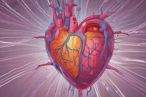
Heart Wall
- Made up of three layers: epicardium, myocardium, and endocardium.
Epicardium
- Outermost layer.
- Surrounds the heart muscle as a protective layer.
- Outer surface has mesothelial cells.
- Inner layer is fused to the myocardium.
- The outer surface of the epicardium is located near the pericardium.
- The visceral layer of the serous pericardium forms part of the epicardium
Myocardium
- Second and thickest layer of the heart wall.
- Fibers of the cardiac muscle are arranged into bundles (fascicles) bound by connective tissue.
- Layer varies in thickness, ventricles have a thicker layer than the atrium.
- The left ventricle is two to three times thicker than the right one.
Endocardium
- Innermost layer.
- Thinnest in the ventricles.
- Thickest in the atria.
- Directly connected to the cardiac appendages of the heart.
- Lined with endothelium, continuous with blood vessels of the heart.
The Cardiac Cycle
The Cardiac Cycle:
- The sequence of mechanical and electrical events that occur with every heartbeat.
Blood Flow through the Heart:
- Oxygenated blood flows from the lungs, through the pulmonary veins, into the left atrium.
- Deoxygenated blood flows from organs and tissues via the superior and inferior vena cava into the right atrium.
- Blood flows from the atria into the ventricles.
- The left ventricle pumps oxygenated blood via the aorta to organs and tissues.
- The right ventricle pumps deoxygenated blood via the pulmonary arteries back to the lungs.
Phases of each Heartbeat:
- Systole: the heart contracts and pumps blood out of the ventricles.
- Diastole: the heart relaxes, and ventricles fill with blood.
The cardiac cycle graph:
- Used to express events during one cardiac cycle.
Seven phases of the Cardiac Cycle
- Atrial Contraction.
- Isovolumetric Contraction.
- Rapid Ventricular Ejection.
- Reduced Ventricular Ejection.
- Isovolumetric Relaxation.
- Rapid Ventricular Filling.
- Reduced Ventricular filling (Diastasis).
Atrial Contraction
- SA node firing → Depolarization signal propagates through walls of atria → right/left atrium contraction → Pumps blood into ventricles
- Pressure increases in atria → a wave on a right atrial pressure curve
- ECG reads a P Wave
- Slight increase in Ventricle volume → ventricular pressure slightly increases.
Isovolumetric Contraction.
- Part of Ventricle Systole.
- Ventricle pressure exceeds atrial pressure → Atrioventricular Valves close → First Heart Sound (S1)
- Aortic and Pulmonary valves are both closed → Blood volumes in ventricles remain the same, ( isovolumetric)
- Ventricular depolarization → Ventricular contraction = QRS complex on ECG
- Ventricular pressure = increase
- Atrial pressure = slight increase (C wave)
- Ventricle Pressure becomes higher than Pressure with in the Aorta and Pulmonary Arteries → Aortic and Pulmonary Valves open
Rapid Ventricular Ejection
- Sudden ejection of a large amount of blood from the ventricles
- Pressure from the left ventricle is equally transmitted to the aorta = both ventricular and aortic pressures reach their maximum
- Volume of blood within the left ventricle decreases sharply
- ECG = ST segment; period between ventricular depolarization and ventricular repolarization
- Ventricular pressure decreases.
- Returns Atrioventricular Valves to their neutral position
- Pressure within the left and right atrium decrease = X descent on the left and right atrial pressure curves
- Atria Start to accumulate blood from the lungs and systematic Circulation.
Reduced Ventricular Ejection.
- Start of the T wave (ventricular repolarization)
- Blood is still moving out of the Ventricle but at a slow rate.
- Ventricle and aortic pressure starts to decrease.
- Atria is Collecting Blood → Atrial Pressure Starts to Increase.
- All are part of Ventricle Systole
Isovolumetric Relaxation
- Beginning of Ventricular Diastole = End of T wave on ECG
- Relaxed Ventricles → Ventricular pressure continues to fall
- Aortic pressure is higher than left ventricle pressure → Blood starts to flow backward toward the heart → the Dicrotic notch (small dip in aortic pressure graph)
- Once the left Ventricle falls enough → Aortic Valve Closes → Second Heart Sound
- The Aortic Valve Closes before the Pulmonic Valve
- All Valves are Closed → No blood is entering or leaving the Ventricles → Volume within the ventricles remains the same = Isovolumetric Ventricular Relaxation
- The Atria are filling with blood → V wave is going up.
- From here There are no ECG changes, Until next cardiac cycle starts.
- Volumes of blood within the ventricles remain the same until the Ventricular Pressure is lower than the Atrial Pressure
Rapid Ventricular Filling
- Atrial pressure exceeds the Ventricular pressure → Atrioventricular Valves open → Ventricles start to fill rapidly with blood from atria.
- Artia pressures decreases = Y descent
Reduced Ventricular Filling (Diastsis)
- The ventricles get 90% of the blood during rapid and reduced ventricular filling (passive ventricular filling)
- The Ventricles get the remaining 10% during the atrial Contraction
- 10% is received during the next cycle → Atrial Contraction.
- Aortic pressure also falls.
Pacemaker Cells
- Cardiac muscle cells that spontaneously create an action potential = automaticity.
- Influenced by sympathetic and parasympathetic nervous systems.
- Cells with the fastest rate of depolarization determine the heart rhythm.
Conducting System of the Atria
Sinoatrial Node (SA node)
- Group of pacemaker cells in the wall of the right atrium.
- Cells set the heart rate.
- The depolarization wave moves fast through pacemaker cells but slowly through atrial and ventricular myocytes.
- Pacemaker cells have short action potentials and short refractory periods.
- The SA node has a firing rate of 60-100 depolarizations per minute at rest.
Bachmann’s Bundle/Atrial Internodal Tracts
- Pacemaker bundles connecting the SA node to spots in the right and left atria.
- Sends the depolarization wave to myocytes in both atria.
Atrioventricular Node (AV Node)
- Receives the depolarization wave from the SA node and sends it to the ventricles.
- Located at the base of the right atrium, near the septum.
- Conduction velocity slows down here, allowing ventricles time to fill with blood before contracting.
- AV nodal cells have small diameters, increasing resistance to electrical flow.
- They use slower opening calcium ion channels.
Conducting System of the Ventricle
- His-purkinje system
- This Conduction system conducts the depolarization wave really quickly
- Allowing the heart to contract in a coordinated way
Bundle of His
- Located in the superior septum.
- Divides into right and left bundle branches.
Left and Right Bundle Bindles
Perkinje Fibers
- Spread throughout the ventricle.
Electrocardiogram (ECG)
- Visualizes the electricity flowing through the heart.
- A 12-lead ECG shows how the depolarization wave moves during each heartbeat.
- A standard ECG uses 10 electrodes: four limb electrodes and six precordial electrodes around the chest.
- These make 12 leads, illustrating the movement of positive charge on the outside of heart cells.
- The ECG tracing shows a positive deflection when a depolarization wave moves towards an electrode and a negative deflection when it moves away.
- Leads II, III, and aVF are "inferior" leads near the inferior wall of the heart, supplied by the right coronary artery.
- Leads I and aVL, along with chest leads V5 and V6, are "lateral" leads near the lateral wall, supplied by the left circumflex artery.
- V1 and V2 are "septal" leads nearest to the interventricular septum.
- V3 and V4 are "anterior" leads nearest to the anterior wall.
- Both septal and anterior regions are supplied by the left anterior descending artery.
Normal Sinus Rhytm
- Measures changes in time on the X-axis (one small box is 0.04 s).
- Voltage on the Y-axis (each small box is 0.1 mV).
- Zero is the "isoelectric line."
- Positive or negative deflections occur when there is a direction away from the isoelectric line.
Depolarization
- Starts in the SA node, then goes through atrial intranodal tracts (Bachmann's bundle) over to the left atrium.
- Overall direction is in the same direction as the lead II vector → positive deflection = P wave.
- Signal is carried from the SA node to the AV node where it is delayed → no depolarization → flat line.
PR Interval
- The interval from the beginning of the P wave through the flat portion.
- Signal goes through the Bundle of His and into left bundle branch and right bundle branches.
Q Wave
- Signal goes through the slow myocytes in the interventricular septum in a direction that’s slightly away from the lead II vector, creating a tiny negative deflection on the ECG
R Wave
- Depolarization wave goes into the purkinje fibers; the largest vectors are the ones in the left Ventricle: apex of the heart depolarizes first
SV Wave
- Wave of depolarization moves towards the top of the ventricles (away from lead II) → negative deflection on the ECG.
QRS Complex
- Ventricular depolarization.
Isoelectric Line
- After tissue depolarizes there is no change in electrical activity
J Point
- Exact point at which it his that isoelectric Line
ST Segment
- Time when their is no net change in electrical activity
T Wave
- Ventricles Repolarization
- Moves in an overall direction upward.
- A wave of negative charge, so it’s a positive deflection on the ECG.
- Spread out over time because repolarization is a slower process/ occurs slightly different times for each cardiomyocyte
- Atrial repolarization doesn’t usually show up at all on the ECG; the small vectors get lost in larger vectors that get created by the QRS complex.
ECG Intervals
- PR interval
- QRS complex
- QT Interval
PR Interval
- Beginning of the p-wave → Beginning of the QRS complex
- Represents the time from the beginning of atrial depolarization to the beginning of ventricular depolarization.
- Normal = 0.12-0.20 seconds
- It is caused by a deviation in the normal depolarization pathway from the SA node to the ventricles.
- Occurs in - Ectopic Atrial Focus/ First Degree Heart Block where the electrical signal travels more slowly through the AV node → PR interval lengthens.
QRS Complex
- Depolarization of the Ventricles
- Normal < 100 Milliseconds
- Abnormal = Change in pathway of the AV node to ventricles ie Ectopic Ventricular Focus
QT Interval
- Beginning of the QRS complex → End of the T-wave.
- Represents Ventricular Systole, Depolarization → Repolarization.
- QTc is a correction of the interval, Accounting for the Heart Rate.
What can affect The QT interval?
- Hypercalcemia shortens the QT while hypocalcemia lengthens it.
- Medication( amiodarone prolongs it)
- Genetic Mutations ie Interited Long-QT syndromes ( i.e LQT1, LQT2, LQT3)
- Complication of Long QT → Ventricular Tachycardia “torsade de pointes” → can lead to sudden cardiac death.
RR Interval
- From One R wave peak to the Next
- Calculate the Heart Rate
- Determines Regularity
Head and Neck Blood Supply
- The head and neck derive blood from the common carotid and subclavian arteries
- Left Common Carotid: Originates directly from the aorta.
- External Carotid Artery: Supplies external structures of the head and face; eight branches.
- Superior Thyroid Artery: Supplies the superior portion of the thyroid gland.
- Ascending Pharyngeal Artery: Supplies the pharynx and soft palate (smallest branch).
- Lingual Artery: Supplies tongue muscles and the floor of the mouth.
- Facial Artery: Supplies muscles and skin of the face.
- Occipital Artery: Supplies the posterior scalp region.
- Posterior Auricular Artery: Supplies the parotid gland, ear, scalp, and muscles around the ear.
- Maxillary Artery: The larger terminal branch supplies the mandible, teeth, and muscles.
- Superficial Temporal Artery: Supplies the temporal region of the scalp
Internal Cartoid Artery
- Supplies the brain; (anterior circulation); branches within the cranial cavity
- Inferior Hypophyseal Artery: Supplies the posterior pituitary gland.
- Superior Hypophyseal Artery: Supplies the hypothalamus and the anterior pituitary gland.
- Ophthalmic Artery: Supplies structures in the orbit.
- Posterior Communicating Artery: Joins the posterior cerebral artery; part of the Circle of Willis.
- Anterior Choroidal Artery: Supplies parts of the midbrain and visual pathway.
- Anterior Cerebral Artery: Supplies the medial cerebral hemisphere, and the Circle of Willis
- Middle Cerebral Artesy: Largest terminal branch; supplies the majority of the lateral surface of the cerebral hemispheres
Right Common Carotid
- Arising from the brachiocephalic trunk
- Internal and External Artery Branches are identical to the left
- Left Subclavian Artery
- Originates directly from the arch of the aorta; branches superiorly and into limbs
- Left Vertebral Artery: Courses through transverse foramina of the cervical vertebrae
- Merges w. right to form the basilar artery for the superior spinal cord, brainstem, cerebellum, and posterior brain
- Basilar Artery: Ascends along the ventral surface of the pons
- Gives rise to the posterior circulation of the brain; divides into cerebral arteries
- Thyrocervical Trunk: Supplies thyroid/parathyroid glands, larynx, and pharynx.
- Costocervical trunk: Arches posteriorly towards the neck of the first rib
- Divides into two terminal branches; supplies posterior neck/thorax muscles
Right Subclavian Artery
- Originates from the brachiocephalic trunk
- Branches and Target areas are identical to the left subclavian artery
Cerebrovascular Disease
Cerebrovascular accident or stroke Stroke occurs when the blood supply to part of the brain is interrupted preventing brain tissue from getting oxygen and nutrients. Brain cells begin to die in minutes. There are two main types – a blocked artery which causes an ischemic stroke or a leaking or burst blood vessel which leads to a hemorrhagic stroke Ischemic strokes are the most common type. Ischemic strokes can be further divided into thrombotic or embolic. Thrombotic strokes are usually caused by atherosclerotic plaques which are fatty deposits that gradually build up in the artery’s wall. If they get big enough to block the blood vessels, they can cause a stroke. Embolic strokes are caused by clots that travel through the bloodstream from another source and lodge in the arteries of the brain. Emboli could break off from a plaque in the common carotid artery and travel through the pathways we looked at earlier to ultimately block one of the smaller arteries in the brain. The brain tissue that is downstream from the location of the lodged clot will be deprived of oxygen and can die within minutes. The symptoms of a stroke depend on which area of the brain has been affected; however, the common symptoms of a stroke can be remembered with the word FAST. This acronym stands for Facial drooping, Arm weakness, Speech difficulties, and Time to call emergency services. A stroke is a medical emergency and prompt treatment is crucial. Early action can reduce brain damage and other complications.
Cerebral Circulation
- Human brain receives about 15% of the total blood pumped by the heart
- Cerebral circulation formed by the following:
- 2 Internal Carotid Arteries
- 2 Vertebral Arteries Anatomoses between these two arteries and their branches give rise to the Circle of Willis Internal carotid arteries Form the anterior part of the cerebral vascular system Ascend on both sides of the neck and enter the cranial vault via the carotid canal Inside the cranial cavity runs through the cavernous sinus(dural venous sinus) Emerges from the cavernous sinus and divides into the following:
- Anterior cerebral artery
- Middle cerebral artery
- Other smaller branches
- 4 parts of the Internal Carotid Artery
- Cervical part: extends from common carotid to the carotid canal
- Petrous part: extends from the carotid canal to the foramen lacerum
- Cavernous part: part of the ICA within the cavernous sinus
- Cerebral part: after it exits from the cavernous sinus
- Ends by bifurcating into two major terminal branches: the anterior cerebral artery and middle cerebral artery
- Branches of the ICA
- Ophthalmic artery
- Enters the orbit
- Gives off the central retinal artery
- Ophthalmic artery
- main blood supply to the retina
- Posterior communicating artery
- Connects the middle cerebral and posterior cerebral artery in the Circle of Willis
- Anterior choroidal artery
- Enters the lateral ventricle of the brain
- Ends in the choroid plexus
- Posterior communicating artery
Anterior Cerebral Arterty
Supply the anteromedial surface of the cerebral cortex Arches over the corpus callosum Supplies the following:
- Medial frontal lobe
- Parietal lobe (through numerous small cortical branches)
- Frontal lobe – supero-lateral portion
- The two anterior cerebral arteries on each side are connected through the anterior communicating artery creates the anterior Circle of Willis
Middle Cerebral Artery
Supplies the majority of the lateral cerebral cortex Travels along the lateral sulcus Branches:
- Central Arteries
- Supply the deeper structures of the brain
- Often referred to as lenticulostriate branches of the middle cerebral artery
- Supplies the following:
- Lateral surface of the temporal lobe
- Part of the infero-lateral surface of the frontal and parietal lobes
- Anterior and middle cerebral arteries are terminal arteries: there is no collateral circulation in areas supplied by them -any blockage in these arteries prevent blood from reaching that part of the brain completely
Vertebral Arteries
Arise from the subclavian arteries Form the “vertebro-basilar system” Ascend through the transverse foramina of the cervical vertebrae to enter the skull through the foramen magnum Pierce the dura mater and enter the subarachnoid space Merge with each other to form the basilar artery where the pons and medulla meet Branches
Anterior Spinal Artery
- Supply rostral anterior two-thirds of the spinal cord
Posterior Inferior Cerebella
- Supplies inferior cerebellum
- Medial branch Moves posterior
- Lateral branch
- Supplies inferior cerebellum
Posterior Artery
- Supplies posterior to the spinal cord
Basilar Artery( Branches)
- Ascends along anterior pons Branches: Supplies the following inferior cerebellum, pons and middle cerebellar peduncle Labyrinthine arteries - Supplies the inner ear Pontine arteries - Supply the pons Superior cerebellar arteries - Supplies the following Superior cerebellum Pineal gland a part of the pons, art of the third ventricle Divides into a pair of terminal branches: Posterior cerebral arteries Form the left and right posterior communicating arteries which merge with the internal carotid arteriesclose the posterior circle of willis The Posterior cerebral arteries supply the posterior and inferior surfaces of the cerebral cortex
Circle of Willis
- Anterior communicating artery connects the two anterior cerebral arteries
- Posterior communicating arteries connect the posterior cerebral arteries with the internal carotid arteries
- Located in the subarachnoid space at the base of the brain, right in front of the midbrain
- Creates a collateral arterial circulation blockage of one of the arteries can be compensated by other arteries to Maintain cerebral perfusion
Dural Venous Sinuses(Veinous Drainage)
- Starts as a series of small superficial veins drain into dural venous sinuses, which are venous channels located between the two layers of dura maer. Superior sagittal sinus
- Runs along the longitudinal fissure Drains to the confluence of sinuses, along with the straight sinus and occipital sinuses empties into the transverse sinuses Transverse sinuses drain to the sigmoid sinuses Sigmoid sinuses drain to the internal jugular veins
Layers of Blood Vessels
- 3 layers: Tunica Intima, Tunica Media, Tunica Externa
Tunica Intima
- Closest to the lumen and blood
- Three layers of its own: endothelium, subendothelium, and internal elastic membrane
Endothelium
- Direct contact with the lumen
- Endothelial cells Absorption of nutrients and oxygen from the blood Disposal of waste into the blood
Subendothelium
- Lies between the endothelium and the internal elastic membrane
- Connective tissue - Supports the endothelium
Internal Elastic Membrane
- Single layer of elastic connective tissue
- Supports the endothelium and subendothelium
- Elasticity allows for the blood vessels to adjust to the pressure of fluctuations in the blood and the recoil of elastic fibers
- Aid in the propulsion of blood through our vasculature
Comparison Between Vein and Artery
- Is comparable to that of an artery, having the same layers that are relatively the same thickness
- Veins have valves are folds of the tunica intima that protrude into the vessel’s lumen
Tunica Media
- Consists of smooth muscle, elastic fibers and connective tissue to varying degrees depending on which artery
- Smooth muscle tissue within blood vessels is under involuntary control by the sympathetic and parasympathetic nervous systems
- Elastic lamellae
Difference between arteries and veins
- Veins: more elastic fibers than smooth muscle
- Arteries: more smooth muscle than veins
- External elastic membrane is what Separates the tunica media from the external elastic membrane.
- Tunica Externa Outermost Layer
Tunica Externa
- Primarily composed of connective tissue Supports the vessel
- Anchors vessels to surrounding structures
Types of Arterioles
Elastic Arteries
- Loads of collagen and elastic fibers within the tunica media
- Allows for the vessels to expand with blood and propel the blood through the circulation by recoil of the elastic fibers
- Examples: aorta, pulmonary arteries which Propels large volumes of blood
Muscular Arteries
- Lots of smooth muscle in the tunica media; controls diameter of blood vessels
- Receive blood from the elastic arteries
- Can either constrict or dilate, Control blood flow to certain areas of the body
- Feeds blood into arterioles which are Smallers
Arterioles
- Much smaller than muscular and elastic arteries
- Are the final vessels which blood travels through before it enters capillaries
- Smaller tunica media with 2 smooth muscle layers
- Play a huge role in controlling vascular resistance
Capillaries
- Smallest blood vessels, erythrocytes travel in a single file due to the small diameter of the vessels
- Allows for nutrients and oxygen to leave red blood cells and enter tissue
- Allows red blood cells and blood to pick up carbon dioxide and waste materials
- Continuous Capillaries - Contain no breaks or holes within their walls
-
Tight Junctions Bind Edothelial Cells
- Fenestrated Capillaries- Allow for greater exchange of waste and nutrients between the blood and surrounding tissue- found in Kidneys,small intestine, and endocrine glands-
Types of Capillaries
Continuous Capillaries
- Contain no breaks or holes within their walls
- Endothelial cells of continuous capillaries are tightly bound by tight junctions
Fenestrated Capillaries
- Allow for greater exchange of waste and nutrients between the blood and surrounding tissue
- i.e. Kidneys, endocrine glands, small intestine
Discontinuous Capillaries
- Release blood into a tissue; exclusively found in the liver
Types of Veins
- Venules
- Medium Veins
- Large Veins Vasa Vasorum
Venules
- the smallest type of vein- receive blood from the capillaries this is where the thin is thin Walled and irregular shaped
Medium Veins
- thin walled and irregular in shape- Composed of other small and medium viens coming together
Large Viens
- thick muscled and Contains longitudinally arranged smooth muscle fibers
Vasa Vasorum
- Small blood vessels is within the walls of larger blood vessel- Provides a Blood Supply
Atherosclerosis
Once lipids and cholesterol within the blood manage to get through and under the tunica intima of a blood vessel Once under the tunica intima the fats can oxidize and attract a release Of products of solid plaque build Up in vessels leading lumen to narrow the blood vessel Wall if the lumen of the blood vessel becomes too narrow blood supply can be limited to what structure is being supplied by The occluded vessel
Veins of the Head and Neck
- Internal Jugular Vein
- Subclavian Vein
- Brachiocephalic Vein
Internal Jugular Vein
- Paired vessel extending from the base of the skull to the sternal end of the clavicle
- Travels within the fibrous carotid sheath lateral to the carotid artery The veins primary role is Drain The Brain
- Formed by the union of the sigmoid and inferior petrosal sinus extends down towards the neck - Drain venous blood from the majority of the skull, brain, oral cavity, and superficial structures of the head and neck
Tributaries
- Sigmoid Sinus: Collects from regions of the brain; drains to the internal jugular vein.
- Inferior Petrosal Sinus: Drains the middle cranial fossa, brain, and inner ear; drains through the jugular foramen into the internal jugular vein.
- Meningeal Veins: Drain the dura mater; travel with meningeal arteries.
- Pharyngeal Venous Plexus: Drains pharynx regions; a network of veins surrounding the pharynx.
- Common Facial Vein: Formed by the union of the facial vein, drains superficial structures of the face.
- Superior and Middle Thyroid Veins: Drains the thyroid gland regions.
Subclavian Vein
- Main veins of the Neck, tucked beneath the Clavicle
- Continues the axillary vein and forms and drains into the brachiocephalic veins
Tributaries
- Anterior Jugular Vein drains structures of the anterior compartment of the neck - formation of the submandibular viens.
- External Jugular vein - drains structures of the head including the sculp and face - formation of the auricular vein joining the posterior brach.
Brachiocephalic Vein
Aka Inominate Viens Forms the confluence of the internal jugular vein and subclavian Veins - just posterior to thesternoclacicular joint additional tributes vertebral Viens from a vernous pleaus supplies the soboccibital muscles . travels through transvers foramina. Internal Thoracic Vein
- Drains the uooer thorax, chest wall and breats
- Formed as continaitons by the musculophornric and upper epigastric viens.
Cardiac Afterload
Ventricular wall stress during systole, the amount of resistance that the ventricles must overcome during systole Factors that affect cardiac afterload:
- Systemic vascular resistance
- Aortic pressure
- Valve diseases
Systemic Vascular Resistance
- Resistance is the systemic blood vessels
Studying That Suits You
Use AI to generate personalized quizzes and flashcards to suit your learning preferences.
Description
Explore the coordinated function of cardiomyocytes through gap junctions and T-tubules. Understand the impact of drugs on muscle contraction and the roles of ion transporters during muscle relaxation. Understand the cardiac cycle, coronary arteries and ventricular filling.